
3.10: Malaria
- Page ID
- 38788

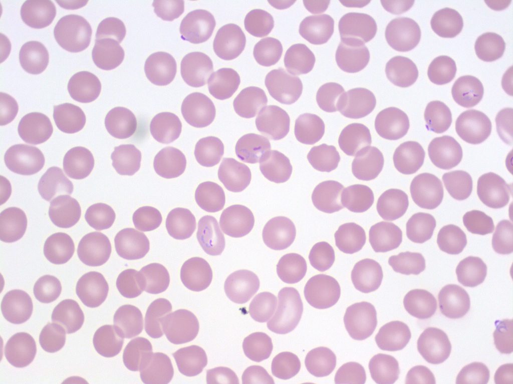
- An image from a peripheral blood smear showing multiple malarial rings (Plasmodium falciparum) inside red blood cells. From MLS Collection, University of Alberta, https://doi.org/10.7939/R3891263S
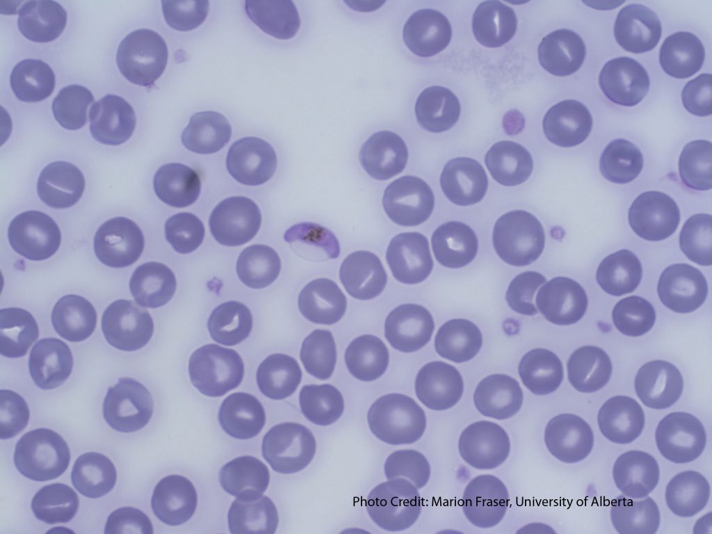
- An image of a peripheral blood smear showing a crescent-shaped gametocyte (characteristic of Plasmodium falciparum). From MLS Collection, University of Alberta, https://doi.org/10.7939/R30R9MK4V
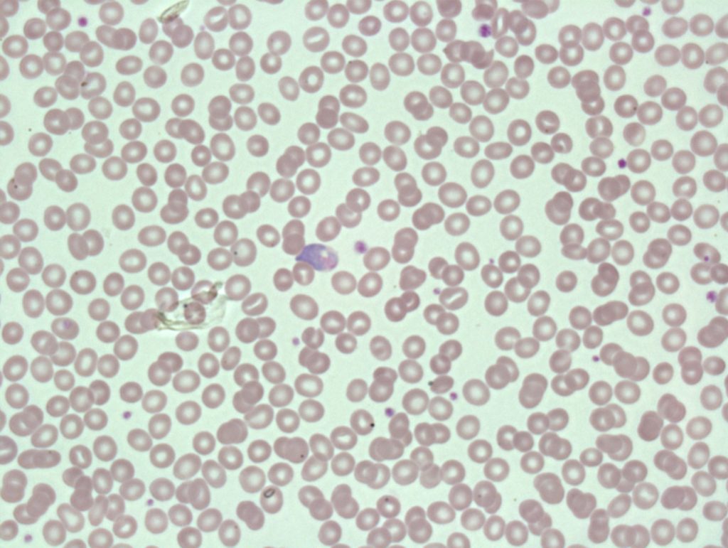
- An image of a peripheral blood smear showing malaria at the gametocyte stage in the center. 60x oil immersion. From MLS Collection, University of Alberta, https://doi.org/10.7939/R37941903
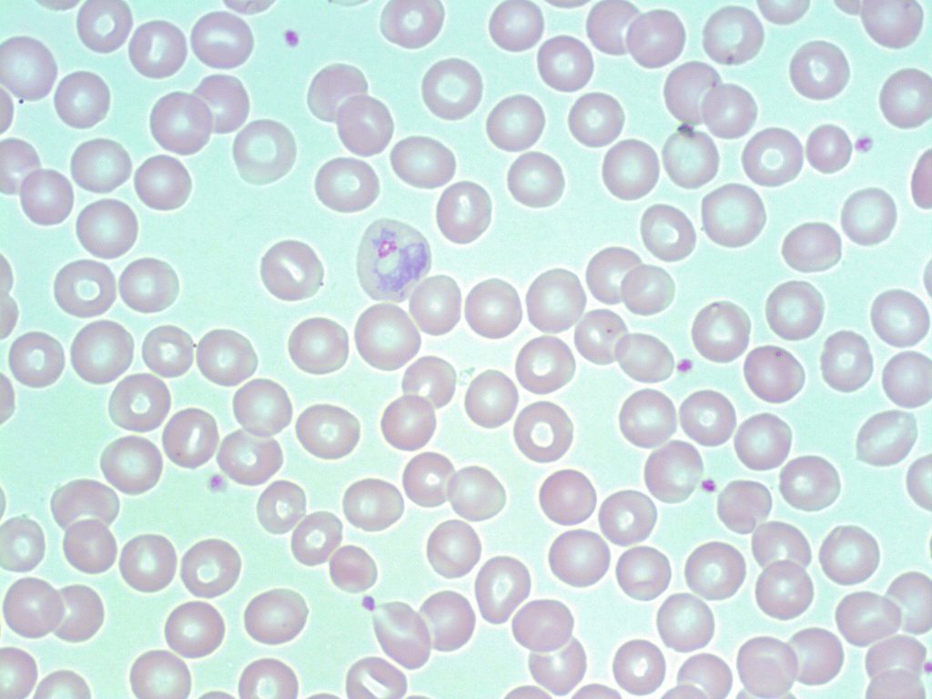
- An image of a peripheral blood smear showing a malarial parasite at the trophozoite stage in a red blood cell. 100x oil immersion. From MLS Collection, University of Alberta, https://doi.org/10.7939/R3WS8J22C
Appearance:
The morphology of malarial parasites seen in the red blood cell varies depending on the stage of maturation and species present. Malaria can appear as rings, trophozoites, schizonts, and gametocytes. Ring forms appear as a pale blue ring with a pink/purple chromatin dot, and more than one may be present in a single red blood cell. Malarial parasites are most often seen intracellular to the red blood cell with various forms.1
Parasites can be visualized using the Giemsa stain during the screening of thin and thick smears.1
Note 1: Banana shaped gametocytes seen are characteristically in Plasmodium falciparum infections.
Note 2: Malarial rings may be confused with platelets when the appear on top of a red blood cell. Platelets may be differentiated by a showing a slight clearing or halo around the platelet.2
Organisms:1
The malarial parasite is spread to humans by the female Anopheles sp. mosquito.
Malaria parasites:1,2
Plasmodium falciparum
Plasmodium vivax
Plasmodium ovale
Plasmodium malariae
Plasmodium knowlesi
References:
1. Keohane EM. Extrinsic defects leading to increased erythrocyte destruction – nonimmune causes. In: Rodak’s hematology clinical applications and principles. St. Louis, Missouri: Saunders; 2015. p. 394-410.
2. Rodak BF, Carr JH. Microorganisms. In: Clinical hematology atlas. 5th ed. St. Louis, Missouri: Elsevier Inc.; 2017. p. 195-202.