
4.3: Sideroblastic Anemia
- Page ID
- 38795

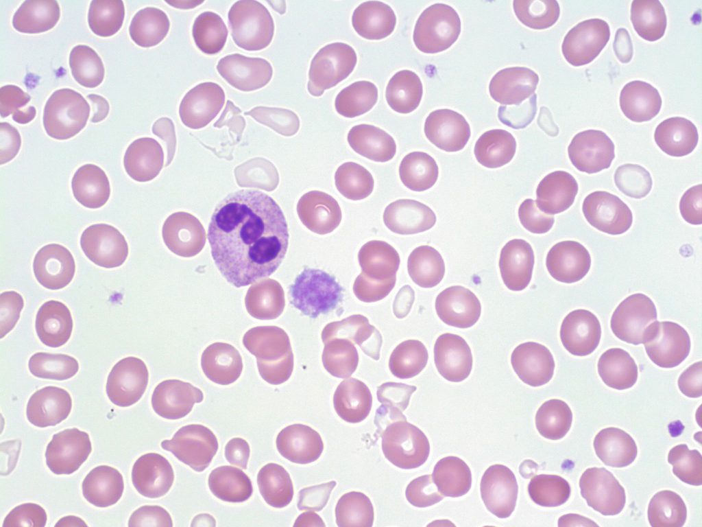
- A peripheral blood smear picture showing a dimorphic population of red blood cells: hypochromic, microcytic and normochromic, normocytic red cells. Dimorphissm is commonly seen in Sideroblastic Anemia cases. 50x oil immersion. From MLS Collection, University of Alberta, https://doi.org/10.7939/R3P844B3X
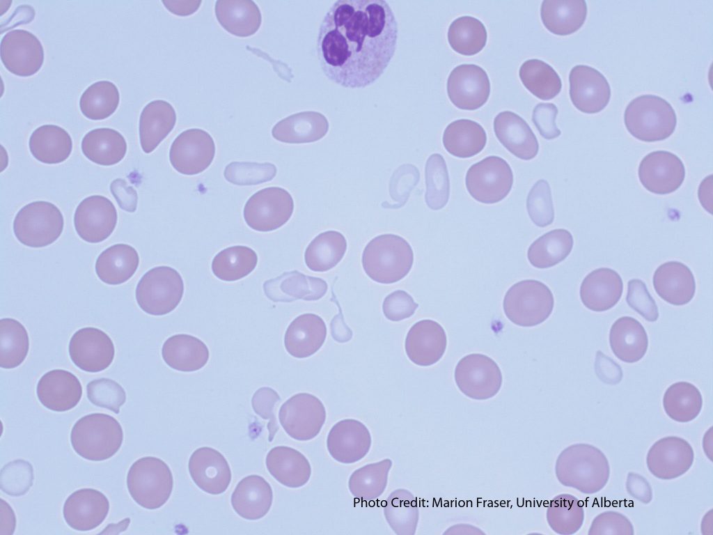
- A peripheral blood smear picture showing a dimorphic RBC population and tear cells in sideroblastic anemia. From MLS Collection, University of Alberta, https://doi.org/10.7939/R3CF9JN85
Cause(s): Development of sideroblastic anemia can be due to hereditary or acquired causes that lead to abnormal heme synthesis.1,2
Hereditary: Sex-linked or autosomal recessive mutations
Acquired: Idiopathic, MDS and other malignancies, drugs, lead toxicity
Laboratory Features of Sideroblastic Anemia:1-3
|
CBC: RBC: Decreased WBC: Variable PLT: Variable Hb: Decreased MCV, MCH, MCHC: Normal to Decreased (as they are averages of the RBC appearance) RDW: Increased RETIC: Decreased |
PBS: Dimorphic population (Normochromic/Normocytic alongside Hypochromic/Microcytic ) Tears Schistocytes Pappenheimer bodies Basophilic stippling |
BM: M:E Ratio: Decreased Erythroid hyperplasia (ineffective erythropoiesis) Ringed sideroblasts Macrophages have increased iron (Increased iron stores) |
|
Iron Studies: Serum Iron: Increased Ferritin: Increased Transferrin: Normal to Decreased Transferrin Saturation: Increased TIBC: Normal to Decreased |
Other Tests: Bilirubin: Increased Haptoglobin: Decreased LD: Increased Prussian blue stain of BM shows increased iron levels and ringed sideroblasts |
References:
1. McKenzie SB. Anemias of disordered iron metabolism and heme synthesis. In: Clinical laboratory hematology. 3rd ed. New Jersey: Pearson; 2015. p. 198-230.
2. Doig K. Disorders of iron kinetics and heme metabolism. In: Rodak’s hematology clinical applications and principles. 5th ed. St. Louis, Missouri: Saunders; 2015. p. 297-313.
3. Finnegan K. Iron metabolism and hypochromic anemias. In: Clinical hematology and fundamentals of hemostasis. 5th ed. Philadelphia: F.A. Davis Company; 2009. p. 117-37.