
6.5: Low Back Pain
- Page ID
- 14815

ACR – Neurologic – Low Back Pain
Case
Low back pain. No red flags
Clinical:
History – This 49 year old male had a 4 week history of low back pain.
Symptoms – Low back pain, limited range of motion, muscle spasm.
Physical – No neurological symptoms or signs. No red flags.
DDx:
Back spasms
Intervertebral disc herniation
Fibromyalgia
Muscle injury
Imaging Recommendation
ACR – Neurologic – Low Back Pain, Acute, Subactue, or Chronic, Variant 1
No Imaging Recommended
Lumbar spine x-rays were ordered, regardless.
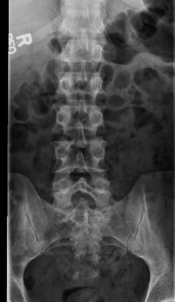
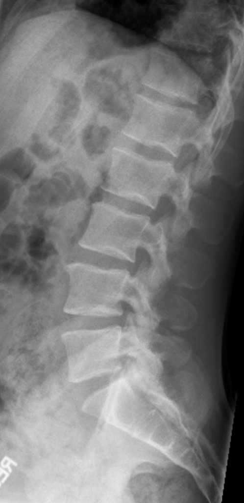
ODIN Link for Lumbar Spine x-ray images, Figure 6.7A and B: mistr.usask.ca/odin/?caseID=20150311180052507
Imaging Assessment
Findings:
A minor lumbar scoliosis, convex to the right, was seen. Minor facet degenerative changes were present. Nil else.
Interpretation:
No significant abnormalities, age appropriate degenerative changes
Diagnosis:
Low back pain, no red flags
Discussion:
Low back pain (LBP), with or without radiculopathy, is one of the leading causes of years lived with disability and the third ranking cause of disability-adjusted life years. It is the second most common reason for a physician visit and affects 80% – 85% of people over their lifetime.
The American College of Physicians and the American Pain Society classify LBP into the following broad categories: nonspecific LBP, back pain potentially associated with radiculopathy or spinal stenosis, and back pain potentially associated with another specific spinal cause.
Additionally, guidelines from the American College of Physicians and the American Pain Society emphasize a focused history and physical examination, reassurance, initial pain management medications if necessary, and consideration of physical therapies without routine imaging in patients with nonspecific LBP. Duration of symptoms also helps guide treatment algorithms in patients with acute, subacute, or chronic LBP.
Additionally, assessment of psychosocial risk factors when obtaining patient history is a strong predictor of patients who are predisposed to developing chronic disabling LBP problems.
Although there is great variability in the definition of acute and subacute LBP, for the purposes of this discussion, we will use the Institute for Clinical Systems Improvement definitions of 0–6 weeks to definite acute LBP, 6–12 weeks for subacute LBP, and >12 weeks to define chronic LBP.
Uncomplicated acute LBP and/or radiculopathy is a benign, self-limited, condition that does not warrant any imaging studies. Imaging is considered in those patients who have had up to 6 weeks of medical management and physical therapy that resulted in little or no improvement in their back pain. It is also considered for those patients presenting with red flags raising suspicion for a serious underlying condition, such as cauda equina syndrome (CES), malignancy, fracture, or infection.
Red Flags:
| Possible Underlying Condition | Red Flag |
| Malignancy or Infection | Known, previous malignancy
Unexplained weight loss Intravenous drug use Chemotherapy Prolonged corticosteroid use |
| Fracture | Fall or lifting in an elderly patient
History of significant trauma Prolong corticosteroid use |
| Major Neurologic Compromise (Cauda equina, paralysis,) | Loss of anal sphincter tone
Fecal or urinary incontinence Urinary retention Saddle anesthesia Progressive weakness in the lower limbs |
Attributions
Figure 6.7A AP x-ray of lower spine. by Dr. Brent Burbridge MD, FRCPC, University Medical Imaging Consultants, College of Medicine, University of Saskatchewan is used under a CC–BY-NC-SA 4.0 license.
Figure 6.7B Lateral x-ray of lower spine. by Dr. Brent Burbridge MD, FRCPC, University Medical Imaging Consultants, College of Medicine, University of Saskatchewan is used under a CC-BY-NC-SA 4.0 license.