
14.2: Acromioclavicular Joint Separation
- Page ID
- 14881

Acromioclavicular (AC) Joint Separation/Dislocation
Case
Acromioclavicular joint dislocation
Clinical:
History – 21 year old female injured her shoulder while wrestling.
Symptoms – This patient complained of a deformed, painful, end of her right collar bone.
Physical – There was swelling and tenderness of the region of the acromioclavicular joint.
DDx:
Acromioclavicular Joint Separation
Clavicle Fracture
Acromion Fracture
Hematoma
Imaging Recommendation
ACR – MSK – Acute Shoulder Pain, Variant 1
Shoulder X-ray
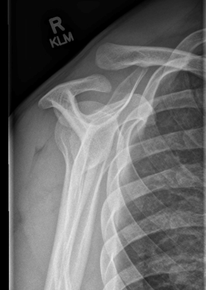
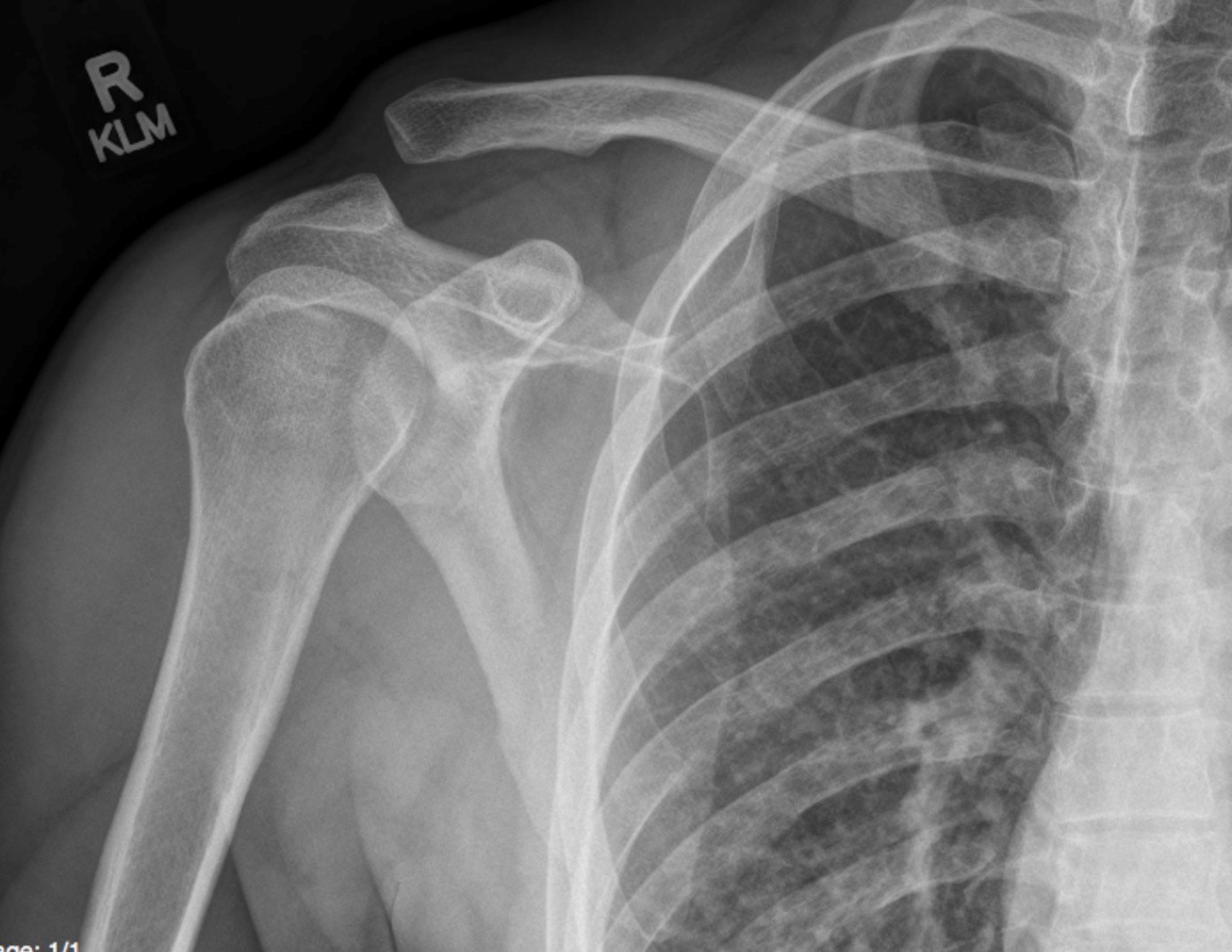
ODIN Link to AC Joint Separation images, Figure 14.2A and B: mistr.usask.ca/odin/?caseID=20150209202015857
Imaging Assessment
Findings:
The lateral clavicle was displaced cranially and the acromioclavicular joint was widened. The coracoclavicular distance was also widened.
Interpretation:
Acromioclavicular joint dislocation, Type 3.
Diagnosis:
Acromioclavicular joint dislocation
Discussion:
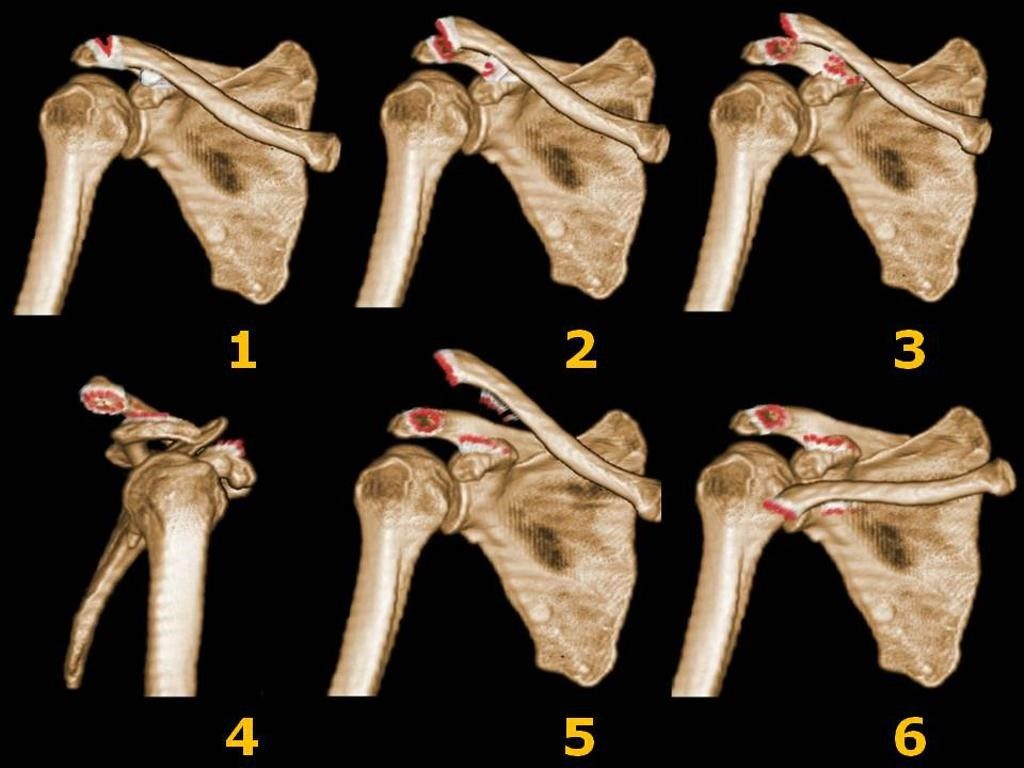
Acromioclavicular joint injuries can be graded on the 6-point Rockwood scale:
| Type | AC Joint | CC Joint | Reducibility | Treatment |
|---|---|---|---|---|
| I | Sprain | Normal | NA | Conservative |
| II | Torn | Sprain – CC distance <25% of the contralateral side | Reducible | Conservative |
| III | Torn | Torn – CC distance increased 25 – 100 % of the contralateral side | Reducible or Non-Reducible | Conservative or Surgical |
| IV | Torn | Torn – Posterior displacement of clavicle into the trapezius muscle | Not Reducible | Surgery |
| V | Torn | Torn – CC distance > 100% of the contralateral side with the clavicle protruding through the delto-trapezial fascia | Not Reducible | Surgery |
| VI | Torn | Torn – Clavicle caudal to the subacromial or subcoracoid | Not Reducible | Surgery |
X-ray findings may include:
- Minor injuries of this joint space usually involve only the joint capsule and the acromioclavicular ligament.
- With more severe injuries the coracoclavicular ligament may be torn leading to a more displaced clavicle and a wider coracoclavicular distance.
- Severe injuries can involve the coracoclavicular ligament, the deltoid muscle and the trapezius muscle.
Attributions
Figure 14.2A X-ray of the right shoulder with AC joint separation by Dr. Brent Burbridge MD, FRCPC, University Medical Imaging Consultants, College of Medicine, University of Saskatchewan is used under a CC-BY-NC-SA 4.0 license.
Figure 14.2B X-ray of the right shoulder with AC joint separation by Dr. Brent Burbridge MD, FRCPC, University Medical Imaging Consultants, College of Medicine, University of Saskatchewan is used under a CC-BY-NC-SA 4.0 license.
Figure 14.3 Acromioclavicular injury classification. Courtesy of Dr. Roberto Schubert, Radiopaedia.org, RID: 19124. Originally published at https://radiopaedia.org/cases/rockwood-classification-system-of-acromioclavicular-joint-injuries under a Creative Commons Attribution-Non-commercial-Share Alike 3.0 License.