
14.6: Hand and Wrist Fractures
- Page ID
- 14885

ACR – MSK – Acute Hand and Wrist Trauma
Case 1
Scaphoid Fracture
Clinical:
History – This patient fell and jammed her wrist while dancing.
Symptoms – Aching pain in the wrist at the base of the thumb.
Physical – There was point tenderness in the anatomic snuff box. No deformity seen.
DDx:
Thumb fracture
Scaphoid fracture
Wrist, soft tissue injury
Imaging Recommendation
ACR – MSK – Hand and Wrist Trauma, Variant 1
X-rays with a scaphoid view
ODIN Link for Scaphoid Bone Fracture images (X-rays and CT), Figure 14.9A and B: mistr.usask.ca/odin/?caseID=20150311232713868

Imaging Assessment
Findings:
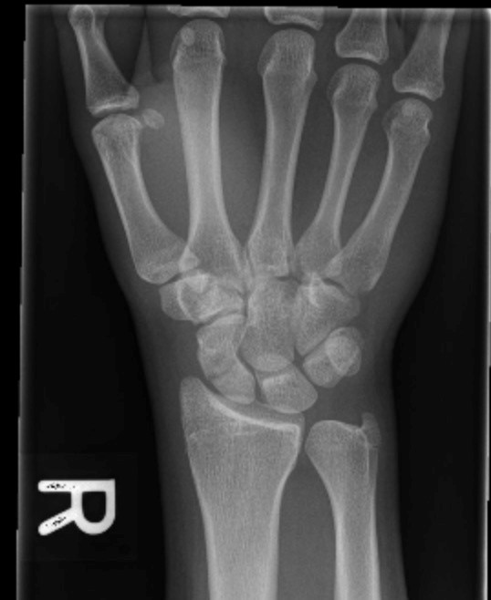
X-rays – The x-rays demonstrated a transverse scaphoid fracture. The patient was casted. Follow-up x-rays 4 weeks later were suspicious for non-union. CT was requested.
CT – The CT images revealed two complications of the scaphoid fracture: a) proximal scaphoid sclerosis secondary to avascular necrosis; b) widening of the scapho-lunate distance consistent with ligamentous injury.
Interpretation:
Scaphoid fracture with non-union, avascular necrosis, and ligamentous injury.
Diagnosis:
Scaphoid fracture with complications
Discussion:
The primary mechanism of injury is a fall on the outstretched hand with an extended, radially deviated wrist, which results in extreme dorsiflexion at the wrist and compression to the radial side of the hand. Forces are transmitted from the hand proximally to the arm through the scaphoid.
An additional fourth radiographic projection (scaphoid view)—an elongated PA view with approximately 30° of cephalad beam angulation and the wrist positioned in 10°–15° of ulnar deviation—is routinely recommended whenever there is clinical suspicion of a scaphoid fracture.
X-ray findings may include:
- A lucent fracture line. This is usually in the waist of the bone.
- Widened joint spaces consistent with inter-carpal ligament tears.
- A step deformity in the cortex of the scaphoid bone due to a fracture.
Scaphoid fractures are notoriously difficult to see on initial radiographs (regardless of the views) and are radiographically occult in up to 20% of cases. Standard practice in patients with clinically suspected scaphoid fractures, but normal initial radiographs, is to apply a cast and to repeat the clinical evaluation and radiographs in 10–14 days when resorption at the fracture line may make previously occult fractures visible. If the repeat radiographs are still normal or equivocal at that time and there continues to be a strong clinical suspicion of scaphoid fracture, imaging with a second modality – bone scintigraphy, CT, or MRI – may be indicated.
There is less evidence favouring either scintigraphy vs. CT in this scenario, although a recent meta-analysis found that MRI was superior to scintigraphy although many hospitals still perform CT or scintigraphy, and the choice of modality often depends on local preferences, expertise, and imaging modalities.
Case 2
Fifth Metacarpal Fracture
ACR – MSK – Acute Hand and Wrist Trauma
Clinical:
History – This patient accidentally punched a cement wall during a bar fight.
Symptoms – Painful hand.
Physical – The dorsum of the hand was mildly swollen. There was point tenderness overlying the distal fifth metacarpal.
DDx:
Fifth Metacarpal fracture
Other fractures
Fifth Metacarpal dislocation
Soft Tissue injury
Imaging Recommendation
ACR – MSK – Hand and Wrist Trauma, Variant 1
X-rays
ODIN Link for Fifth Metacarpal Fracture images, figure 14.10A and B: mistr.usask.ca/odin/?caseID=20161129152213974

Imaging Assessment
Findings:
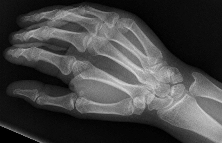
The neck of the fifth metacarpal was fractured. There was significant angulation of the fracture, 80 degrees. No intra-articular abnormality.
Interpretation:
Fifth metacarpal neck fracture. Orthopedic Surgery consultation was warranted due to the angulation of the fracture.
Diagnosis:
Fifth Metacarpal Neck Fracture
Discussion:
Fifth metacarpal neck fractures occur frequently in the general population and account for around 20% of all fractures of the hand.
X-ray findings may include:
- A fracture of the neck of the bone.
- A measurement of the fracture angle should be performed.
- There may be comminution and extension of the fracture into the joint space.
- Overlying soft tissue swelling is common in acute injuries.
Clinical evaluations on patients with these fractures take into consideration shortening, rotation and angular deviation during flexion.
The closed reduction technique most used is the Jahss maneuver, which improves the degree of angulation of the distal fragment of the fracture. The decision to implement surgical treatment depends on clinical, radiographic parameters, and also on the patient’s age, profession, activity level and handedness.
Attributions
Figure 14.9A X-ray of the wrist displaying a scaphoid bone fracture by Dr. Brent Burbridge MD, FRCPC, University Medical Imaging Consultants, College of Medicine, University of Saskatchewan is used under a CC-BY-NC-SA 4.0 license.
Figure 14.9B CT scan of the wrist displaying a scaphoid bone fracture by Dr. Brent Burbridge MD, FRCPC, University Medical Imaging Consultants, College of Medicine, University of Saskatchewan is used under a CC-BY-NC-SA 4.0 license.
Figure 14.10A X-ray of the hand and wrist, displaying a neck of fifth metacarpal fracture by Dr. Brent Burbridge MD, FRCPC, University Medical Imaging Consultants, College of Medicine, University of Saskatchewan is used under a CC-BY-NC-SA 4.0 license.
Figure 14.10B X-ray of the hand and wrist displaying a neck of fifth metacarpal fracture with the fracture angle measured by Dr. Brent Burbridge MD, FRCPC, University Medical Imaging Consultants, College of Medicine, University of Saskatchewan is used under a CC-BY-NC-SA 4.0 license.