
2.3: Agglutination
- Page ID
- 38766

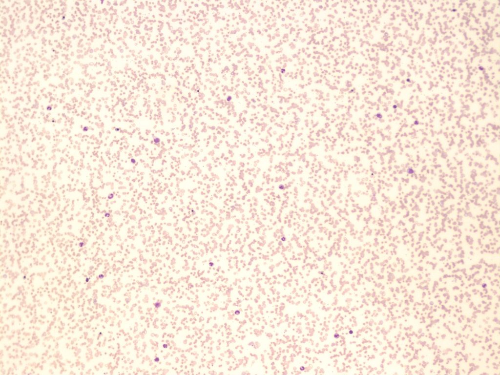
- A peripheral blood smear demonstrating agglutination of red blood cells. 10x magnification. From MLS Collection, University of Alberta, https://doi.org/10.7939/R3C824V90
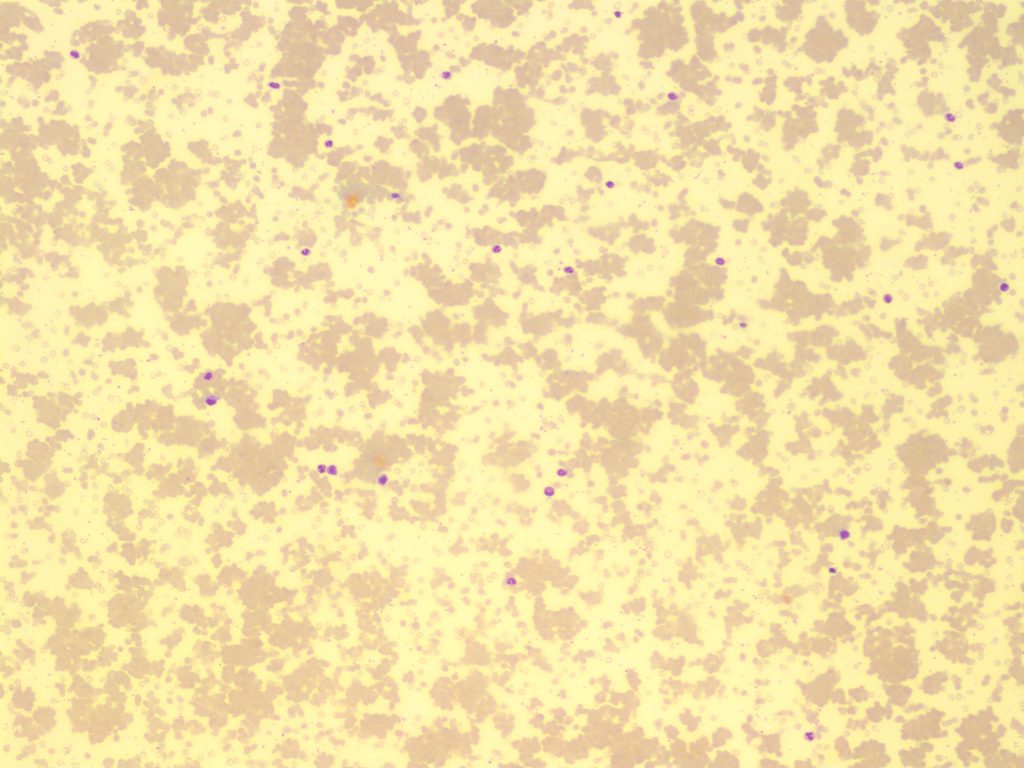
- A peripheral blood smear demonstrating severe agglutination of red blood cells. 10x magnification. From MLS Collection, University of Alberta, https://doi.org/10.7939/R34J0BC72
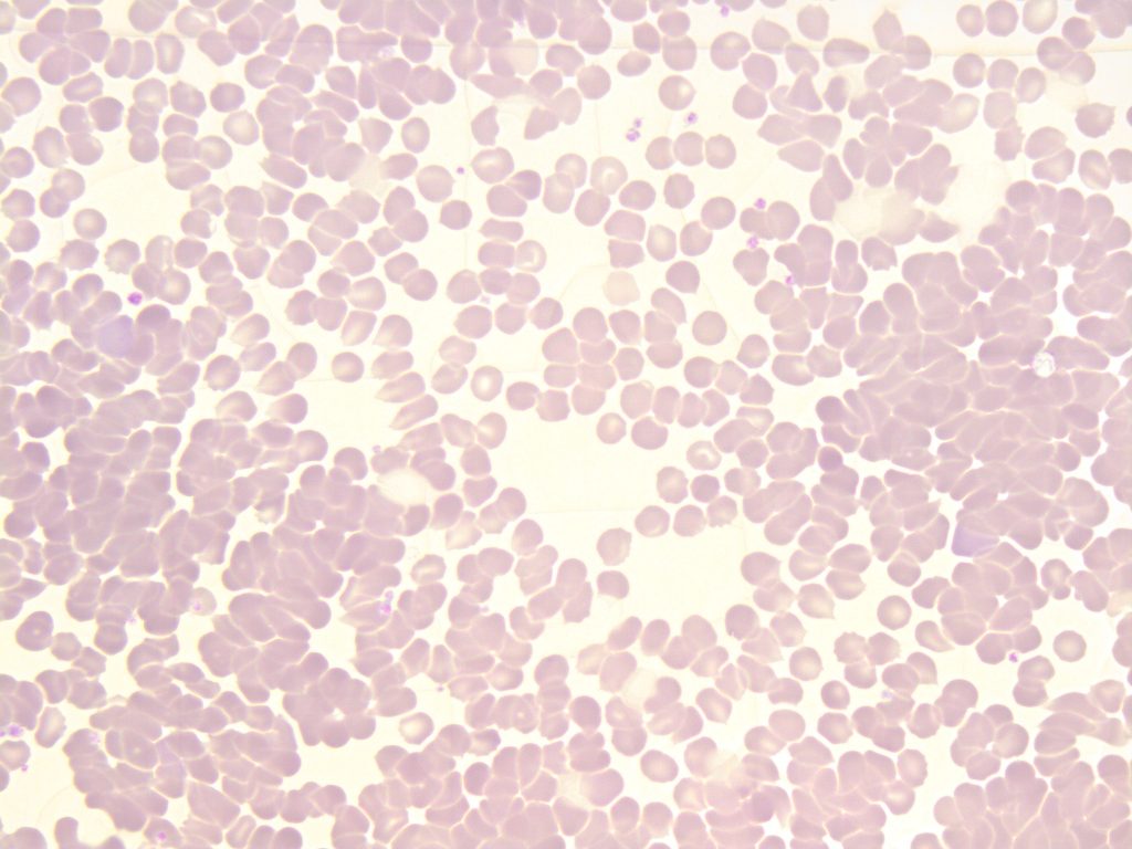
- A peripheral blood smear demonstrating autoagglutination of red blood cells. 50x oil immersion. From MLS Collection, University of Alberta, https://doi.org/10.7939/R3599ZH26
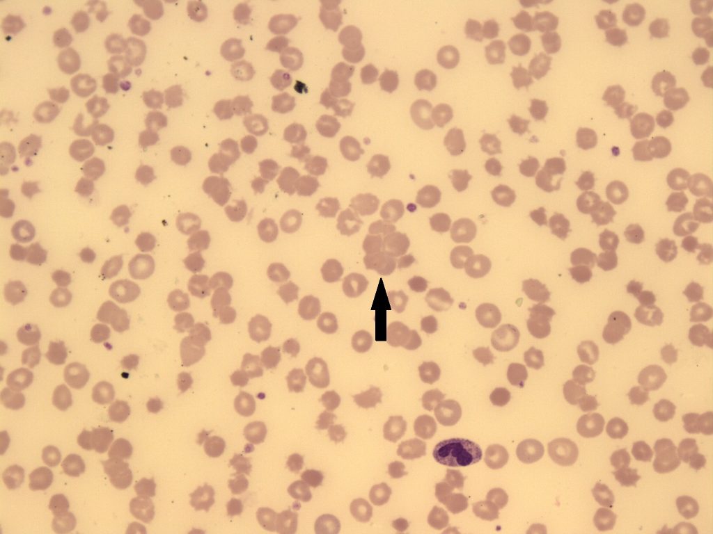
- An image from a peripheral blood smear showing agglutination of red blood cells. The arrow points to a cluster of red blood cells. 50x magnification. From MLS Collection, University of Alberta, https://doi.org/10.7939/R3SB3XD80
Cell Description:
This alteration of RBC distribution presents as irregular and random grape-like clusters or clumps. Agglutination is differentiated from Rouleaux by the lack of linear chains or “coin staking”. 1-4
The outlines of individual cells may not be evident.
Cell Formation:
Agglutination is caused by the formation of antibody-antigen complexes and occurs at room temperatures. Auto-agglutination is produced as a result of a complex formed between the patient’s own RBC antigens and antibodies, mediated by cold-reacting antibodies. Agglutination can be reversed when the blood sample is warmed to 37°C.1,2,5
Associated Disease/Clinical States: 2-4
Cold Hemagglutinin Disease
Paroxysmal Cold Hemoglobinuria
Cold Autoimmune Hemolytic Anemia
Note: Formation is NOT reversed with the addition of saline.5
References:
1. Hemolytic anemia: membrane defects. In: Clinical laboratory hematology. 3rd ed. New Jersey: Pearson; 2015. p. 317-33.
2. Jones KW. Evaluation of cell morphology and introduction to platelet and white blood cell morphology. In: Clinical hematology and fundamentals of hemostasis. 5th ed. Philadelphia: F.A. Davis Company; 2009. p. 93-116.
3. Ford J. Red blood cell morphology. Int J Lab Hematol [Internet]. 2013 Mar 9 [cited 2018 Jul 12];35(3):351–7. Available from: https://doi.org/10.1111/ijlh.12082
4. Turgeon ML. Normal erythrocyte lifecycle and physiology. In: Clinical hematology: theory and procedures. 4th ed. Philadelphia, PA: Lippincott Williams & Wilkins; 1999. p. 71-98.
5. Rodak BF, Carr JH. Variations in shape and distribution of erythrocytes. In: Clinical hematology atlas. 5th ed. St. Louis, Missouri: Elsevier Inc.; 2017. p. 93-106.