
2.2: Acanthocytes (Spur Cells)
- Page ID
- 38765

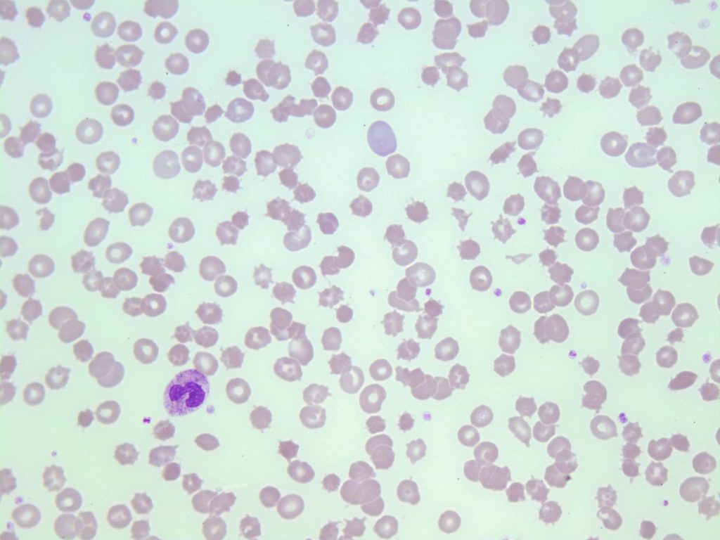
- An image of a peripheral blood smear containing some acanthocytes. 50x oil immersion. From MLS Collection, University of Alberta, https://doi.org/10.7939/R3MP4W32X
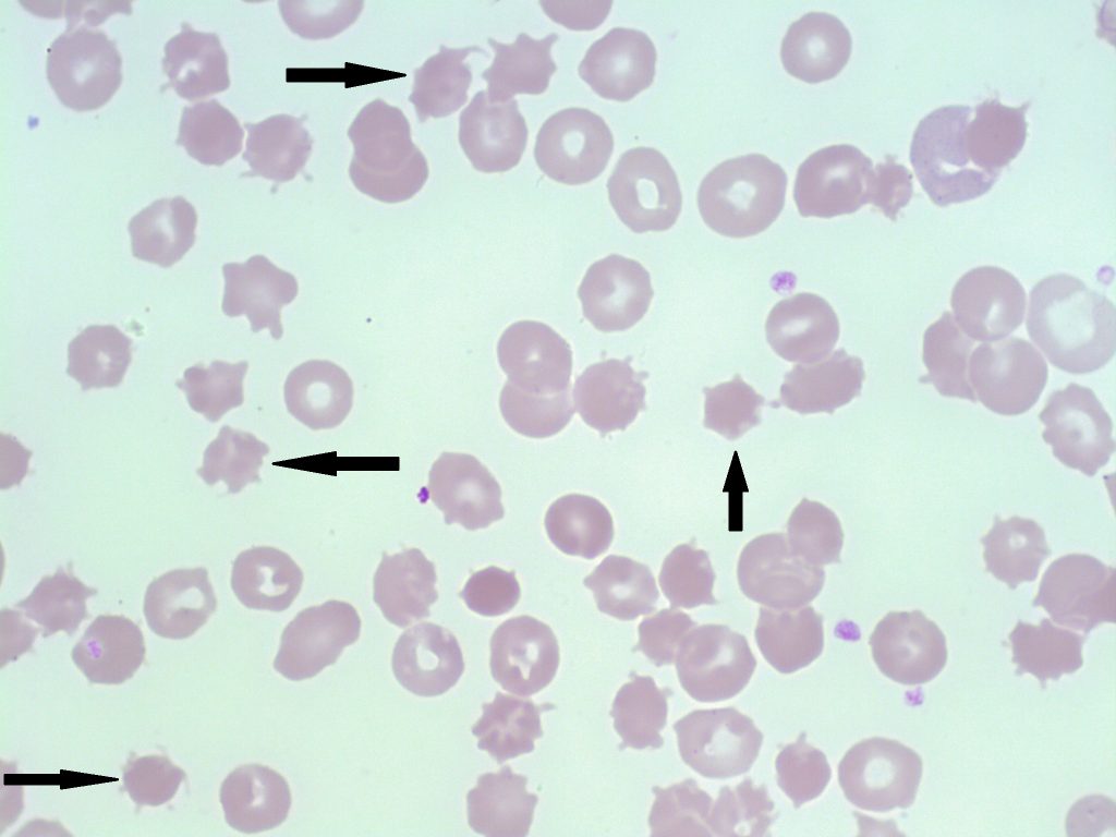
- An image of a peripheral blood smear containing some acanthocytes (examples shown with arrows). 50x oil immersion. From MLS Collection, University of Alberta, https://doi.org/10.7939/R3MP4W32X
Cell Description:
Red blood cells appear small and dense, lacking an area of central pallor with multiple spiky projections (spicules) of varying lengths protruding from the membrane. Projections are irregularly distributed around the cell membrane.1-3
Cell Formation:
Acanthocyte formation occurs as a result of either hereditary or acquired membrane defects. Defects that cause an imbalance between the membrane cholesterol and lipid content affect the RBC’s ability to deform resulting in more rigid plasma membrane. Red blood cells are then remodelled in circulation, resulting in an acanthocyte.1,3,4
Associated Disease/Clinical States: 1-5
Abetalipoproteinemia (Inherited)
Lecithin-cholesterol acyltransferase (LCAT) Deficiency
Liver Disease
Post-splenectomy
Pyruvate Kinase (PK) Deficiency
References:
1. Cochran-Black D. Hemolytic anemia: membrane defects. In: Clinical laboratory hematology. 3rd ed. New Jersey: Pearson; 2015. p. 317-33.
2. Manchanda N. Anemias: red blood morphology and approach to diagnosis. In: Rodak’s hematology clinical applications and principles. 5th ed. St. Louis, Missouri: Saunders; 2015. p. 284-96.
3. Turgeon ML. Normal erythrocyte lifecycle and physiology. In: Clinical hematology: theory and procedures. 4th ed. Philadelphia, PA: Lippincott Williams & Wilkins; 1999. p. 71-98
4. Harmening D. The red blood cell: structure and function. In: Clinical hematology and fundamentals of hemostasis. 5th ed. Philadelphia: F.A. Davis Company; 2009. p. 759-792.
5. Rodak BF, Carr JH. Variations in shape and distribution of erythrocytes. In: Clinical hematology atlas. 5th ed. St. Louis, Missouri: Elsevier Inc.; 2017. p. 93-106.