
2.4: Myofascial Release
- Page ID
- 59116

A Look at Fascial Anatomy
Andreas Vesalius (1514-1564) is often considered to be the first anatomist and is best remembered for publishing the famous anatomy text, De humani corporis fabrica in 1543. If you look at these early illustrations, they present the fascia and muscles as one continuous soft tissue structure. Fast forward to the 20th century (texts we study) most omit fascial tissue in order to depict muscles in a cleaner fashion. Some recent anatomy textbooks have made an effort to include this ‘forgotten tissue’ in their depictions and descriptions.
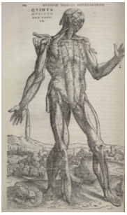
Image from De humani corporis fabrica circa 1543
An example of this is the Functional Atlas of the Human Fascial System by Carla Stecco, an Orthopedic surgeon and a professor of human anatomy at the University of Padua in Italy, the same University that once employed Andreas Vesalius in the early 1500’s. Another example is Anatomy Trains by Thomas Myers, in this book Myers presents conceptual ‘myofascial meridians’, recent systematic review confirmed a number of these continuous soft tissue structures (Wilke et al., 2016; Wilke et al., 2019).
To better understand myofascial release, there is a need to clarify the definition of fascia and how it interacts with various other structures: muscles, nerves, vessels.
Fascia has Been Used as an Ambiguous Term
Inconsistent definitions in the literature has led to confusion for researchers and therapists. A definition put forth by the Fascial Research Society hopes to provide some guidance. These researchers suggest making the distinction between A Fascia and The Fascial System (Schleip et al., 2019).
A Fascia – “A fascia is a sheath, a sheet, or any other dissectible aggregations of connective tissue that forms beneath the skin to attach, enclose, and separate muscles and other internal organs.”
The Fascial System – “The fascial system consists of the three-dimensional continuum of soft, collagen-containing, loose and dense fibrous connective tissues that permeate the body. It incorporates elements such as adipose tissue, adventitiae and neurovascular sheaths, aponeuroses, deep and superficial fasciae, epineurium, joint capsules, ligaments, membranes, meninges, myofascial expansions, periostea, retinacula, septa, tendons, visceral fasciae, and all the intramuscular and intermuscular connective tissues including endo-/peri-/epimysium.”
Myofascial Release in Various Forms Stimulates Mechanoreceptors
Ascribing a patient’s pain solely to a tissue-driven pain problem is often an oversimplification of a complex process. This insight provides us with an opportunity to re-frame our clinical models. When it comes to myofascial release a biopsychosocial framework helps put into context the interconnected and multidirectional interaction between a number of proposed mechanisms of action, including but not limited to affective touch, contextual factors, neurological factors, and mechanical factors.
Neurologically myofascial release may be used to stimulate mechanoreceptors, which in turn, trigger tonus changes in skeletal muscle fibers. Furthermore, input from sensory neurons may prevent the spinal cord from amplifying nociceptive signaling.
Myofascial Release in Various Forms Influences Tissue and Cell Physiology
Researchers have investigated the effect of soft-tissue massage on cellular signaling and tissue remodeling; this is referred to as mechanotherapy. Geoffrey Bove, a researcher at the University of New England, has conducted research examining the effect of modelled manual therapy on repetitive motion disorders and the development of fibrosis. One study published in the Journal of Neurological Sciences showed soft-tissue massage prevented the deposition of collagen and transforming growth factor beta 1 (TGF beta 1) in the nerves and connective tissues of the forearm (Bove et al., 2016). This was recently followed up by a study published in the prestigious journal Pain showing that by attenuating the inflammatory response (with modelled massage) in the early stages of an injury, they were able to prevent the development of neural fibrosis (Bove et al., 2019). This is potentially impactful in postoperative rehabilitation because TGF-β1 plays a key role in tissue remodeling and fibrosis.
Furthermore, a recent joint research effort between Timothy Butterfield of the University of Kentucky and researchers at Colorado State University demonstrated that modelled massage enhanced satellite cell numbers (Miller et al., 2018; Hunt et al., 2019). This was in addition to earlier research from Butterfield and his collaborators at the University of Kentucky, which proposes the idea that mechanical stimulation prompts a phenotype change of pro-inflammatory M1 macrophages into anti-inflammatory M2 macrophages (Waters-Banker et al., 2014). Another group of researchers at The University of Arizona propose that mechanical stimulation can trigger fibroblasts to express anti-inflammatory cytokines (Zein-Hammoud & Standley, 2015; Zein-Hammoud & Standley, 2019). Taken together the increase in satellite cell numbers and reduction in inflammatory signaling may play a role in tissue remodeling and improve the body’s ability to respond to subsequent rehabilitation.
Does Myofascial Release Break Adhesions?
As a result of aging, injury or trauma the neuromuscular system undergoes remodeling, which involves muscles, fascia, and the central and peripheral nervous system. If there has been significant adaptations this may impair the body’s ability to respond to subsequent rehabilitation (Zullo et al., 2020). Traditionally when soft tissue structures have a reduced ability to glide adhesions are blamed. Currently there is a paucity of research to support the claim that manual therapy can break mature adhesions. However, in the developmental phase manual therapy may be able to attenuate the development of post-surgical adhesions (Bove et al., 2017). In the remodeling phase the mechanisms by which myofascial release interrupts the sequelae of pathological healing is most likely not in a single unified response.
Myofascial Release is a treatment approach that stimulates mechanoreceptors and influences tissue and cell physiology. Clinically this translates into improved proprioception, increased range of motion and pain management.
References and Sources
Barbe, M. F., Harris, M. Y., Cruz, G. E., Amin, M., Billett, N. M., Dorotan, J. T., Day, E. P., Kim, S. Y., & Bove, G. M. (2021). Key indicators of repetitive overuse-induced neuromuscular inflammation and fibrosis are prevented by manual therapy in a rat model. BMC musculoskeletal disorders, 22(1), 417. https://doi.org/10.1186/s12891-021-04270-0
Behm, D. G., & Wilke, J. (2019). Do Self-Myofascial Release Devices Release Myofascia? Rolling Mechanisms: A Narrative Review. Sports medicine (Auckland, N.Z.), 49(8), 1173–1181. doi:10.1007/s40279-019-01149-y
Bialosky, J. E., Beneciuk, J. M., Bishop, M. D., Coronado, R. A., Penza, C. W., Simon, C. B., & George, S. Z. (2018). Unraveling the Mechanisms of Manual Therapy: Modeling an Approach. The Journal of orthopaedic and sports physical therapy, 48(1), 8–18. doi:10.2519/jospt.2018.7476
Bove, G. M., Harris, M. Y., Zhao, H., & Barbe, M. F. (2016). Manual therapy as an effective treatment for fibrosis in a rat model of upper extremity overuse injury. Journal of the neurological sciences, 361, 168–180. doi:10.1016/j.jns.2015.12.029
Bove, G. M., Chapelle, S. L., Hanlon, K. E., Diamond, M. P., & Mokler, D. J. (2017). Attenuation of postoperative adhesions using a modeled manual therapy. PloS one, 12(6), e0178407. doi:10.1371/journal.pone.0178407
Bove, G. M., Delany, S. P., Hobson, L., Cruz, G. E., Harris, M. Y., Amin, M., … Barbe, M. F. (2019). Manual therapy prevents onset of nociceptor activity, sensorimotor dysfunction, and neural fibrosis induced by a volitional repetitive task. Pain, 160(3), 632–644. doi:10.1097/j.pain.0000000000001443
Chaitow, L. (2017). What’s in a name: Myofascial Release or Myofascial Induction?. Journal of bodywork and movement therapies, 21(4), 749–751. doi:10.1016/j.jbmt.2017.09.008
Dischiavi, S. L., Wright, A. A., Hegedus, E. J., & Bleakley, C. M. (2018). Biotensegrity and myofascial chains: A global approach to an integrated kinetic chain. Medical hypotheses, 110, 90–96. https://doi.org/10.1016/j.mehy.2017.11.008
França, M., Sinhorim, L., Martins, D. F., Schleip, R., Machado-Pereira, N., de Souza, G. M., Horewicz, V. V., & Santos, G. M. (2020). Manipulation of the Fascial System Applied During Acute Inflammation of the Connective Tissue of the Thoracolumbar Region Affects Transforming Growth Factor-β1 and Interleukin-4 Levels: Experimental Study in Mice. Frontiers in physiology, 11, 587373. https://doi.org/10.3389/fphys.2020.587373
Hilliard, B. A., Amin, M., Popoff, S. N., & Barbe, M. F. (2021). Force dependent effects of chronic overuse on fibrosis-related genes and proteins in skeletal muscles. Connective tissue research, 62(1), 133–149. https://doi.org/10.1080/03008207.2020.1828379
Hunt, E. R., Confides, A. L., Abshire, S. M., Dupont-Versteegden, E. E., & Butterfield, T. A. (2019). Massage increases satellite cell number independent of the age-associated alterations in sarcolemma permeability. Physiological reports, 7(17), e14200. doi:10.14814/phy2.14200
Miller, B. F., Hamilton, K. L., Majeed, Z. R., Abshire, S. M., Confides, A. L., Hayek, A. M., … Dupont-Versteegden, E. E. (2018). Enhanced skeletal muscle regrowth and remodelling in massaged and contralateral non-massaged hindlimb. The Journal of physiology, 596(1), 83–103. doi:10.1113/JP275089
Plikus, M. V., Wang, X., Sinha, S., Forte, E., Thompson, S. M., Herzog, E. L., Driskell, R. R., Rosenthal, N., Biernaskie, J., & Horsley, V. (2021). Fibroblasts: Origins, definitions, and functions in health and disease. Cell, 184(15), 3852–3872. https://doi.org/10.1016/j.cell.2021.06.024
Schleip, R., Hedley, G., & Yucesoy, C. A. (2019). Fascial nomenclature: Update on related consensus process. Clinical anatomy (New York, N.Y.), 32(7), 929–933. doi:10.1002/ca.23423
Schleip, R., & Klingler, W. (2019). Active contractile properties of fascia. Clinical anatomy (New York, N.Y.), 32(7), 891–895. doi:10.1002/ca.23391
Schleip, R., Gabbiani, G., Wilke, J., Naylor, I., Hinz, B., Zorn, A., … Klingler, W. (2019). Fascia Is Able to Actively Contract and May Thereby Influence Musculoskeletal Dynamics: A Histochemical and Mechanographic Investigation. Frontiers in physiology, 10, 336. doi:10.3389/fphys.2019.00336
Stecco, A., Stern, R., Fantoni, I., De Caro, R., & Stecco, C. (2016). Fascial Disorders: Implications for Treatment. PM & R: the journal of injury, function, and rehabilitation, 8(2), 161–168. doi:10.1016/j.pmrj.2015.06.006
Waters-Banker, C., Dupont-Versteegden, E. E., Kitzman, P. H., & Butterfield, T. A. (2014). Investigating the mechanisms of massage efficacy: the role of mechanical immunomodulation. Journal of athletic training, 49(2), 266–273. doi:10.4085/1062-6050-49.2.25
Wilke, J., Krause, F., Vogt, L., & Banzer, W. (2016). What Is Evidence-Based About Myofascial Chains: A Systematic Review. Archives of physical medicine and rehabilitation, 97(3), 454–461. doi:10.1016/j.apmr.2015.07.023
Wilke, J., Schleip, R., Yucesoy, C. A., & Banzer, W. (2018). Not merely a protective packing organ? A review of fascia and its force transmission capacity. Journal of applied physiology (Bethesda, Md. : 1985), 124(1), 234–244. doi:10.1152/japplphysiol.00565.2017
Wilke, J., & Krause, F. (2019). Myofascial chains of the upper limb: A systematic review of anatomical studies. Clinical anatomy (New York, N.Y.), 32(7), 934–940. doi:10.1002/ca.23424
Wilke, J., Debelle, H., Tenberg, S., Dilley, A., & Maganaris, C. (2020). Ankle Motion Is Associated With Soft Tissue Displacement in the Dorsal Thigh: An in vivo Investigation Suggesting Myofascial Force Transmission Across the Knee Joint. Frontiers in physiology, 11, 180. https://doi.org/10.3389/fphys.2020.00180
Wong, R., Geyer, S., Weninger, W., Guimberteau, J. C., & Wong, J. K. (2016). The dynamic anatomy and patterning of skin. Experimental dermatology, 25(2), 92–98. doi:10.1111/exd.12832
Zein-Hammoud, M., & Standley, P. R. (2015). Modeled Osteopathic Manipulative Treatments: A Review of Their in Vitro Effects on Fibroblast Tissue Preparations. The Journal of the American Osteopathic Association, 115(8), 490–502. doi:10.7556/jaoa.2015.103
Zein-Hammoud, M., & Standley, P. R. (2019). Optimized Modeled Myofascial Release Enhances Wound Healing in 3-Dimensional Bioengineered Tendons: Key Roles for Fibroblast Proliferation and Collagen Remodeling. Journal of manipulative and physiological therapeutics, 42(8), 551–564. https://doi.org/10.1016/j.jmpt.2019.01.001
Zügel, M., Maganaris, C. N., Wilke, J., Jurkat-Rott, K., Klingler, W., Wearing, S. C., … Hodges, P. W. (2018). Fascial tissue research in sports medicine: from molecules to tissue adaptation, injury and diagnostics: consensus statement. British journal of sports medicine, 52(23), 1497. doi:10.1136/bjsports-2018-099308
Zullo, A., Fleckenstein, J., Schleip, R., Hoppe, K., Wearing, S., & Klingler, W. (2020). Structural and Functional Changes in the Coupling of Fascial Tissue, Skeletal Muscle, and Nerves During Aging. Frontiers in physiology, 11, 592. https://doi.org/10.3389/fphys.2020.00592