
Preface
- Page ID
- 41536


Figure 0.1: This record player stand is used as a metaphor
Why study histology and embryology?
This home-built stand for a record player is going to serve as a metaphor. Measurements were made to ensure there would be enough space for records. It is built out of wooden dowels, and used some floor tiles to create a smooth surface. Why those two? Actually, those building materials were re-used, they were left over from previous projects. Sure, a trip to the hardware store could have meant more appropriate materials, but would the hassle have been worth it? Currently, there is a Covid-19 pandemic, the answer is no. This is why you should study embryology, many instructions and materials are re-used during human development. Some things should make sense in this book, such as the instructions which guide even-spacing between teeth. But some things won’t make sense initially, such as why is enamel made by epithelial cells, when it looks like bone tissue? Why can a piece of pig heart be used to surgically repair gingival tissue? What is the philtrum for? These are questions that can be better answered by studying where they came from (embryology) rather than studying their appearance in adults (anatomy). Imagine not knowing that flies develop from maggots, you might believe in spontaneous generation after observing the appearance of maggots on rotting food. Then why study histology? The answer to that is simple: embryos are tiny, you need a microscope to see what is going on. But more importantly, things happen in the oral cavity you can’t see, but you should understand conceptually. For instance, with a basic understanding of histology you will understand why a pocket depth over 3mm is considered unhealthy. You can conceptualize what makes the linea alba appear white in some patients. You can explain what causes perikymata. You are on your own, however, on how to pronounce perikymata.
Author bios:
Laird C. Sheldahl, Ph.D. is the lead author and illustrator of this textbook. He has a Ph.D. in physiology and pharmacology. He currently teaches anatomy and physiology, as well as histology and embryology for dental hygiene, at Mt. Hood Community College in Gresham, OR. His Ph.D. thesis studied a morphogen involved in the formation of the head, including neural crest cell migration. This morphogen, a Wnt, is re-used during development of the teeth, which in turn involves neural crest cells. To study this process in frogs (their embryos are not inside uteruses, which makes them much easier to study) he did a lot of microscopy, which has helped considerably in the writing of this textbook. He does not have a background in dental hygiene, and is therefore very happy to have expert collaborators.
Student editors
Amen Mohammed
Conventions used in this eBook
There are two types of links found in this eBook. Internal links will bring you to the section of the book where that word is defined. These links should be in blue, regular text, such as junctional epithelium or calcium hydroxyapatite. Use the back button to return to your original spot. Other links in the book will take you to an external website, such as Wikipedia. These links should be a purple monospace font, such as this: external link. We don’t have control over how they appear on your eReader, however. External links are for further reading if you are interested, but are not required reading to understand the material we present. The basic format of each chapter is as follows:
| chapter outline | |
|---|---|
| Philosophy | Where we try to provide a road-map for what I will be covering, and why we are covering it. |
| Physiology | Where we cover histology and embryology. |
| Clinical significance | Where we cover why the histology or embryology is relevant to your practice. |
Table 0.1: Basic format of this textbook
Figure 0.2: Example of an animated image
Animated images
The image in Fig. 0.2 is an animated .gif file, it should be cycling through a series of changes. The authors wish to take advantage of the things that can be done in an eBook, but not a print book.
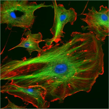
Figure 0.3 : Example of an image created by someone else. Image credit: Fluorescent cells by NIH image is in the Public Domain CC0
Other images
Images that we have not created have their sources listed in the figure legend, such as Fig. 0.3. This is to keep in line with the Creative Commons rules for using other people’s work the way they have asked. This may be of use to teachers, students can ignore this information.
Course objectives
| Course objective | Textbook chapter(s) |
|---|---|
| 1. Identify core concepts of cell biology and general histology related to the face and oral cavity | 1,2 |
| 2. Demonstrate an understanding of the role of epithelial tissues, underlying connective tissues and neural crest cells in the face and oral cavity | 3,4,5 |
| 3. Identify the basic patterns of early human developmental biology related to formation of the face and oral cavity | 6,7 |
| 4. Describe the major steps of amelogenesis and enamel structure | 8 |
| 5. Describe the major steps of odontogenesis and formation of the dentin-pulp complex | 9 |
| 6. Describe the major steps of development of tooth roots and the periodontium | 10 |
| 7. Identify mesenchymal-epithelial relationships in tooth development and tooth eruption. | 8, 9, 11 |
Table 0.2: Course objective (for instructor use)
Copyright information

Except where otherwise noted, the material in this book is licensed under Creative Commons Attribution Non-Commercial Share Alike 4.0 International. Feel free to distribute, edit or use portions of this book as long as you adhere to the creative commons rules– you must attribute the authors (unless explicitly labelled as in the public domain), do not use contents found in this text for commercial purposes (unless explicitly labeled as something other than NC), and mark any material with the same license associated with it.