
9.7C: Neck Muscles
- Page ID
- 7562
\( \newcommand{\vecs}[1]{\overset { \scriptstyle \rightharpoonup} {\mathbf{#1}} } \)
\( \newcommand{\vecd}[1]{\overset{-\!-\!\rightharpoonup}{\vphantom{a}\smash {#1}}} \)
\( \newcommand{\id}{\mathrm{id}}\) \( \newcommand{\Span}{\mathrm{span}}\)
( \newcommand{\kernel}{\mathrm{null}\,}\) \( \newcommand{\range}{\mathrm{range}\,}\)
\( \newcommand{\RealPart}{\mathrm{Re}}\) \( \newcommand{\ImaginaryPart}{\mathrm{Im}}\)
\( \newcommand{\Argument}{\mathrm{Arg}}\) \( \newcommand{\norm}[1]{\| #1 \|}\)
\( \newcommand{\inner}[2]{\langle #1, #2 \rangle}\)
\( \newcommand{\Span}{\mathrm{span}}\)
\( \newcommand{\id}{\mathrm{id}}\)
\( \newcommand{\Span}{\mathrm{span}}\)
\( \newcommand{\kernel}{\mathrm{null}\,}\)
\( \newcommand{\range}{\mathrm{range}\,}\)
\( \newcommand{\RealPart}{\mathrm{Re}}\)
\( \newcommand{\ImaginaryPart}{\mathrm{Im}}\)
\( \newcommand{\Argument}{\mathrm{Arg}}\)
\( \newcommand{\norm}[1]{\| #1 \|}\)
\( \newcommand{\inner}[2]{\langle #1, #2 \rangle}\)
\( \newcommand{\Span}{\mathrm{span}}\) \( \newcommand{\AA}{\unicode[.8,0]{x212B}}\)
\( \newcommand{\vectorA}[1]{\vec{#1}} % arrow\)
\( \newcommand{\vectorAt}[1]{\vec{\text{#1}}} % arrow\)
\( \newcommand{\vectorB}[1]{\overset { \scriptstyle \rightharpoonup} {\mathbf{#1}} } \)
\( \newcommand{\vectorC}[1]{\textbf{#1}} \)
\( \newcommand{\vectorD}[1]{\overrightarrow{#1}} \)
\( \newcommand{\vectorDt}[1]{\overrightarrow{\text{#1}}} \)
\( \newcommand{\vectE}[1]{\overset{-\!-\!\rightharpoonup}{\vphantom{a}\smash{\mathbf {#1}}}} \)
\( \newcommand{\vecs}[1]{\overset { \scriptstyle \rightharpoonup} {\mathbf{#1}} } \)
\( \newcommand{\vecd}[1]{\overset{-\!-\!\rightharpoonup}{\vphantom{a}\smash {#1}}} \)
\(\newcommand{\avec}{\mathbf a}\) \(\newcommand{\bvec}{\mathbf b}\) \(\newcommand{\cvec}{\mathbf c}\) \(\newcommand{\dvec}{\mathbf d}\) \(\newcommand{\dtil}{\widetilde{\mathbf d}}\) \(\newcommand{\evec}{\mathbf e}\) \(\newcommand{\fvec}{\mathbf f}\) \(\newcommand{\nvec}{\mathbf n}\) \(\newcommand{\pvec}{\mathbf p}\) \(\newcommand{\qvec}{\mathbf q}\) \(\newcommand{\svec}{\mathbf s}\) \(\newcommand{\tvec}{\mathbf t}\) \(\newcommand{\uvec}{\mathbf u}\) \(\newcommand{\vvec}{\mathbf v}\) \(\newcommand{\wvec}{\mathbf w}\) \(\newcommand{\xvec}{\mathbf x}\) \(\newcommand{\yvec}{\mathbf y}\) \(\newcommand{\zvec}{\mathbf z}\) \(\newcommand{\rvec}{\mathbf r}\) \(\newcommand{\mvec}{\mathbf m}\) \(\newcommand{\zerovec}{\mathbf 0}\) \(\newcommand{\onevec}{\mathbf 1}\) \(\newcommand{\real}{\mathbb R}\) \(\newcommand{\twovec}[2]{\left[\begin{array}{r}#1 \\ #2 \end{array}\right]}\) \(\newcommand{\ctwovec}[2]{\left[\begin{array}{c}#1 \\ #2 \end{array}\right]}\) \(\newcommand{\threevec}[3]{\left[\begin{array}{r}#1 \\ #2 \\ #3 \end{array}\right]}\) \(\newcommand{\cthreevec}[3]{\left[\begin{array}{c}#1 \\ #2 \\ #3 \end{array}\right]}\) \(\newcommand{\fourvec}[4]{\left[\begin{array}{r}#1 \\ #2 \\ #3 \\ #4 \end{array}\right]}\) \(\newcommand{\cfourvec}[4]{\left[\begin{array}{c}#1 \\ #2 \\ #3 \\ #4 \end{array}\right]}\) \(\newcommand{\fivevec}[5]{\left[\begin{array}{r}#1 \\ #2 \\ #3 \\ #4 \\ #5 \\ \end{array}\right]}\) \(\newcommand{\cfivevec}[5]{\left[\begin{array}{c}#1 \\ #2 \\ #3 \\ #4 \\ #5 \\ \end{array}\right]}\) \(\newcommand{\mattwo}[4]{\left[\begin{array}{rr}#1 \amp #2 \\ #3 \amp #4 \\ \end{array}\right]}\) \(\newcommand{\laspan}[1]{\text{Span}\{#1\}}\) \(\newcommand{\bcal}{\cal B}\) \(\newcommand{\ccal}{\cal C}\) \(\newcommand{\scal}{\cal S}\) \(\newcommand{\wcal}{\cal W}\) \(\newcommand{\ecal}{\cal E}\) \(\newcommand{\coords}[2]{\left\{#1\right\}_{#2}}\) \(\newcommand{\gray}[1]{\color{gray}{#1}}\) \(\newcommand{\lgray}[1]{\color{lightgray}{#1}}\) \(\newcommand{\rank}{\operatorname{rank}}\) \(\newcommand{\row}{\text{Row}}\) \(\newcommand{\col}{\text{Col}}\) \(\renewcommand{\row}{\text{Row}}\) \(\newcommand{\nul}{\text{Nul}}\) \(\newcommand{\var}{\text{Var}}\) \(\newcommand{\corr}{\text{corr}}\) \(\newcommand{\len}[1]{\left|#1\right|}\) \(\newcommand{\bbar}{\overline{\bvec}}\) \(\newcommand{\bhat}{\widehat{\bvec}}\) \(\newcommand{\bperp}{\bvec^\perp}\) \(\newcommand{\xhat}{\widehat{\xvec}}\) \(\newcommand{\vhat}{\widehat{\vvec}}\) \(\newcommand{\uhat}{\widehat{\uvec}}\) \(\newcommand{\what}{\widehat{\wvec}}\) \(\newcommand{\Sighat}{\widehat{\Sigma}}\) \(\newcommand{\lt}{<}\) \(\newcommand{\gt}{>}\) \(\newcommand{\amp}{&}\) \(\definecolor{fillinmathshade}{gray}{0.9}\)Cervical muscles are those associated with the front of the neck; vertebral muscles are associated with the vertebral column.
- Outline the neck muscles and their movements
Key Points
- Numerous muscles contribute to the processes of speaking and swallowing.
- These muscles can be divided into suprahyoid and ingrahyoid groups based on their locations relative to the hyoid bone.
- The hyoid bone, located beneath the mandible, acts as a key attachment point for muscles involved in speaking and swallowing.
- Numerous muscles contribute to both the stabilization and fine movements of the head and neck.
Key Terms
- suprahyoid muscles: A group of muscles located above the hyoid bone, responsible for its elevation which widens the esophagus.
- hyoid bone: A -shaped bone which sits below the mandible
and in front of the esophagus, facilitating the wide range of movements associated with speaking and swallowing. - infrahyoid muscles: A group of muscles located below the hyoid bone, responsible for its depression which narrows the esophagus.
Muscles of the neck play important roles in mastication (chewing), swallowing, speaking and supporting and moving the head. All muscles found in the neck are paired, meaning they exist to both the left and right side of the spine. The muscles involved in chewing have been discussed previously so only those involved in swallowing and support will be discussed below.
Muscles Involved in Swallowing and Speaking
Located to the anterior of the neck, these muscles are split into two based on their location relative to the hyoid bone. The U-shaped hyoid bone sits below the mandible and in front of the esophagus, providing a level of protection and facilitating the wide range of muscle activity required for speaking and swallowing.
Suprahyoid Muscles
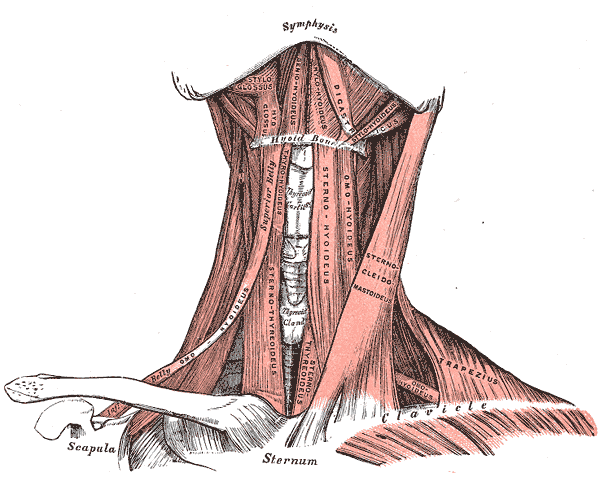
Suprahyoid and infrahyoid muscles of the neck: Suprahyoid and infrahyoid muscle groups are named based on their location relative to the hyoid bone. The hyoid bone sits below the mandible and in front of the esophagus, providing a level of protection but also facilitating the wide range of muscle activity required for speaking and swallowing.
The four suprahyoid muscles found above the hyoid bone act in concert to elevate the hyoid bone, assisting with swallowing by widening the esophagus.
- Stylohyoid: The most superior of the suprahyoid muscles, the stylohyoid originates from the skull and attaches to the hyoid bone.
- Digastric: The digastric muscle is split into two parts that are connected by a tendon attached to the hyoid bone. The anterior section originates from the mandible and the posterior section from the skull.
- Mylohyoid: The mylohyoid is a broad flat muscle which forms the floor of the oral cavity. It originates from the mandible and attaches to the hyoid bone.
- Geniohyoid: The deepest of the suprahyoid muscles, the geniohyoid muscle originates from the mandible and attaches to the hyoid bone.
Infrahyoid Muscles
The four infrahyoid muscles found below the hyoid bone act in concert to depress the hyoid bone during swallowing and speaking, compressing the esophagus.
- Sternohyoid: A superficial muscle which originates from the sternum and attaches onto the hyoid bone.
- Omohyoid: Located laterally to the sternohyoid, the omohyoid muscle is split in two parts attached by a tendon. The inferior region originates from the scapula, joins the superior region, and attaches to the hyoid bone.
- Sternothyroid: Sitting deeper than the sternohyoid, the sternothyroid originates from the sternum and attaches to the thyroid cartilage associated with the hyoid bone.
- Thyrohyoid: A short continuation of the sternothyroid muscle, the thyrohyoid originates from the thyroid cartilage and attaches to the hyoid bone.
Muscles of the Back and Neck
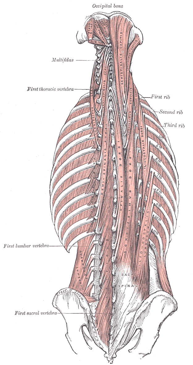
Muscles of the back and neck: Muscles of the back and neck play an important role in maintaining posture and the movement of the head and neck.
The muscles of the back and neck are responsible for maintaining posture and facilitating movement of the head and neck. They are divided into three layers.
Superficial Layer
Two muscles in the superficial layer are responsible for rotation of the head.
- Splenius Capitis: A thick rectangular muscle, the most superior of the neck muscles.
- Attachments: Originates from the upper spine and attaches to the skull.
- Actions: Rotates and extend the head and neck.
- Splenius Cervicis: A small triangular-shaped muscle located immediately below the splenius capitis.
- Attachments: Originates from the spine and attaches several vertebrae higher.
- Actions: Rotate and extend the head and neck.
Intermediate Layer
Three columnar muscles in the intermediate layer are responsible for flexion and extension of the neck as well as posture maintenance. All three originate from a common tendon associated with the pelvis and can be divided into thoracic, cervicis, and capitis regions.
- Iliocostalis: The most laterally located of the three intermediate muscles.
- Attachments: Originates from the common tendon and attaches to the ribs and lower neck.
- Actions: Extends and controls abduction and adduction of the spine and neck.
- Longissimus: Located between the iliocostalis and spinalis muscles, this is the largest of the intermediate layer muscles.
- Attachments: Originates from the common tendon and attaches to the lower ribs, the spine, and the skull.
- Actions: Extends and controls abduction and adduction of the spine and neck.
- Spinalis: The most medially located and smallest of the three intermediate layer muscles.
- Attachments: Originates from the common tendon and attaches to the upper spine and skull.
- Actions: Extends and flexes to control abduction and adduction of the spine and neck.
Deep Layer
Two muscles in the deep layer are responsible for maintenance of posture and rotation of the neck.
- Semispinalis: The semispinalis is the most superficial of the deep muscles.
- Attachments: A broad origin on the upper regions of the spine, with each origin attaching several vertebrae higher or to the skull.
- Actions: Extends and rotates the head and maintains posture.
- Multifidus: The multifidus is located underneath the semispinalis muscle and is key in maintaining posture.
- Attachments: A broad origin up the length of the spine, with each origin attaching several vertebrae higher.
- Actions: Maintains posture through the spine.
Other Muscles That Act on the Neck
Several other muscles act on the head and neck. Below are three with a larger impact.
- Trapezius: The trapezius is the most superficial muscle of the back and forms a broad flat triangle.
- Attachments: The trapezius originates from the skull and spine of the upper back and neck. It attaches to the clavicle and scapula.
- Actions: The superior region supports the arm and elevates and rotates the scapula. It controls adduction, abduction and rotation of the head, the intermediate region retracts the scapula, and the inferior region rotates and depresses the scapula.
- Sternocleidomastoid: A thick rectangular muscle that is responsible for many movements within the neck.
- Attachments: Dual-headed, the sternocleidomastoid originates from the clavicle and the sternum and attaches to the mandible.
- Actions: Abduction, adduction, extension, flexion, and rotation of the neck depending on intra and inter-muscle contractions.
- Platysma: A broad sheet of muscle arising from the fascia covering the pectorals.
- Attachments: Originates from the fascia covering the pectorals and attaches to various locations within the mandible and dermis of the face and neck.
- Actions: Depresses the mandible and angles the lip and mouth, wrinkling the skin upon the neck flexing.
KEY MOVEMENTS
- Extension (tilting head backwards): Produced by the semispinalis, splenus capitis, longissimus, trapezius (superior fibers), and sternocleidomastoid (posterior fibers).
- Flexion (tilting head forwards): Produced by the sternocleidomastoid (anterior fibers).
- Abduction (tilting head towards shoulder): Produced by the sternocleidomastoid, longissimus, splenius capitis, semispinalis, and trapezius (superior fibers)
- Adduction (returning head to midline): Produced by the sternocleidomastoid, longissimus, splenius capitis, semispinalis, and trapezius (superior fibers)
- Rotation (rotation head to left or right): Produced by the sternocleidomastoid, longissimus, splenius capitis, semispinalis, and trapezius (superior fibers)
LICENSES AND ATTRIBUTIONS
CC LICENSED CONTENT, SHARED PREVIOUSLY
- Curation and Revision. Authored by: Boundless.com. Provided by: Boundless.com. License: CC BY-SA: Attribution-ShareAlike
CC LICENSED CONTENT, SPECIFIC ATTRIBUTION
- Depressor supercilii muscle. Provided by: Wikipedia. Located at: en.Wikipedia.org/wiki/Depress...ercilii_muscle. License: CC BY-SA: Attribution-ShareAlike
- Levator anguli oris. Provided by: Wikipedia. Located at: en.Wikipedia.org/wiki/Levator_anguli_oris. License: CC BY-SA: Attribution-ShareAlike
- Nasalis muscle. Provided by: Wikipedia. Located at: en.Wikipedia.org/wiki/Nasalis_muscle. License: CC BY-SA: Attribution-ShareAlike
- Levator palpebrae superioris muscle. Provided by: Wikipedia. Located at: en.Wikipedia.org/wiki/Levator...erioris_muscle. License: CC BY-SA: Attribution-ShareAlike
- Levator labii superioris alaeque nasi muscle. Provided by: Wikipedia. Located at: en.Wikipedia.org/wiki/Levator...ue_nasi_muscle. License: CC BY-SA: Attribution-ShareAlike
- Occipitofrontalis muscle. Provided by: Wikipedia. Located at: en.Wikipedia.org/wiki/Occipitofrontalis_muscle. License: CC BY-SA: Attribution-ShareAlike
- Dilator naris muscle. Provided by: Wikipedia. Located at: en.Wikipedia.org/wiki/Dilator_naris_muscle. License: CC BY-SA: Attribution-ShareAlike
- Superior tarsal muscle. Provided by: Wikipedia. Located at: en.Wikipedia.org/wiki/Superior_tarsal_muscle. License: CC BY-SA: Attribution-ShareAlike
- Depressor septi nasi muscle. Provided by: Wikipedia. Located at: en.Wikipedia.org/wiki/Depress...ti_nasi_muscle. License: CC BY-SA: Attribution-ShareAlike
- Levator labii superioris. Provided by: Wikipedia. Located at: en.Wikipedia.org/wiki/Levator_labii_superioris. License: CC BY-SA: Attribution-ShareAlike
- Buccinator muscle. Provided by: Wikipedia. Located at: en.Wikipedia.org/wiki/Buccinator_muscle. License: CC BY-SA: Attribution-ShareAlike
- Procerus muscle. Provided by: Wikipedia. Located at: en.Wikipedia.org/wiki/Procerus_muscle. License: CC BY-SA: Attribution-ShareAlike
- Zygomaticus major muscle. Provided by: Wikipedia. Located at: en.Wikipedia.org/wiki/Zygomaticus_major_muscle. License: CC BY-SA: Attribution-ShareAlike
- Zygomaticus minor muscle. Provided by: Wikipedia. Located at: en.Wikipedia.org/wiki/Zygomaticus_minor_muscle. License: CC BY-SA: Attribution-ShareAlike
- Risorius. Provided by: Wikipedia. Located at: en.Wikipedia.org/wiki/Risorius. License: CC BY-SA: Attribution-ShareAlike
- Depressor anguli oris muscle. Provided by: Wikipedia. Located at: en.Wikipedia.org/wiki/Depressor_anguli_oris_muscle. License: CC BY-SA: Attribution-ShareAlike
- Mentalis. Provided by: Wikipedia. Located at: en.Wikipedia.org/wiki/Mentalis. License: CC BY-SA: Attribution-ShareAlike
- Depressor labii inferioris muscle. Provided by: Wikipedia. Located at: en.Wikipedia.org/wiki/Depressor_labii_inferioris_muscle. License: CC BY-SA: Attribution-ShareAlike
- Orbicularis oris muscle. Provided by: Wikipedia. Located at: en.Wikipedia.org/wiki/Orbicularis_oris_muscle. License: CC BY-SA: Attribution-ShareAlike
- Boundless. Provided by: Boundless Learning. Located at: www.boundless.com//physiology...bicularis-oris. License: CC BY-SA: Attribution-ShareAlike
- Boundless. Provided by: Boundless Learning. Located at: www.boundless.com//physiology...ontalis-muscle. License: CC BY-SA: Attribution-ShareAlike
- buccinator. Provided by: Wiktionary. Located at: en.wiktionary.org/wiki/buccinator. License: CC BY-SA: Attribution-ShareAlike
- Buccinator. Provided by: Wikipedia. Located at: en.Wikipedia.org/wiki/File:Buccinator.jpg. License: CC BY-SA: Attribution-ShareAlike
- Gray378. Provided by: Wikipedia. Located at: en.Wikipedia.org/wiki/File:Gray378.png. License: CC BY-SA: Attribution-ShareAlike
- Temporalis muscle. Provided by: Wikipedia. Located at: en.Wikipedia.org/wiki/Temporalis_muscle. License: CC BY-SA: Attribution-ShareAlike
- Lateral pterygoid muscle. Provided by: Wikipedia. Located at: en.Wikipedia.org/wiki/Lateral_pterygoid_muscle. License: CC BY-SA: Attribution-ShareAlike
- Table of muscles of the human body. Provided by: Wikipedia. Located at: en.Wikipedia.org/wiki/Table_of_muscles_of_the_human_body. License: CC BY-SA: Attribution-ShareAlike
- Medial pterygoid muscle. Provided by: Wikipedia. Located at: en.Wikipedia.org/wiki/Medial_pterygoid_muscle. License: CC BY-SA: Attribution-ShareAlike
- Masseter muscle. Provided by: Wikipedia. Located at: en.Wikipedia.org/wiki/Masseter_muscle. License: CC BY-SA: Attribution-ShareAlike
- masseter. Provided by: Wiktionary. Located at: en.wiktionary.org/wiki/masseter. License: CC BY-SA: Attribution-ShareAlike
- lateral pterygoid. Provided by: Wikipedia. Located at: en.Wikipedia.org/wiki/lateral%20pterygoid. License: CC BY-SA: Attribution-ShareAlike
- Boundless. Provided by: Boundless Learning. Located at: www.boundless.com//physiology/definition/temporalis. License: CC BY-SA: Attribution-ShareAlike
- Buccinator. Provided by: Wikipedia. Located at: en.Wikipedia.org/wiki/File:Buccinator.jpg. License: CC BY-SA: Attribution-ShareAlike
- Gray378. Provided by: Wikipedia. Located at: en.Wikipedia.org/wiki/File:Gray378.png. License: CC BY-SA: Attribution-ShareAlike
- Gray1024. Provided by: Wikipedia. Located at: en.Wikipedia.org/wiki/File:Gray1024.png. License: CC BY-SA: Attribution-ShareAlike
- Musculuspterygoideuslateralis. Provided by: Wikipedia. Located at: en.Wikipedia.org/wiki/File:Musculuspterygoideuslateralis.png. License: CC BY-SA: Attribution-ShareAlike
- Gray382. Provided by: Wikipedia. Located at: en.Wikipedia.org/wiki/File:Gray382.png. License: CC BY-SA: Attribution-ShareAlike
- Suprahyoid muscles. Provided by: Wikipedia. Located at: en.Wikipedia.org/wiki/Suprahyoid_muscles. License: CC BY-SA: Attribution-ShareAlike
- Omohyoid muscle. Provided by: Wikipedia. Located at: en.Wikipedia.org/wiki/Omohyoid_muscle. License: CC BY-SA: Attribution-ShareAlike
- Platysma muscle. Provided by: Wikipedia. Located at: en.Wikipedia.org/wiki/Platysma_muscle. License: CC BY-SA: Attribution-ShareAlike
- Sternocleidomastoid muscle. Provided by: Wikipedia. Located at: en.Wikipedia.org/wiki/Sternocleidomastoid_muscle. License: CC BY-SA: Attribution-ShareAlike
- Scalene muscles. Provided by: Wikipedia. Located at: en.Wikipedia.org/wiki/Scalene_muscles. License: CC BY-SA: Attribution-ShareAlike
- Sternothyroid muscle. Provided by: Wikipedia. Located at: en.Wikipedia.org/wiki/Sternothyroid_muscle. License: CC BY-SA: Attribution-ShareAlike
- Longus capitis muscle. Provided by: Wikipedia. Located at: en.Wikipedia.org/wiki/Longus_capitis_muscle. License: CC BY-SA: Attribution-ShareAlike
- Stylohyoid muscle. Provided by: Wikipedia. Located at: en.Wikipedia.org/wiki/Stylohyoid_muscle. License: CC BY-SA: Attribution-ShareAlike
- Scalenus medius. Provided by: Wikipedia. Located at: en.Wikipedia.org/wiki/Scalenus_medius. License: CC BY-SA: Attribution-ShareAlike
- Rectus capitis anterior muscle. Provided by: Wikipedia. Located at: en.Wikipedia.org/wiki/Rectus_capitis_anterior_muscle. License: CC BY-SA: Attribution-ShareAlike
- Scalenus anterior. Provided by: Wikipedia. Located at: en.Wikipedia.org/wiki/Scalenus_anterior. License: CC BY-SA: Attribution-ShareAlike
- Digastric muscle. Provided by: Wikipedia. Located at: en.Wikipedia.org/wiki/Digastric_muscle. License: CC BY-SA: Attribution-ShareAlike
- Longus colli muscle. Provided by: Wikipedia. Located at: en.Wikipedia.org/wiki/Longus_colli_muscle. License: CC BY-SA: Attribution-ShareAlike
- Thyrohyoid muscle. Provided by: Wikipedia. Located at: en.Wikipedia.org/wiki/Thyrohyoid_muscle. License: CC BY-SA: Attribution-ShareAlike
- Geniohyoid muscle. Provided by: Wikipedia. Located at: en.Wikipedia.org/wiki/Geniohyoid_muscle. License: CC BY-SA: Attribution-ShareAlike
- Sternohyoid muscle. Provided by: Wikipedia. Located at: en.Wikipedia.org/wiki/Sternohyoid_muscle. License: CC BY-SA: Attribution-ShareAlike
- Mylohyoid muscle. Provided by: Wikipedia. Located at: en.Wikipedia.org/wiki/Mylohyoid_muscle. License: CC BY-SA: Attribution-ShareAlike
- Scalenus posterior. Provided by: Wikipedia. Located at: en.Wikipedia.org/wiki/Scalenus_posterior. License: CC BY-SA: Attribution-ShareAlike
- Rectus capitis lateralis muscle. Provided by: Wikipedia. Located at: en.Wikipedia.org/wiki/Rectus_capitis_lateralis_muscle. License: CC BY-SA: Attribution-ShareAlike
- Infrahyoid muscles. Provided by: Wikipedia. Located at: en.Wikipedia.org/wiki/Infrahyoid_muscles. License: CC BY-SA: Attribution-ShareAlike
- suprahyoid muscles. Provided by: Wikipedia. Located at: en.Wikipedia.org/wiki/suprahyoid%20muscles. License: CC BY-SA: Attribution-ShareAlike
- Boundless. Provided by: Boundless Learning. Located at: www.boundless.com//physiology...n/longus-colli. License: CC BY-SA: Attribution-ShareAlike
- platysma muscle. Provided by: Wikipedia. Located at: en.Wikipedia.org/wiki/platysma%20muscle. License: CC BY-SA: Attribution-ShareAlike
- Buccinator. Provided by: Wikipedia. Located at: en.Wikipedia.org/wiki/File:Buccinator.jpg. License: CC BY-SA: Attribution-ShareAlike
- Gray378. Provided by: Wikipedia. Located at: en.Wikipedia.org/wiki/File:Gray378.png. License: CC BY-SA: Attribution-ShareAlike
- Gray1024. Provided by: Wikipedia. Located at: en.Wikipedia.org/wiki/File:Gray1024.png. License: CC BY-SA: Attribution-ShareAlike
- Musculuspterygoideuslateralis. Provided by: Wikipedia. Located at: en.Wikipedia.org/wiki/File:Musculuspterygoideuslateralis.png. License: CC BY-SA: Attribution-ShareAlike
- Gray382. Provided by: Wikipedia. Located at: en.Wikipedia.org/wiki/File:Gray382.png. License: CC BY-SA: Attribution-ShareAlike
- image389.gif. Provided by: Anatomy of the Human Body. Located at: http://www.bartleby.com/107/illus389.html. License: Public Domain: No Known Copyright
- image386.gif. Provided by: Anatomy of the Human Body. Located at: http://www.bartleby.com/107/illus386.html. License: Public Domain: No Known Copyright