
9.7: Appendicular Muscles of the Pelvic Girdle and Lower Limbs
- Page ID
- 63432
\( \newcommand{\vecs}[1]{\overset { \scriptstyle \rightharpoonup} {\mathbf{#1}} } \)
\( \newcommand{\vecd}[1]{\overset{-\!-\!\rightharpoonup}{\vphantom{a}\smash {#1}}} \)
\( \newcommand{\id}{\mathrm{id}}\) \( \newcommand{\Span}{\mathrm{span}}\)
( \newcommand{\kernel}{\mathrm{null}\,}\) \( \newcommand{\range}{\mathrm{range}\,}\)
\( \newcommand{\RealPart}{\mathrm{Re}}\) \( \newcommand{\ImaginaryPart}{\mathrm{Im}}\)
\( \newcommand{\Argument}{\mathrm{Arg}}\) \( \newcommand{\norm}[1]{\| #1 \|}\)
\( \newcommand{\inner}[2]{\langle #1, #2 \rangle}\)
\( \newcommand{\Span}{\mathrm{span}}\)
\( \newcommand{\id}{\mathrm{id}}\)
\( \newcommand{\Span}{\mathrm{span}}\)
\( \newcommand{\kernel}{\mathrm{null}\,}\)
\( \newcommand{\range}{\mathrm{range}\,}\)
\( \newcommand{\RealPart}{\mathrm{Re}}\)
\( \newcommand{\ImaginaryPart}{\mathrm{Im}}\)
\( \newcommand{\Argument}{\mathrm{Arg}}\)
\( \newcommand{\norm}[1]{\| #1 \|}\)
\( \newcommand{\inner}[2]{\langle #1, #2 \rangle}\)
\( \newcommand{\Span}{\mathrm{span}}\) \( \newcommand{\AA}{\unicode[.8,0]{x212B}}\)
\( \newcommand{\vectorA}[1]{\vec{#1}} % arrow\)
\( \newcommand{\vectorAt}[1]{\vec{\text{#1}}} % arrow\)
\( \newcommand{\vectorB}[1]{\overset { \scriptstyle \rightharpoonup} {\mathbf{#1}} } \)
\( \newcommand{\vectorC}[1]{\textbf{#1}} \)
\( \newcommand{\vectorD}[1]{\overrightarrow{#1}} \)
\( \newcommand{\vectorDt}[1]{\overrightarrow{\text{#1}}} \)
\( \newcommand{\vectE}[1]{\overset{-\!-\!\rightharpoonup}{\vphantom{a}\smash{\mathbf {#1}}}} \)
\( \newcommand{\vecs}[1]{\overset { \scriptstyle \rightharpoonup} {\mathbf{#1}} } \)
\( \newcommand{\vecd}[1]{\overset{-\!-\!\rightharpoonup}{\vphantom{a}\smash {#1}}} \)
\(\newcommand{\avec}{\mathbf a}\) \(\newcommand{\bvec}{\mathbf b}\) \(\newcommand{\cvec}{\mathbf c}\) \(\newcommand{\dvec}{\mathbf d}\) \(\newcommand{\dtil}{\widetilde{\mathbf d}}\) \(\newcommand{\evec}{\mathbf e}\) \(\newcommand{\fvec}{\mathbf f}\) \(\newcommand{\nvec}{\mathbf n}\) \(\newcommand{\pvec}{\mathbf p}\) \(\newcommand{\qvec}{\mathbf q}\) \(\newcommand{\svec}{\mathbf s}\) \(\newcommand{\tvec}{\mathbf t}\) \(\newcommand{\uvec}{\mathbf u}\) \(\newcommand{\vvec}{\mathbf v}\) \(\newcommand{\wvec}{\mathbf w}\) \(\newcommand{\xvec}{\mathbf x}\) \(\newcommand{\yvec}{\mathbf y}\) \(\newcommand{\zvec}{\mathbf z}\) \(\newcommand{\rvec}{\mathbf r}\) \(\newcommand{\mvec}{\mathbf m}\) \(\newcommand{\zerovec}{\mathbf 0}\) \(\newcommand{\onevec}{\mathbf 1}\) \(\newcommand{\real}{\mathbb R}\) \(\newcommand{\twovec}[2]{\left[\begin{array}{r}#1 \\ #2 \end{array}\right]}\) \(\newcommand{\ctwovec}[2]{\left[\begin{array}{c}#1 \\ #2 \end{array}\right]}\) \(\newcommand{\threevec}[3]{\left[\begin{array}{r}#1 \\ #2 \\ #3 \end{array}\right]}\) \(\newcommand{\cthreevec}[3]{\left[\begin{array}{c}#1 \\ #2 \\ #3 \end{array}\right]}\) \(\newcommand{\fourvec}[4]{\left[\begin{array}{r}#1 \\ #2 \\ #3 \\ #4 \end{array}\right]}\) \(\newcommand{\cfourvec}[4]{\left[\begin{array}{c}#1 \\ #2 \\ #3 \\ #4 \end{array}\right]}\) \(\newcommand{\fivevec}[5]{\left[\begin{array}{r}#1 \\ #2 \\ #3 \\ #4 \\ #5 \\ \end{array}\right]}\) \(\newcommand{\cfivevec}[5]{\left[\begin{array}{c}#1 \\ #2 \\ #3 \\ #4 \\ #5 \\ \end{array}\right]}\) \(\newcommand{\mattwo}[4]{\left[\begin{array}{rr}#1 \amp #2 \\ #3 \amp #4 \\ \end{array}\right]}\) \(\newcommand{\laspan}[1]{\text{Span}\{#1\}}\) \(\newcommand{\bcal}{\cal B}\) \(\newcommand{\ccal}{\cal C}\) \(\newcommand{\scal}{\cal S}\) \(\newcommand{\wcal}{\cal W}\) \(\newcommand{\ecal}{\cal E}\) \(\newcommand{\coords}[2]{\left\{#1\right\}_{#2}}\) \(\newcommand{\gray}[1]{\color{gray}{#1}}\) \(\newcommand{\lgray}[1]{\color{lightgray}{#1}}\) \(\newcommand{\rank}{\operatorname{rank}}\) \(\newcommand{\row}{\text{Row}}\) \(\newcommand{\col}{\text{Col}}\) \(\renewcommand{\row}{\text{Row}}\) \(\newcommand{\nul}{\text{Nul}}\) \(\newcommand{\var}{\text{Var}}\) \(\newcommand{\corr}{\text{corr}}\) \(\newcommand{\len}[1]{\left|#1\right|}\) \(\newcommand{\bbar}{\overline{\bvec}}\) \(\newcommand{\bhat}{\widehat{\bvec}}\) \(\newcommand{\bperp}{\bvec^\perp}\) \(\newcommand{\xhat}{\widehat{\xvec}}\) \(\newcommand{\vhat}{\widehat{\vvec}}\) \(\newcommand{\uhat}{\widehat{\uvec}}\) \(\newcommand{\what}{\widehat{\wvec}}\) \(\newcommand{\Sighat}{\widehat{\Sigma}}\) \(\newcommand{\lt}{<}\) \(\newcommand{\gt}{>}\) \(\newcommand{\amp}{&}\) \(\definecolor{fillinmathshade}{gray}{0.9}\)- Identify the major appendicular muscles of the pelvic girdle and lower limb
- Identify the movement caused by these muscles
The appendicular muscles of the lower body position and stabilize the pelvic girdle, which serves as a foundation for the lower limbs. Comparatively, there is much more movement at the pectoral girdle than at the pelvic girdle. There is very little movement of the pelvic girdle because of its connection with the sacrum at the base of the axial skeleton. The pelvic girdle is less range of motion because it was designed to stabilize and support the body.
Muscles of the Thigh
What would happen if the pelvic girdle, which attaches the lower limbs to the torso, were capable of the same range of motion as the pectoral girdle? For one thing, walking would expend more energy if the heads of the femurs were not secured in the acetabula of the pelvis. The body’s center of gravity is in the area of the pelvis. If the center of gravity were not to remain fixed, standing up would be difficult as well. Therefore, what the leg muscles lack in range of motion and versatility, they make up for in size and power, facilitating the body’s stabilization, posture, and movement.
Gluteal and Pelvic Muscles That Move the Femur
Most muscles that insert on the femur (the thigh bone) and move it, originate on the pelvic girdle (Table \(\PageIndex{1}\)).
- The psoas major (the "p" is silent when pronouncing) and iliacus make up the iliopsoas group. These muscles originate on the posterior, interior of the pelvis and the vertebral column, pass over the pubis and insert into lesser trochanter of the femur. When they pass the inguinal ligament their fibers blend together so in the thigh the joined muscle is called the iliopsoas. Their anterior position relative to the hip joint makes them strong hip flexors (Figure \(\PageIndex{1}\)).
- Some of the largest and most powerful muscles in the body are the gluteal muscles or gluteal group. From superficial to deep as well as largest to smallest these three muscles are the gluteus maximus, gluteus medius, and gluteus minimus. Gluteus maximus is the prime mover of hip extension as well as a lateral rotator. The gluteus medius and minimus both abduct and medially rotate the hip (Figure \(\PageIndex{1}\)). The gluteus maximus has two insertion points: one in the posterior femur and one on the superior, lateral tibia via that iliotibial (IT) band.


- Also in the gluteal compartment, the tensor fasciae latae (TFL) is a thick, squarish muscle in the superior aspect of the lateral thigh. It acts as a synergist of the gluteus medius in abducting the thigh. It also helps stabilize the lateral aspect of the knee by pulling on the iliotibial tract (band), making it taut (Figure \(\PageIndex{1}\)).
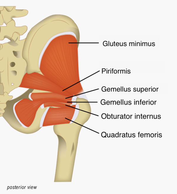
- Deep to the gluteal muscles, the piriformis, superior gemellus, obturator externus, inferior gemellus, obturator internus, and quadratus femoris laterally rotate the femur at the hip (Figure \(\PageIndex{2}\)).
Muscles of the Medial Compartment
Six muscles are located in the medial compartment of the thigh (Figure \(\PageIndex{3}\)). Most of these muscles perform adduction at the hip joint, and each performs other actions as well. The adductor longus, adductor brevis, gracilis, and pectineus also flex the hip joint. Adductor magnus, depending on which fibers are activated, can flex, extend, and laterally rotate the thigh. Gracilis is the only one of this groups of muscles that does not insert into the femur, it inserts into the posterior, medial aspect of the superior tibia which also allows it to cause flexion at the knee joint. The only muscle in this compartment that does not adduct is the obturator externus, this muscle's action is to laterally rotate the hip.


| Muscle | Origin | Insertion | Movement | Image(s) |
|---|---|---|---|---|
| Iliopsoas group | ||||
| Iliacus | Iliac fossa; iliac crest; lateral sacrum | Lesser trochanter of femur | Flexes hip |  |
| Psoas major | Lumbar vertebrae (L1 - L5); thoracic vertebrae (T12) | Lesser trochanter of femur | Flexes hip |  |
| Gluteal Group | ||||
| Gluteus maximus | Dorsal ilium; sacrum; coccyx | Gluteal tuberosity of femur; iliotibial tract | Extend and laterally rotates hip |  |
| Gluteus medius | Lateral surface of ilium | Greater trochanter of femur | Abducts and medially rotates hip |  |
| Gluteus minimus | External surface of ilium | Greater trochanter of femur | Abducts and medially rotates hip |  |
| Tensor fascia lata | Anterior aspect of iliac crest; anterior superior iliac spine | Iliotibial tract | Abducts and medially rotates hip |  |
| Deep Gluteal Muscles | ||||
| Inferior gemellus | Ischial tuberosity | Greater trochanter of femur | Laterally rotates hip; maintains posture by stabilizing hip joint |  |
| Obturator externus | Outer surfaces of obturator membrane, pubis, and ischium; margins of obturator foramen | Trochanteric fossa of posterior femur | Laterally rotates hip; maintains posture by stabilizing hip joint |  |
| Obturator internus | Inner surface of obturator membrane; greater sciatic notch; margins of obturator foramen | Greater trochanter in front of piriformis | Laterally rotates hip; maintains posture by stabilizing hip joint |  |
| Piriformis | Anterolateral surface of sacrum | Greater trochanter of femur | Laterally rotates hip; maintains posture by stabilizing hip joint |  |
| Quadratus femoris | Ischial tuberosity | Trochanteric crest of femur | Laterally rotates hip; maintains posture by stabilizing hip joint | 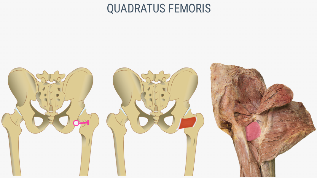 |
| Superior gemellus | Ischial spine | Greater trochanter of femur | Laterally rotates hip; maintains posture by stabilizing hip joint |  |
| Medial Muscles | ||||
| Adductor brevis | Body of pubis; inferior ramus of pubis | Linea aspera above adductor longus | Adducts and flexes hip | 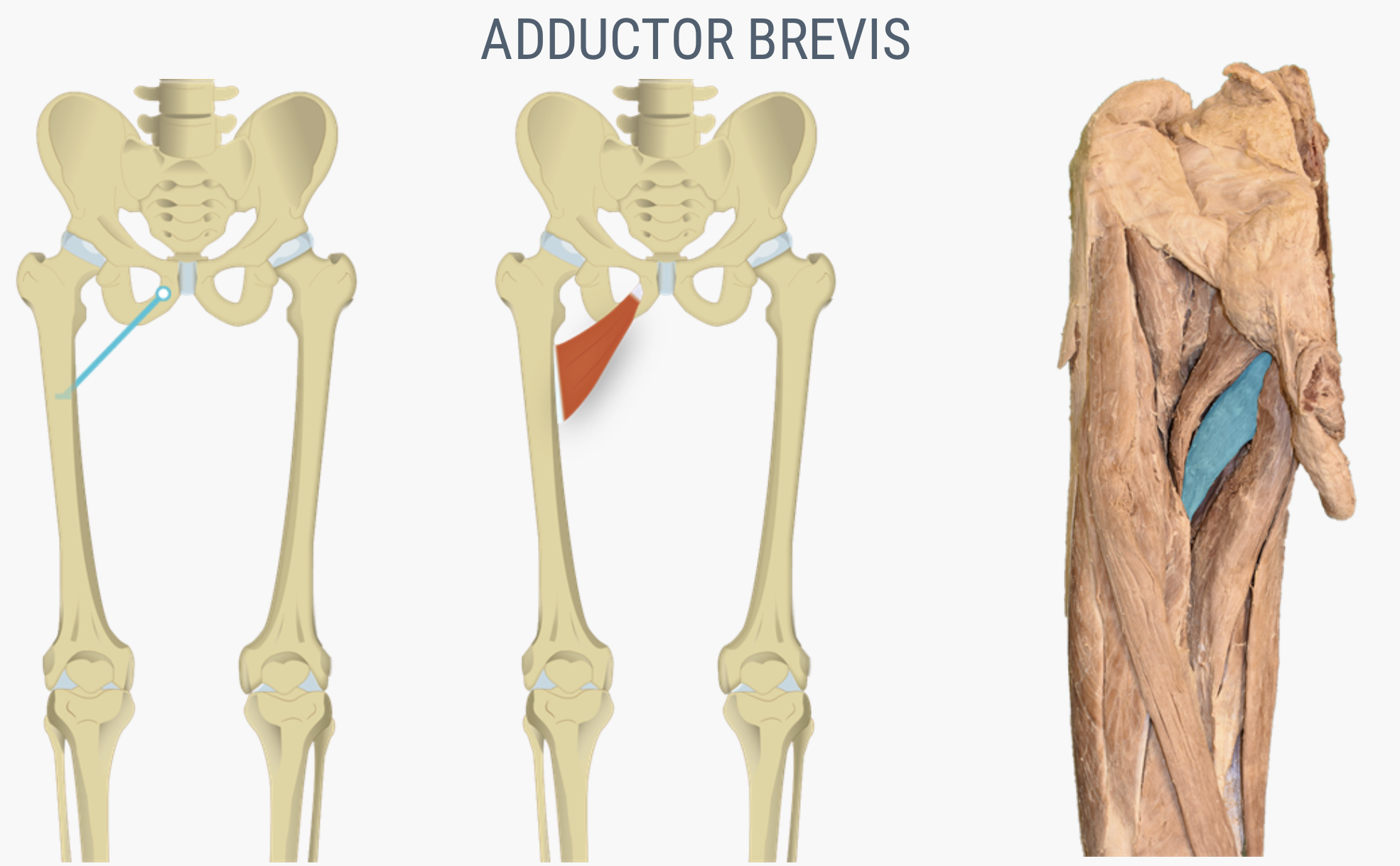 |
| Adductor longus | Pubis near pubic symphysis | Linea aspera | Adducts and flexes hip | 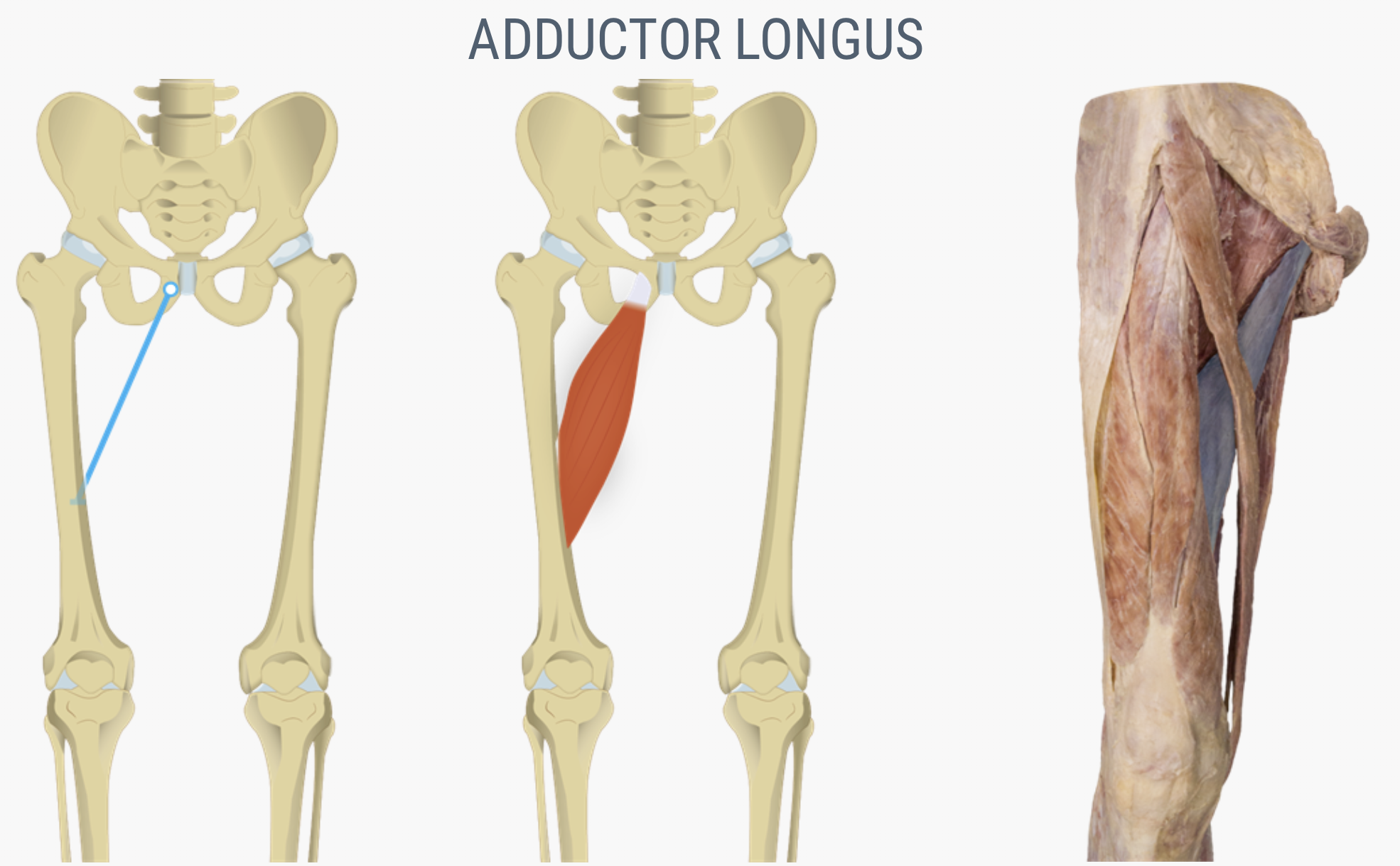 |
| Adductor magnus | Ischial rami; pubic rami; ischial tuberosity | Linea aspera; adductor tubercle of femur |
Adducts hip Posterior fibers: extends and laterally rotates hip Anterior fibers: flex hip |
 |
| Gracilis | Inferior ramus; body of pubis; ischial ramus | Medial surface of tibia | Adducts and flexes hip; flexes knee |  |
| Pectineus | Pectineal line of pubis | Lesser trochanter to linea aspera of posterior aspect of femur | Adducts and flexes hip |  |
Muscles of the Anterior Compartment
The muscles of the anterior compartment of the thigh flex the thigh and extend the leg. This compartment contains the quadriceps femoris group, which actually comprises four muscles that extend and stabilize the knee (Figure \(\PageIndex{4}\)). The rectus femoris is on the anterior aspect of the thigh, the vastus lateralis is on the lateral aspect of the thigh, the vastus medialis is on the medial aspect of the thigh, and the vastus intermedius is between the vastus lateralis and vastus medialis and deep to the rectus femoris. The tendon common to all four is the quadriceps tendon (patellar tendon), which inserts into the patella and continues below it as the patellar ligament. The patellar ligament attaches to the tibial tuberosity.


In addition to the quadriceps femoris, the sartorius is a band-like muscle that extends from the anterior superior iliac spine to the medial side of the proximal tibia (Figure \(\PageIndex{4}\)). This versatile muscle flexes the leg at the knee and flexes, abducts, and laterally rotates the leg at the hip. This muscle allows us to sit cross-legged.
| Muscle | Origin | Insertion | Movement | Image(s) |
|---|---|---|---|---|
| Anterior Compartment of Thigh: Quadricepts Femoris Group | ||||
| Rectus femoris | Anterior inferior iliac spine; superior margin of acetabulum | Patella; tibial tuberosity | Moves lower leg out in front of body, as when kicking; assists in raising knee |  |
| Sartorius | Anterior superior iliac spine | Medial aspect of proximal tibia | Moves back of lower legs up and back toward the buttocks, as when kneeling; assists in moving thigh diagonally upward and outward as when mounting a bike |  |
| Vastus intermedius | Proximal femur shaft | Patella; tibial tuberosity | Moves lower leg out in front of body, as when kicking | 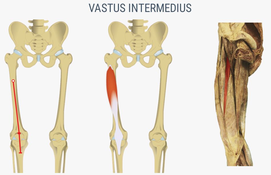 |
| Vastus lateralis | Greater trochanter; intertrochanteric line; linea apera | Patella; tibial tuberosity | Moves lower leg out in front of body, as when kicking | 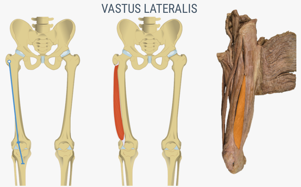 |
| Vastus medialis | Linea aspera; intertrochanteric line | Patella; tibial tuberosity | Moves lower leg out in front of body, as when kicking |  |
| Posterior Compartment of Thigh: Hamstring Group | ||||
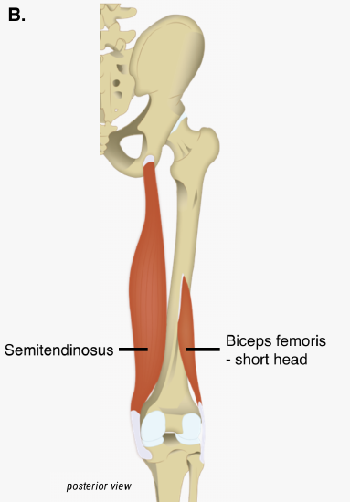
| Biceps femoris | Ischial tuberosity; linea aspera; distal femur | Head of fibula; lateral condyle of tibia | Moves back of lower legs up and back toward the buttocks, as when kneeling; moves thigh down and back; twists the thigh (and lower leg) outward |  |
| Semimembranosus | Ischial tuberosity | Medial condyle of tibia; lateral condyle of femur | Moves back of lower legs up and back toward the buttocks, as when kneeling; moves thigh down and back; twists the thigh (and lower leg) outward |  |
| Semitendinosus | Ischial tuberosity | Upper tibial shaft | Moves back of lower legs up and back toward the buttocks, as when kneeling; moves thigh down and back; twists the thigh (and lower leg) outward |  |
Muscles of the Posterior Compartment
The posterior compartment of the thigh includes muscles that flex the leg and extend the thigh. The three long muscles on the back of the knee are the hamstring group, which flexes the knee and extends the hip. These are the biceps femoris (long head), semitendinosus, and semimembranosus. The biceps femoris also has a short head that is not considered part of the hamstrings because it only flexes the knee and does not extend the hip (Figure \(\PageIndex{5}\)). The tendons of these muscles form the popliteal fossa, the diamond-shaped space at the back of the knee.


Muscles That Move the Feet and Toes
The muscles that move the ankle, foot, and toes and are located in the leg are called the crural muscles. Similar to the thigh muscles, the muscles of the leg are divided by fascia into compartments, although the leg has three: anterior, lateral, and posterior (Table \(\PageIndex{3}\)).
Muscles of the Anterior Compartment
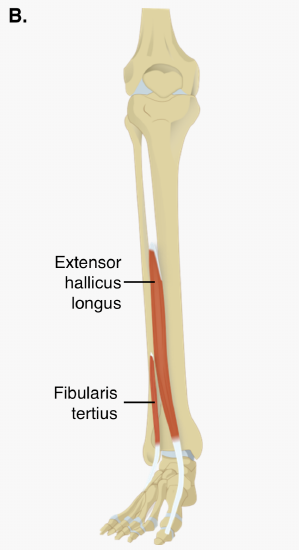
The muscles in the anterior compartment of the leg: the tibialis anterior, a long and thick muscle on the lateral surface of the tibia, the extensor hallucis longus, deep under it, and the extensor digitorum longus, lateral to it, all contribute to raising the front of the foot when they contract. Some, but not all, people have a third muscle in this group called the fibularis tertius (tertius="third"), a small muscle that originates on the anterior surface of the fibula, is associated with the extensor digitorum longus and sometimes fused to it (Figure \(\PageIndex{6}\)).


| Prime Mover | Origin | Insertion | Movement | Image(s) |
|---|---|---|---|---|
| Anterior Compartment of Leg | ||||
| Extensor digitorum longus | Lateral condyle of tibia; proximal portion of fibula; interosseous membrane | Middle and distal phalanges of toes 2 - 5 | Extends toes 2-4; dorsiflexion of ankle |  |
| Extensor hallucis longus | Anteromedial fibula shaft; interosseous membrane | Distal phalanx hallux (big toe) | Extends the big toe; dorsiflexion of ankle | 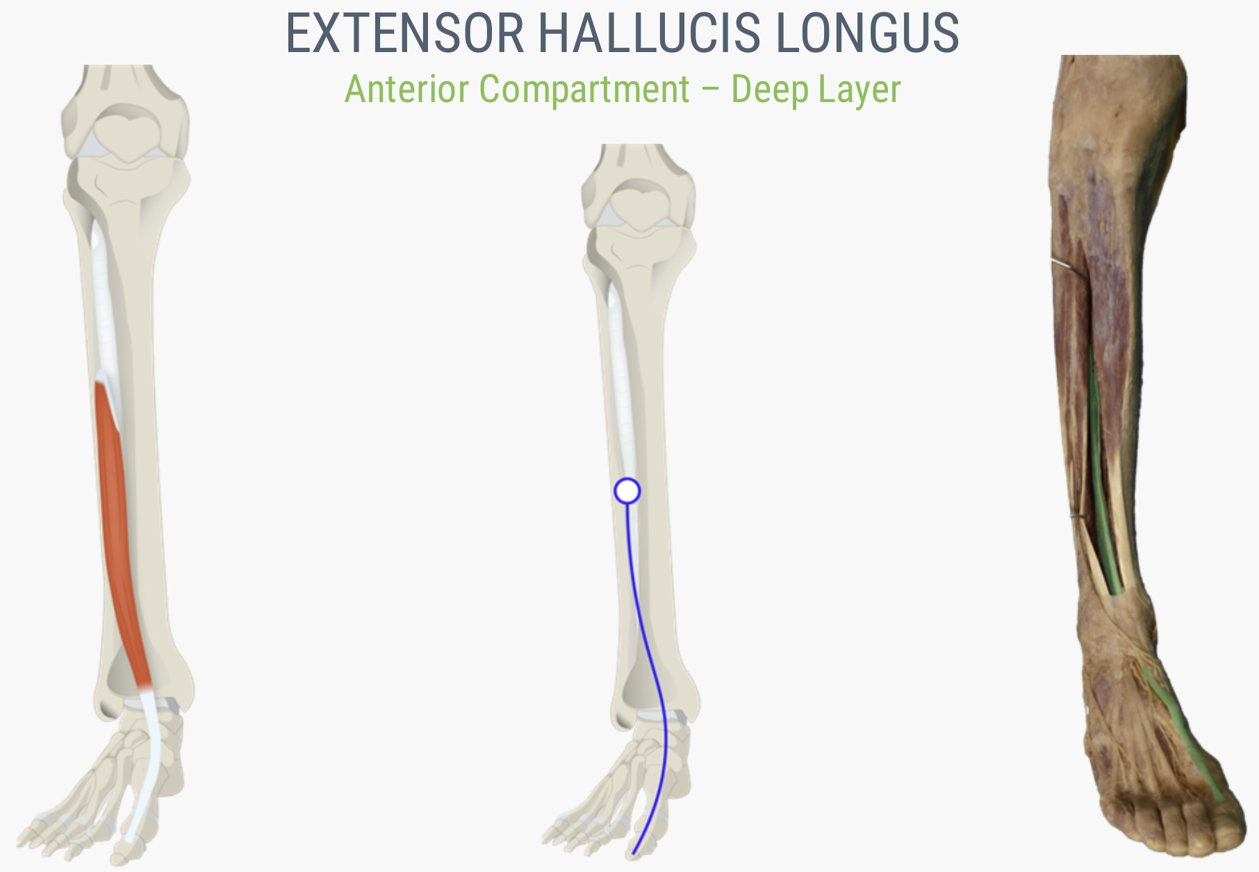 |
| Tibialis anterior | Lateral condyle and upper tibial shaft; interosseous membrane | Interior surface of medial cuneiform; first metatarsal bone | Dorsiflexion of ankle; inversion of intertarsal |  |
| Lateral Compartment of Leg | ||||
| Fibularis (peroneus) brevis | Distal fibula shaft | Proximal end of fifth metatarsal | Eversion of intertarsal; plantarflexion of ankle |  |
| Fibularis (peroneus) longus | Upper portion of lateral fibula | First metatarsal; medial cuneiform | Eversion of intertarsal, plantarflexion of ankle |  |
| Posterior Compartment of Leg: Superficial Muscles | ||||
| Gastrocnemius | Medial and lateral condyles of femur | Posterior calcaneus | Plantarflexion of ankle; flexion of knee |  |
| Soleus | Superior tibia; fibula; interosseous membrane | Posterior calcaneus | Plantarflexion of ankle |  |
| Posterior Compartment of Leg: Deep Muscles | ||||
| Flexor digitorum longus | Posterior tibia | Distal phalanges of toes 2 - 5 | Flexion of digits 2-5; plantarflexion of ankle |  |
| Flexor hallucis longus | Midshaft of fibula; interosseous membrane | Distal phalanx of hallux (big toe) | Flexes big toe; plantarflexion of ankle |  |
| Tibialis posterior | Superior tibia; fibula; interosseous membrane | Tarsals and metatarsals 2 - 4 | Plantarflexion of ankle; inversion of intertarsal |  |


The foot also has intrinsic muscles, which originate and insert within it (similar to the intrinsic muscles of the hand). These muscles primarily provide support for the foot and its arch, and contribute to movements of the toes. The principal support for the longitudinal arch of the foot is a deep fascia called plantar aponeurosis, which runs from the calcaneus bone to the toes (inflammation of this tissue is the cause of “plantar fasciitis,” which can affect runners.
Explore the muscles of the lower limb in this 3D rending of a human leg model.
Concept Review
The pelvic girdle attaches the legs to the axial skeleton. The hip joint is where the pelvic girdle and the leg come together. The hip is joined to the pelvic girdle by many muscles. In the gluteal region, the psoas major and iliacus form the iliopsoas. The large and strong gluteus maximus, gluteus medius, and gluteus minimus extend and abduct the femur. Along with the gluteus maximus, the tensor fascia lata muscle forms the iliotibial tract. The lateral rotators of the femur at the hip are the piriformis, obturator internus, obturator externus, superior gemellus, inferior gemellus, and quadratus femoris. On the medial part of the thigh, the adductor longus, adductor brevis, and adductor magnus adduct the thigh and medially rotate it. The pectineus muscle adducts and flexes the femur at the hip.
The thigh muscles that move the femur, tibia, and fibula are divided into medial, anterior, and posterior compartments. The medial compartment includes the adductors, pectineus, and the gracilis. The anterior compartment comprises the quadriceps femoris, quadriceps tendon, patellar ligament, and the sartorius. The quadriceps femoris is made of four muscles: the rectus femoris, the vastus lateralis, the vastus medius, and the vastus intermedius, which together extend the knee. The posterior compartment of the thigh includes the hamstrings: the biceps femoris, semitendinosus, and the semimembranosus, which all flex the knee.
The muscles of the leg that move the foot and toes are divided into anterior, lateral, superficial- and deep-posterior compartments. The anterior compartment includes the tibialis anterior, the extensor hallucis longus, the extensor digitorum longus, and the fibularis (peroneus) tertius. The lateral compartment houses the fibularis (peroneus) longus and the fibularis (peroneus) brevis. The superficial posterior compartment has the gastrocnemius, soleus, and plantaris; and the deep posterior compartment has the popliteus, tibialis posterior, flexor digitorum longus, and flexor hallucis longus.
Review Questions
Q. The large muscle group that attaches the leg to the pelvic girdle and produces extension of the hip joint is the ________ group.
A. gluteal
B. obturator
C. adductor
D. abductor
- Answer
-
Answer: A
Q. Which muscle produces movement that allows you to cross your legs?
A. the gluteus maximus
B. the piriformis
C. the gracilis
D. the sartorius
- Answer
-
Answer: D
Q. What is the largest muscle in the lower leg?
A. soleus
B. gastrocnemius
C. tibialis anterior
D. tibialis posterior
- Answer
-
Answer: B
Q. The vastus intermedius muscle is deep to which of the following muscles?
A. biceps femoris
B. rectus femoris
C. vastus medialis
D. vastus lateralis
- Answer
-
Answer: B
Critical Thinking Questions
Q. Which muscles form the hamstrings? How do they function together?
- Answer
-
A. The biceps femoris, semimembranosus, and semitendinosus form the hamstrings. The hamstrings flex the leg at the knee joint.
Q. Which muscles form the quadriceps? How do they function together?
- Answer
-
A. The rectus femoris, vastus medialis, vastus lateralis, and vastus intermedius form the quadriceps. The quadriceps muscles extend the leg at the knee joint.
Glossary
- adductor brevis
- muscle that adducts and medially rotates the thigh
- adductor longus
- muscle that adducts, medially rotates, and flexes the thigh
- adductor magnus
- muscle with an anterior fascicle that adducts, medially rotates and flexes the thigh, and a posterior fascicle that assists in thigh extension
- anterior compartment of the leg
- region that includes muscles that dorsiflex the foot
- anterior compartment of the thigh
- region that includes muscles that flex the thigh and extend the leg
- biceps femoris
- hamstring muscle
- calcaneal tendon
- (also, Achilles tendon) strong tendon that inserts into the calcaneal bone of the ankle
- extensor digitorum brevis
- muscle that extends the toes
- extensor digitorum longus
- muscle that is lateral to the tibialis anterior
- extensor hallucis longus
- muscle that is partly deep to the tibialis anterior and extensor digitorum longus
- femoral triangle
- region formed at the junction between the hip and the leg and includes the pectineus, femoral nerve, femoral artery, femoral vein, and deep inguinal lymph nodes
- fibularis brevis
- (also, peroneus brevis) muscle that plantar flexes the foot at the ankle and everts it at the intertarsal joints
- fibularis longus
- (also, peroneus longus) muscle that plantar flexes the foot at the ankle and everts it at the intertarsal joints
- fibularis tertius
- small muscle that is associated with the extensor digitorum longus
- flexor digitorum longus
- muscle that flexes the four small toes
- flexor hallucis longus
- muscle that flexes the big toe
- gastrocnemius
- most superficial muscle of the calf
- gluteal group
- muscle group that extends, flexes, rotates, adducts, and abducts the femur
- gluteus maximus
- largest of the gluteus muscles that extends the femur
- gluteus medius
- muscle deep to the gluteus maximus that abducts the femur at the hip
- gluteus minimus
- smallest of the gluteal muscles and deep to the gluteus medius
- gracilis
- muscle that adducts the thigh and flexes the leg at the knee
- hamstring group
- three long muscles on the back of the leg
- iliacus
- muscle that, along with the psoas major, makes up the iliopsoas
- iliopsoas group
- muscle group consisting of iliacus and psoas major muscles, that flexes the thigh at the hip, rotates it laterally, and flexes the trunk of the body onto the hip
- iliotibial tract
- muscle that inserts onto the tibia; made up of the gluteus maximus and connective tissues of the tensor fasciae latae
- inferior gemellus
- muscle deep to the gluteus maximus on the lateral surface of the thigh that laterally rotates the femur at the hip
- lateral compartment of the leg
- region that includes the fibularis (peroneus) longus and the fibularis (peroneus) brevis and their associated blood vessels and nerves
- medial compartment of the thigh
- a region that includes the adductor longus, adductor brevis, adductor magnus, pectineus, gracilis, and their associated blood vessels and nerves
- obturator externus
- muscle deep to the gluteus maximus on the lateral surface of the thigh that laterally rotates the femur at the hip
- obturator internus
- muscle deep to the gluteus maximus on the lateral surface of the thigh that laterally rotates the femur at the hip
- patellar ligament
- extension of the quadriceps tendon below the patella
- pectineus
- muscle that abducts and flexes the femur at the hip
- pelvic girdle
- hips, a foundation for the lower limb
- piriformis
- muscle deep to the gluteus maximus on the lateral surface of the thigh that laterally rotates the femur at the hip
- plantar aponeurosis
- muscle that supports the longitudinal arch of the foot
- plantaris
- muscle that runs obliquely between the gastrocnemius and the soleus
- popliteal fossa
- diamond-shaped space at the back of the knee
- popliteus
- muscle that flexes the leg at the knee and creates the floor of the popliteal fossa
- posterior compartment of the leg
- region that includes the superficial gastrocnemius, soleus, and plantaris, and the deep popliteus, flexor digitorum longus, flexor hallucis longus, and tibialis posterior
- posterior compartment of the thigh
- region that includes muscles that flex the leg and extend the thigh
- psoas major
- muscle that, along with the iliacus, makes up the iliopsoas
- quadratus femoris
- muscle deep to the gluteus maximus on the lateral surface of the thigh that laterally rotates the femur at the hip
- quadriceps femoris group
- four muscles, that extend and stabilize the knee
- quadriceps tendon
- (also, patellar tendon) tendon common to all four quadriceps muscles, inserts into the patella
- rectus femoris
- quadriceps muscle on the anterior aspect of the thigh
- sartorius
- band-like muscle that flexes, abducts, and laterally rotates the leg at the hip
- semimembranosus
- hamstring muscle
- semitendinosus
- hamstring muscle
- soleus
- wide, flat muscle deep to the gastrocnemius
- superior gemellus
- muscle deep to the gluteus maximus on the lateral surface of the thigh that laterally rotates the femur at the hip
- tensor fascia lata
- muscle that flexes and abducts the thigh
- tibialis anterior
- muscle located on the lateral surface of the tibia
- tibialis posterior
- muscle that plantar flexes and inverts the foot
- vastus intermedius
- quadricep muscle that is between the vastus lateralis and vastus medialis and is deep to the rectus femoris
- vastus lateralis
- quadricep muscle on the lateral aspect of the thigh
- vastus medialis
- quadricep muscle on the medial aspect of the thigh
Contributors and Attributions
OpenStax Anatomy & Physiology (CC BY 4.0). Access for free at https://openstax.org/books/anatomy-and-physiology