
17.1: Shared Structures
- Page ID
- 52811

Different types of blood vessels vary slightly in their structures, but they share the same general features. Arteries and arterioles have thicker walls than veins and venules because they are closer to the heart and receive blood that is surging at a far greater pressure (Figure 17.1). Each type of vessel has a lumen—a hollow passageway through which blood flows. Arteries have smaller lumens than veins, a characteristic that helps to maintain the pressure of blood moving through the system. Together, their thicker walls and smaller diameters give arterial lumens a more rounded appearance in cross section than the lumens of veins.
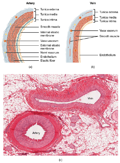
Figure \(\PageIndex{1}\): Structure of Blood Vessels (a) Arteries and (b) veins share the same general features, but the walls of arteries are much thicker because of the higher pressure of the blood that flows through them. (c) A micrograph shows the relative differences in thickness. LM × 160. (Micrograph provided by the Regents of the University of Michigan Medical School © 2012) (CC-BY-4.0, OpenStax, Human Anatomy)
Comparison of Tunics in Arteries and Veins
| Arteries | Veins | |
| General Appearance |
Thick Walls with small lumens |
Thin walls with large lumens Generally appear flattened |
| Tunica Intima | Endothelium usually appears wavy due to constriction of smooth muscle Internal elastic membrane presents in larger vessels |
Endolithelium appears smooth Internal elastic membrane absent |
| Tunica media | Normally the thickest layer in arteries Smooth muscle cells and elastic fibers predominate (the proportions of these vary with distance from the heart" External elastic membrane present in larger vessels |
Normally thinner than the tunica externa Smooth muscle cells and collagenous fibers predeominate Esternal elastic membrane absent |
| Tunica externa | Normally thinner than the tunica media in all but the largest arteries Collagenous and elastic fibers Nervi vasorum and vasa vasorum present |
Normally the thickest layer in veins Collagenous and smooth fibers predominate Some smooth muscle fibers Nervi vasoum and vasa vasorum present |