
2.13: The Electrooculogram
- Page ID
- 41663

\( \newcommand{\vecs}[1]{\overset { \scriptstyle \rightharpoonup} {\mathbf{#1}} } \)
\( \newcommand{\vecd}[1]{\overset{-\!-\!\rightharpoonup}{\vphantom{a}\smash {#1}}} \)
\( \newcommand{\dsum}{\displaystyle\sum\limits} \)
\( \newcommand{\dint}{\displaystyle\int\limits} \)
\( \newcommand{\dlim}{\displaystyle\lim\limits} \)
\( \newcommand{\id}{\mathrm{id}}\) \( \newcommand{\Span}{\mathrm{span}}\)
( \newcommand{\kernel}{\mathrm{null}\,}\) \( \newcommand{\range}{\mathrm{range}\,}\)
\( \newcommand{\RealPart}{\mathrm{Re}}\) \( \newcommand{\ImaginaryPart}{\mathrm{Im}}\)
\( \newcommand{\Argument}{\mathrm{Arg}}\) \( \newcommand{\norm}[1]{\| #1 \|}\)
\( \newcommand{\inner}[2]{\langle #1, #2 \rangle}\)
\( \newcommand{\Span}{\mathrm{span}}\)
\( \newcommand{\id}{\mathrm{id}}\)
\( \newcommand{\Span}{\mathrm{span}}\)
\( \newcommand{\kernel}{\mathrm{null}\,}\)
\( \newcommand{\range}{\mathrm{range}\,}\)
\( \newcommand{\RealPart}{\mathrm{Re}}\)
\( \newcommand{\ImaginaryPart}{\mathrm{Im}}\)
\( \newcommand{\Argument}{\mathrm{Arg}}\)
\( \newcommand{\norm}[1]{\| #1 \|}\)
\( \newcommand{\inner}[2]{\langle #1, #2 \rangle}\)
\( \newcommand{\Span}{\mathrm{span}}\) \( \newcommand{\AA}{\unicode[.8,0]{x212B}}\)
\( \newcommand{\vectorA}[1]{\vec{#1}} % arrow\)
\( \newcommand{\vectorAt}[1]{\vec{\text{#1}}} % arrow\)
\( \newcommand{\vectorB}[1]{\overset { \scriptstyle \rightharpoonup} {\mathbf{#1}} } \)
\( \newcommand{\vectorC}[1]{\textbf{#1}} \)
\( \newcommand{\vectorD}[1]{\overrightarrow{#1}} \)
\( \newcommand{\vectorDt}[1]{\overrightarrow{\text{#1}}} \)
\( \newcommand{\vectE}[1]{\overset{-\!-\!\rightharpoonup}{\vphantom{a}\smash{\mathbf {#1}}}} \)
\( \newcommand{\vecs}[1]{\overset { \scriptstyle \rightharpoonup} {\mathbf{#1}} } \)
\(\newcommand{\longvect}{\overrightarrow}\)
\( \newcommand{\vecd}[1]{\overset{-\!-\!\rightharpoonup}{\vphantom{a}\smash {#1}}} \)
\(\newcommand{\avec}{\mathbf a}\) \(\newcommand{\bvec}{\mathbf b}\) \(\newcommand{\cvec}{\mathbf c}\) \(\newcommand{\dvec}{\mathbf d}\) \(\newcommand{\dtil}{\widetilde{\mathbf d}}\) \(\newcommand{\evec}{\mathbf e}\) \(\newcommand{\fvec}{\mathbf f}\) \(\newcommand{\nvec}{\mathbf n}\) \(\newcommand{\pvec}{\mathbf p}\) \(\newcommand{\qvec}{\mathbf q}\) \(\newcommand{\svec}{\mathbf s}\) \(\newcommand{\tvec}{\mathbf t}\) \(\newcommand{\uvec}{\mathbf u}\) \(\newcommand{\vvec}{\mathbf v}\) \(\newcommand{\wvec}{\mathbf w}\) \(\newcommand{\xvec}{\mathbf x}\) \(\newcommand{\yvec}{\mathbf y}\) \(\newcommand{\zvec}{\mathbf z}\) \(\newcommand{\rvec}{\mathbf r}\) \(\newcommand{\mvec}{\mathbf m}\) \(\newcommand{\zerovec}{\mathbf 0}\) \(\newcommand{\onevec}{\mathbf 1}\) \(\newcommand{\real}{\mathbb R}\) \(\newcommand{\twovec}[2]{\left[\begin{array}{r}#1 \\ #2 \end{array}\right]}\) \(\newcommand{\ctwovec}[2]{\left[\begin{array}{c}#1 \\ #2 \end{array}\right]}\) \(\newcommand{\threevec}[3]{\left[\begin{array}{r}#1 \\ #2 \\ #3 \end{array}\right]}\) \(\newcommand{\cthreevec}[3]{\left[\begin{array}{c}#1 \\ #2 \\ #3 \end{array}\right]}\) \(\newcommand{\fourvec}[4]{\left[\begin{array}{r}#1 \\ #2 \\ #3 \\ #4 \end{array}\right]}\) \(\newcommand{\cfourvec}[4]{\left[\begin{array}{c}#1 \\ #2 \\ #3 \\ #4 \end{array}\right]}\) \(\newcommand{\fivevec}[5]{\left[\begin{array}{r}#1 \\ #2 \\ #3 \\ #4 \\ #5 \\ \end{array}\right]}\) \(\newcommand{\cfivevec}[5]{\left[\begin{array}{c}#1 \\ #2 \\ #3 \\ #4 \\ #5 \\ \end{array}\right]}\) \(\newcommand{\mattwo}[4]{\left[\begin{array}{rr}#1 \amp #2 \\ #3 \amp #4 \\ \end{array}\right]}\) \(\newcommand{\laspan}[1]{\text{Span}\{#1\}}\) \(\newcommand{\bcal}{\cal B}\) \(\newcommand{\ccal}{\cal C}\) \(\newcommand{\scal}{\cal S}\) \(\newcommand{\wcal}{\cal W}\) \(\newcommand{\ecal}{\cal E}\) \(\newcommand{\coords}[2]{\left\{#1\right\}_{#2}}\) \(\newcommand{\gray}[1]{\color{gray}{#1}}\) \(\newcommand{\lgray}[1]{\color{lightgray}{#1}}\) \(\newcommand{\rank}{\operatorname{rank}}\) \(\newcommand{\row}{\text{Row}}\) \(\newcommand{\col}{\text{Col}}\) \(\renewcommand{\row}{\text{Row}}\) \(\newcommand{\nul}{\text{Nul}}\) \(\newcommand{\var}{\text{Var}}\) \(\newcommand{\corr}{\text{corr}}\) \(\newcommand{\len}[1]{\left|#1\right|}\) \(\newcommand{\bbar}{\overline{\bvec}}\) \(\newcommand{\bhat}{\widehat{\bvec}}\) \(\newcommand{\bperp}{\bvec^\perp}\) \(\newcommand{\xhat}{\widehat{\xvec}}\) \(\newcommand{\vhat}{\widehat{\vvec}}\) \(\newcommand{\uhat}{\widehat{\uvec}}\) \(\newcommand{\what}{\widehat{\wvec}}\) \(\newcommand{\Sighat}{\widehat{\Sigma}}\) \(\newcommand{\lt}{<}\) \(\newcommand{\gt}{>}\) \(\newcommand{\amp}{&}\) \(\definecolor{fillinmathshade}{gray}{0.9}\)Eye movements can be recorded using electrodes placed on the skin near the eyes. This kind of recording is an electrooculogram or EOG. An EOG records eye movement because of a voltage difference between the cornea and the retina (Figure 1). As the eye moves, the vector of this electric field changes with respect to recording electrodes placed on the skin at fixed points.

The human eye has six muscles attached to its exterior surface. These muscles are grouped into three antagonistic pairs that control horizontal, vertical, and torsional movement and position of the eye:
Horizontal axis:
- the medial rectus muscle turns the eye toward the nose (medially) (adducts)
- the lateral rectus muscle turns the eye away from nose (laterally) (abducts)
Vertical axis:
- the superior rectus muscle turns the eye up (elevates) with a slight rotation toward the nose (medially) (adducts)
- the inferior rectus muscle turns the eye down (depresses) with a slight rotation towards the nose (medially) (adducts)
Torsional axis:
- the superior oblique muscle rotates the top of the eye toward the nose (laterally) (abducts) with a slight depression
- the inferior oblique muscle rotates the top of the eye away from the nose (laterally) (abducts) with a slight elevation
These muscles are innervated by motor neurons that have electrical activity with a tonic component that controls the position of the eye, and a phasic component that controls the velocity of eye movement. Even though the eye position commands and the eye velocity commands are linear functions of the firing frequency of the motor neuron, they are separate sets of commands. The eye velocity commands are sent along a direct path from specialized brain formations or fields to the motor neurons. However, the eye position commands are the products of the integration of eye velocity commands sent along an indirect path to a network of neurons that functions as a neural integrator. It is the output of the integrator that provides eye position commands to the motor neurons.
The integration of signals from different groups of neurons in the oculomotor system control five types of eye movement, each with a unique function and distinctive properties. These types are: saccades, pursuit, vestibular ocular reflex (VOR), vergence, and optokinetic reflex.
In this experiment, the subject will perform tasks that will generate electrical activity that will alter the standing voltage between the front and back of the eye that is correlated with horizontal eyeball movement. This movement will be obtained by electrodes placed on the skin near the eye. The record of this electrical activity is known as an electrooculogram (EOG).
These electrical changes are unique to each of four different types of eye movement (saccades, VOR, pursuit, and vergence). In our experiment, we will explore saccades, vestibular ocular reflex (VOR) and pursuit.
Saccades
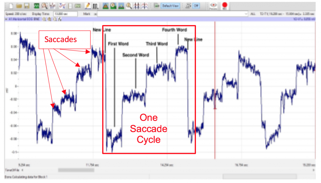
The fovea centralis (focal point) is the region of the retina that sees in detail. Saccadic eye movements rotate both eyes so that image of interest falls on the fovea. You are using saccades at this very moment to point the fovea of your eyes at the words in this sentence. To compensate for the poor vision that occurs during saccades, saccadic movements are quick, with a velocity from 400 degrees to as high as 800 degrees of movement in a second.

Saccades are also accurate because the saccadic system uses an internal estimate of eye position from its neural integrator to guide and stop the saccades. When looking at an object, saccades move the eye in all potential directions in the orbit, but they are interspersed with pauses in eye movement, called fixations (see the trace of eye movement in Figure 2). During fixations, the gaze is directed to a single location, only. In Figure 2, saccadic eye movement results in eye movements that allow the subject to view the different components of the picture, while fixations allow the subject to stop eye movement and focus on a single detailed feature of the picture.
Vestibular Ocular Reflex (VOR)
The vestibular ocular reflex keeps the image of the outside world stationary on the entire retina when the head moves. VOR connects lateral movement of the head in one direction with lateral movement of the eyes in the opposite direction. For example, if you rotate your head to the right as you look at this word, your eyes rotate to the left. VOR is a phasic response that is faster than pursuit because it is a simple central reflex arc that involves only three neurons. The signal that indicates the velocity of head movement originates in the semicircular canals of the ear and goes through an afferent nerve and an interneuron on its way to the motor neurons of the oculomotor muscles. The muscles rotate the eyes at a velocity that matches the velocity of the head, thus, keeping the image stationary on the retina.

The eyes are held on the image through a tonic response along an indirect path through a reverberating neural circuit between the afferent nerve and motor neurons. Without this neural circuit, which is a short-term memory device, the eyes would drift back to center and off the image while the head was still rotated.
Smooth Pursuit Eye Movements
Pursuit eye movement keeps the fovea pointed at a moving target, like a ball rolling on the horizon. Smooth pursuit movements may be vertical or horizontal, and each will produce a different pattern on the EOG. There is an initial delay (latency) in pursuit movement because the signal from the eye, that indicates the object is moving, has to be conducted through many synapses to the brainstem. Initially, when the object starts to move, a saccade helps the fovea catch up to the object until the pursuit movement begins to track the object. Smooth pursuit movements continuously track the moving target through eye movement, only (no head movement). Smooth pursuit movements are slow (up to 30 degrees per minute) and do not require fixations for focusing.
Methods
Set up
Start the Software
- Click on LabScribe3, Click Settings→ Human Muscle→ EOG
- All Students will be a subject in this experiment. Select the first person from your group to be the subject in this experiment.
- Use an alcohol swab to clean and scrub the areas where the electrodes will be placed (Methods Figure 1). Let the areas dry before attaching the electrodes.
- Trim the electrodes and then remove the plastic disk. Apply the electrodes to one of the scrubbed areas. Attach an electrode to each of the other areas.
- Attach the electrodes so that the red lead is located next to the left eye, the black lead is located next to the right eye, and the green ground is located under the right ear on/near the mastoid process.
- Electrode pads can be cut to avoid the hairline lateral to each eye and the hair line in the mastoid process area.
- Drape the leads for the electrodes and have the subject place the iWire electrode box on their lap. There should be no tension on the electrodes.
- The subject should sit quietly with their hands and iWire box on their lap.

Week 12: Electrooculogram Activity (EOG)
Note: for these activities, it is important to observe your subject’s physical eye movements.
Exercise 1: Saccades
Aim: To demonstrate the type of electrical activity that occurs in the muscles as the subject is reading using paragraphs in different settings.
The number of words in each line, the length of each line, and the formatting of the paragraph will affect the shape of the EOG recording.
Control:
- Once the electrode pads and leads are placed
- Subject will close their eyes and move their eyes left to right 4 times, and up and down 4 times and blink your eyes 4times while being recorded. This will provide a baseline and proof that electrodes are in the correct positions.
- Type in Mark box subject’s name, control, once you start Recording, click on Mark to place wording on data screen.
Narrow Normal Saccades
- Procedure for Narrow Normal Paragraph. Select writing sample #4 Narrow Normal Paragraph for the subject to read as his or her EOG is recording.
- The subject should avoid any voluntary movements of his or her head or body during the recording. ALL READING ARE TO BE DONE SILENTLY WITHOUT MOVING ANY FACIAL MUSCLES. Only the subject’s eyes should be moving while he or she is reading. Have subject hold the document at eye level.
- Type “Narrow Normal Saccades” in the Mark box to the right of the Mark button on the LabScribe3 Main window.
- As the subject is focusing on the first word in the paragraph, click Record. Instruct the subject to begin reading as you click on the Mark button to mark the recording.
- After the first cyclic pattern in completed, click on the AutoScale button. Observe the subject’s eyes as he or she is reading.
- Click Stop to halt recording when the subject has finished reading.
- Select Save As in the File menu, type Narrow Normal Paragraph and the subject’s name for the file. Click on the Save button to save the data file to Desktop to your Table’s Folder if desired.
Wide Normal Saccades
- Procedure for Wide Normal Paragraph. Select writing sample #11.
- The subject should avoid any voluntary movements of his or her head or body during the recording. Only the subject’s eyes should be moving while he or she is reading. Have subject hold the document at eye level.
- Type “Wide Normal Saccades” in the Mark box to the right of the Mark button on the LabScribe3 Main window. As the subject is focusing on the first wordin the paragraph, click Record. Click the mark button to mark the start of the reading.
- Click stop and save the recording.
Wide Slow Saccades
- Type Wide Slow Saccades in the Mark box to the right of the Mark button on the LabScribe3 Main window.
- Instruct the same subject: hold the document at eye level and move from word to word in the same paragraph using a slower saccadic movement. FOCUS on each word FOR TWO SECONDS, to do this say the word to yourself twice before moving to the next word. (Example: A,A, neurologist, neurologist, has, has, etc….)
- Record, mark and save the recording: Save as Narrow Slow Saccade and the subject’s name.
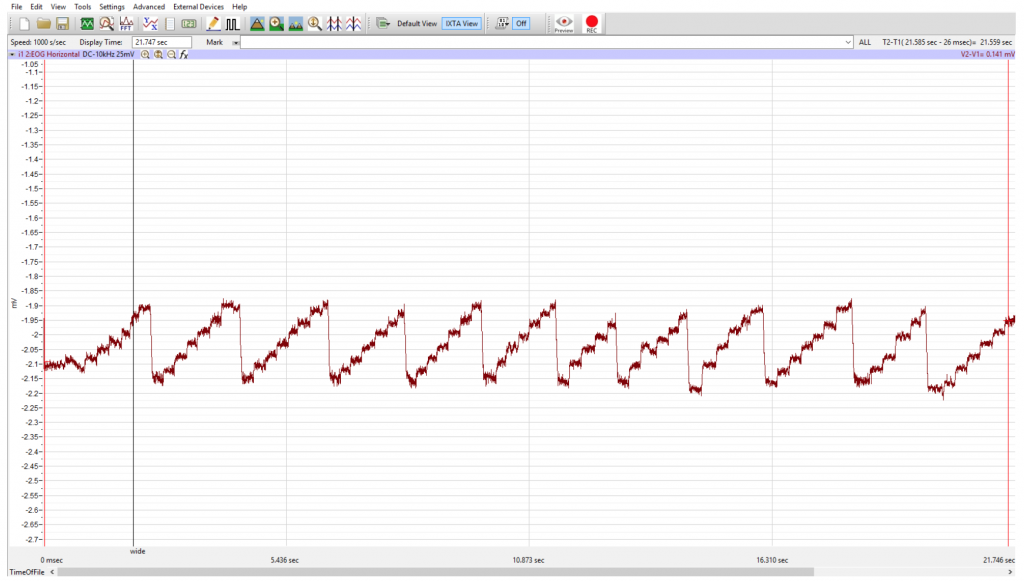
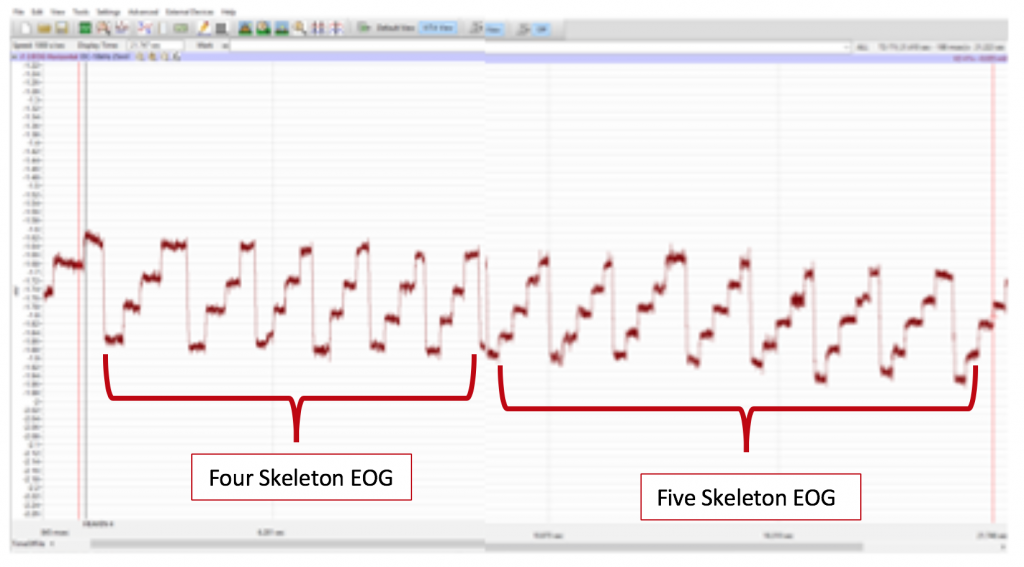
Repeat these same instructions, slowly reading and focusing on each word, for the Spaced Word paragraph (words space far apart in the document), the four skeleton document (each line having the word “skeleton” spaced throughout a line four times, and the five skeleton document (each line having the word “skeleton” space throughout five times).
Exercise 2: Vestibular Ocular Reflex (VOR)
Aim: To demonstrate the type of electrical activity that occurs in the muscles as the subject remains focused on an image or word on a page while rotating his or her head from side to side.
Note: Observe the subject’s eye movements as well as the pattern of the recording.
- Using the Card with VOR printed on it, have the subject focus on the letters VOR as the subject rotates his or her head from side to side.
- Inform the subject to avoid any voluntary movements of his or her BODY during the recording . Only the subject’s HEAD should be moving while he or she is focusing on the VOR card.
- Type “VOR”in the Mark boxto the right of the Mark button on the LabScribe3 Main window.
- As the subject is focusing on the VOR image and begins rotating his or her head first to the left side then to the right side. Continue moving head left to right while keeping their eyes focused on the VOR image. Click Record. Click on the Mark button to mark the recording. Have the subject do this for 15 seconds.
- After the first cyclic EOG pattern in completed: click on the AutoScale button and observe the subject’s eyes as the subject is rotating his or her head.
- When the subject has finished the exercise Click Stop.
- Select Save in the File menu.

Exercise 3: Pursuit Eye Movement
Aim: To demonstrate the type of electrical activity that occurs in the muscles as the subject follows a moving target vs a stationary target.
Use computers to view the computer-generated image of a moving ball:
For Horizontal Smooth Pursuit Test find the file named “Horizontal Ball” located on the P: drive.
- The subject must position their face 12 INCHES in front of the screen, prepare subject to focus on the LEFT SIDE of the computer screen to begin this exercise. Remind the subject that only their eyes will move in this exercise. You will record activity for Four Cycles, a Cycle is watching the dot from the LEFT to RIGHTand BACK TO LEFT. Do this Four Times.
- Type “Horizontal SmoothPursuit” in the Mark boxto the right of the Mark button. As the computer program is started, watch the screen and say “RECORDING” when the dots returns to the VERY LEFT HAND SIDE OF THE SCREEN and the subject begins following the target with his or her eyes.
- CLICK RECORD and Click MARK to place the Mark on the recording screen.
- REMIND the Subject that ONLY the SUBJECT’S EYES should be moving while he or she is following the target.
- Have subject follow the dot “LEFT to RIGHTand BACK TO LEFT” for FOUR CYCLES
After the first cyclic EOG pattern in completed,
- Click on the AutoScale button
- Observe the subject’s eyes as the subject is following the target.
6. When the target has completed FOUR CYCLES. Click STOP to halt recording Select Save in Your Table’s File on the Desktop. Title this “Horizontal Smooth Pursuit and the subject’s name.
Vertical Smooth Pursuit
For Vertical Smooth Pursuit Test find the file named “Vertical Ball” located on the P: drive.
Have COMPUTER UNPLUGED and SET AT EYE LEVEL and 12 INCHES in front of subject prepare subject to focus on the CENTER of the computer screen to begin this exercise. Remind the subject that only their eyes will move in this exercise. You will record activity for Four Cycles, a Cycle is watching the dot from the TOP TO BOTTOM BACK TO TOP. Do this Four Times.
- Type “Vertical SmoothPursuit” in the Mark box to the right of the Mark button.
- As the computer program is started, watch the screen and say “RECORDING” when the dots returns to the VERY TOP OF THE SCREEN and the subject begins following the target with his or her eyes.
- CLICK RECORD and Click MARK to place the Mark on the recording screen.
- REMIND the Subject that ONLY the SUBJECT’S EYES should be moving while he or she is following the target.
- Have subject follow the dot “TOP TO BOTTOM BACK TO TOP ” for FOUR CYCLES
After the first cyclic EOG pattern is completed,
- Click on the AutoScale button
- Observe the subject’s eyes as the subject is following the target.
6. When the target has completed FOUR CYCLES. Click STOP to halt recording Select Save in Your Table’s File on the Desktop. Title this “Vertical Smooth Pursuit and the subject’s name.
