
4.2: Diagnostic Tests
- Page ID
- 150532

\( \newcommand{\vecs}[1]{\overset { \scriptstyle \rightharpoonup} {\mathbf{#1}} } \)
\( \newcommand{\vecd}[1]{\overset{-\!-\!\rightharpoonup}{\vphantom{a}\smash {#1}}} \)
\( \newcommand{\dsum}{\displaystyle\sum\limits} \)
\( \newcommand{\dint}{\displaystyle\int\limits} \)
\( \newcommand{\dlim}{\displaystyle\lim\limits} \)
\( \newcommand{\id}{\mathrm{id}}\) \( \newcommand{\Span}{\mathrm{span}}\)
( \newcommand{\kernel}{\mathrm{null}\,}\) \( \newcommand{\range}{\mathrm{range}\,}\)
\( \newcommand{\RealPart}{\mathrm{Re}}\) \( \newcommand{\ImaginaryPart}{\mathrm{Im}}\)
\( \newcommand{\Argument}{\mathrm{Arg}}\) \( \newcommand{\norm}[1]{\| #1 \|}\)
\( \newcommand{\inner}[2]{\langle #1, #2 \rangle}\)
\( \newcommand{\Span}{\mathrm{span}}\)
\( \newcommand{\id}{\mathrm{id}}\)
\( \newcommand{\Span}{\mathrm{span}}\)
\( \newcommand{\kernel}{\mathrm{null}\,}\)
\( \newcommand{\range}{\mathrm{range}\,}\)
\( \newcommand{\RealPart}{\mathrm{Re}}\)
\( \newcommand{\ImaginaryPart}{\mathrm{Im}}\)
\( \newcommand{\Argument}{\mathrm{Arg}}\)
\( \newcommand{\norm}[1]{\| #1 \|}\)
\( \newcommand{\inner}[2]{\langle #1, #2 \rangle}\)
\( \newcommand{\Span}{\mathrm{span}}\) \( \newcommand{\AA}{\unicode[.8,0]{x212B}}\)
\( \newcommand{\vectorA}[1]{\vec{#1}} % arrow\)
\( \newcommand{\vectorAt}[1]{\vec{\text{#1}}} % arrow\)
\( \newcommand{\vectorB}[1]{\overset { \scriptstyle \rightharpoonup} {\mathbf{#1}} } \)
\( \newcommand{\vectorC}[1]{\textbf{#1}} \)
\( \newcommand{\vectorD}[1]{\overrightarrow{#1}} \)
\( \newcommand{\vectorDt}[1]{\overrightarrow{\text{#1}}} \)
\( \newcommand{\vectE}[1]{\overset{-\!-\!\rightharpoonup}{\vphantom{a}\smash{\mathbf {#1}}}} \)
\( \newcommand{\vecs}[1]{\overset { \scriptstyle \rightharpoonup} {\mathbf{#1}} } \)
\(\newcommand{\longvect}{\overrightarrow}\)
\( \newcommand{\vecd}[1]{\overset{-\!-\!\rightharpoonup}{\vphantom{a}\smash {#1}}} \)
\(\newcommand{\avec}{\mathbf a}\) \(\newcommand{\bvec}{\mathbf b}\) \(\newcommand{\cvec}{\mathbf c}\) \(\newcommand{\dvec}{\mathbf d}\) \(\newcommand{\dtil}{\widetilde{\mathbf d}}\) \(\newcommand{\evec}{\mathbf e}\) \(\newcommand{\fvec}{\mathbf f}\) \(\newcommand{\nvec}{\mathbf n}\) \(\newcommand{\pvec}{\mathbf p}\) \(\newcommand{\qvec}{\mathbf q}\) \(\newcommand{\svec}{\mathbf s}\) \(\newcommand{\tvec}{\mathbf t}\) \(\newcommand{\uvec}{\mathbf u}\) \(\newcommand{\vvec}{\mathbf v}\) \(\newcommand{\wvec}{\mathbf w}\) \(\newcommand{\xvec}{\mathbf x}\) \(\newcommand{\yvec}{\mathbf y}\) \(\newcommand{\zvec}{\mathbf z}\) \(\newcommand{\rvec}{\mathbf r}\) \(\newcommand{\mvec}{\mathbf m}\) \(\newcommand{\zerovec}{\mathbf 0}\) \(\newcommand{\onevec}{\mathbf 1}\) \(\newcommand{\real}{\mathbb R}\) \(\newcommand{\twovec}[2]{\left[\begin{array}{r}#1 \\ #2 \end{array}\right]}\) \(\newcommand{\ctwovec}[2]{\left[\begin{array}{c}#1 \\ #2 \end{array}\right]}\) \(\newcommand{\threevec}[3]{\left[\begin{array}{r}#1 \\ #2 \\ #3 \end{array}\right]}\) \(\newcommand{\cthreevec}[3]{\left[\begin{array}{c}#1 \\ #2 \\ #3 \end{array}\right]}\) \(\newcommand{\fourvec}[4]{\left[\begin{array}{r}#1 \\ #2 \\ #3 \\ #4 \end{array}\right]}\) \(\newcommand{\cfourvec}[4]{\left[\begin{array}{c}#1 \\ #2 \\ #3 \\ #4 \end{array}\right]}\) \(\newcommand{\fivevec}[5]{\left[\begin{array}{r}#1 \\ #2 \\ #3 \\ #4 \\ #5 \\ \end{array}\right]}\) \(\newcommand{\cfivevec}[5]{\left[\begin{array}{c}#1 \\ #2 \\ #3 \\ #4 \\ #5 \\ \end{array}\right]}\) \(\newcommand{\mattwo}[4]{\left[\begin{array}{rr}#1 \amp #2 \\ #3 \amp #4 \\ \end{array}\right]}\) \(\newcommand{\laspan}[1]{\text{Span}\{#1\}}\) \(\newcommand{\bcal}{\cal B}\) \(\newcommand{\ccal}{\cal C}\) \(\newcommand{\scal}{\cal S}\) \(\newcommand{\wcal}{\cal W}\) \(\newcommand{\ecal}{\cal E}\) \(\newcommand{\coords}[2]{\left\{#1\right\}_{#2}}\) \(\newcommand{\gray}[1]{\color{gray}{#1}}\) \(\newcommand{\lgray}[1]{\color{lightgray}{#1}}\) \(\newcommand{\rank}{\operatorname{rank}}\) \(\newcommand{\row}{\text{Row}}\) \(\newcommand{\col}{\text{Col}}\) \(\renewcommand{\row}{\text{Row}}\) \(\newcommand{\nul}{\text{Nul}}\) \(\newcommand{\var}{\text{Var}}\) \(\newcommand{\corr}{\text{corr}}\) \(\newcommand{\len}[1]{\left|#1\right|}\) \(\newcommand{\bbar}{\overline{\bvec}}\) \(\newcommand{\bhat}{\widehat{\bvec}}\) \(\newcommand{\bperp}{\bvec^\perp}\) \(\newcommand{\xhat}{\widehat{\xvec}}\) \(\newcommand{\vhat}{\widehat{\vvec}}\) \(\newcommand{\uhat}{\widehat{\uvec}}\) \(\newcommand{\what}{\widehat{\wvec}}\) \(\newcommand{\Sighat}{\widehat{\Sigma}}\) \(\newcommand{\lt}{<}\) \(\newcommand{\gt}{>}\) \(\newcommand{\amp}{&}\) \(\definecolor{fillinmathshade}{gray}{0.9}\)There are several diagnostic test abbreviations used in various healthcare settings. The tables below list abbreviations followed by key concepts and some example orders to help you understand the terms and the ways in which they could be used. Some abbreviations are very similar, with just a change in a capital or lowercase letter, but this change totally alters the meaning of the abbreviation.
| ABBREVIATION | MEANING |
| Angio | angiography |
| A&P | auscultation and percussion |
| Alb | albumin |
| Alk phos | alkaline phosphatase |
| Ba | barium |
| BaE | barium enema |
| BMR | basal metabolic rate |
| bs | blood sugar |
| BUN | blood urea nitrogen |
Key Concepts
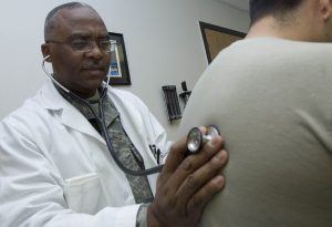
The abbreviation A&P (auscultation and percussion) is a form of assessment in which the healthcare provider auscultates, or listens, to a body part, usually with a stethoscope. In Figure 4.1, the physician is listening to a patient’s lungs with a stethoscope. When performing percussion, the physician uses their hand in a striking movement to a body part, then listens to the sound that occurs. A lower-pitched sound indicates that there could be a mass in that area, whereas a higher-pitched sound indicates air in the space.
What is barium, anyway? Barium is a liquid contrast medium that can be taken orally (barium swallow) or through the rectum (barium enema). It helps visualize the organs when certain types of imaging are performed; for example, X-rays or CT scans.
Examples of diagnostic test orders:
- BaE tomorrow am
- bs to be taken at 0800 and 1600 today
- Coronary Angio to rule out blockage ASAP
Explanation of diagnostic test orders:
- Barium enema tomorrow morning.
- Blood sugar to be taken at 8 o’clock in the morning and 4 o’clock in the afternoon today.
- Coronary angiogram to rule out blockage as soon as possible.
| ABBREVIATION | MEANING |
| C&S | culture and sensitivity test |
| Ca | calcium |
| CBC | complete blood count |
| CO2 | carbon dioxide |
| CSF | cerebrospinal fluid |
| C-spine | cervical spine films |
| CT | computerized tomography |
| CXR | chest X-ray |
| DI | diagnostic imaging |
| diff | differential |
Key Concepts
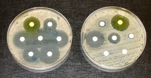
Figure 4.2 is an image of a culture and sensitivity (C&S) test used to help determine which antibiotics the bacteria being tested are sensitive to and which they are resistant to. In the dish on the left, the bacteria are all sensitive to the antibiotic, and in the dish on the right, most of the bacteria are resistant to the antibiotic, so it would not be an effective treatment. A culture and sensitivity test can be done on any specimen if an infection is suspected; for example, wound tissue, stool, or urine.
What is a diff? A diff (differential) is a blood test that measures the percentage of all the different types of blood cells in your blood, such as monocytes, basophils, and eosinophils. It is used to look for infections, allergic reactions, and certain diseases (Whitten, 2021).
Examples of diagnostic test orders:
- Urine for C&S STAT
- CBC with diff
- CXR ASAP
Explanation of diagnostic test orders:
- Urine for culture and sensitivity immediately.
- Complete blood count with differential.
- Chest X-ray as soon as possible.
| ABBREVIATION | MEANING |
| DRE | digital rectal exam |
| ECG | electrocardiogram |
| Echo | echocardiogram |
| EEG | electroencephalogram |
| EMG | electromyogram |
| ERCP | endoscopic retrograde cholangiopancreatography (test of the pancreas and gallbladder) |
| ESR | erythrocyte sedimentation rate |
| ESWL | extracorporeal shock wave lithotripsy |
| ETOH | level of alcohol consumption or ethyl alcohol |
| ETT | exercise tolerance test |
Key Concept
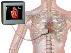
An ECG (electrocardiogram), as discussed in a previous chapter, records the electrical activity of the heart. An Echo (echocardiogram), on the other hand, uses sound waves to create an image of the heart, its structures, and how it is functioning to ensure that it is working correctly.
Examples of diagnostic test orders:
- ECG and Echo @ 0900
- Diagnostic EEG for seizure activity
- Patient to have ETT tomorrow am due to increased angina
Explanation of diagnostic test orders:
- Electrocardiogram and echocardiogram at 9 o’clock in the morning.
- Diagnostic electroencephalogram for seizure activity.
- Patient to have exercise tolerance test tomorrow morning due to increased angina.
| ABBREVIATION | MEANING |
| FBS | fasting blood sugar |
| Fe | iron |
| Ga scan | gallium scan |
| GTT | glucose tolerance test |
| H | hydrogen |
| hCG | human chorionic gonadotropin (pregnancy test) |
| Hct | hematocrit |
| HDL | high density lipoprotein |
| Hg | mercury |
| Hgb | hemoglobin |
Key Concepts
Fasting blood sugar (FBS) is a test performed to determine a person’s blood sugar level after fasting (not eating) for eight hours. This test is usually performed to determine whether or not a person has diabetes (Alberta Health Services, 2022a).
The glucose tolerance test (GTT) measures the body’s ability to process and use certain sugars, such as glucose. During an oral glucose tolerance test, a patient will drink a small amount of a very concentrated sweetened drink, then their glucose level is measured one to three hours later (Alberta Health Services, 2022a). Most of the time, FBS is taken prior to this test, and the results are compared. This test is most commonly used to detect gestational diabetes in pregnant women.
Examples of diagnostic test orders:
- FBS in AM NPO at 2400
- Add Hgb and Fe to routine lab tests
- HCG on arrival to unit
Explanation of diagnostic test orders:
- Fasting blood sugar in the morning and nothing to eat or drink at midnight.
- Add hemoglobin and iron to routine laboratory tests.
- Pregnancy test on arrival to unit.
| ABBREVIATION | MEANING |
| I | iodine |
| IVC | intravenous cholangiogram |
| IVP | intravenous pyelogram |
| K | potassium |
| KUB | kidneys, ureters, bladder |
| lab | laboratory |
| LDL | low-density lipoprotein |
| LFT | liver function test |
| Lymphs | lymphocytes |
| Lytes | electrolytes |
Key Concepts
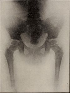
Fig 4.4 is an image of a KUB (kidneys, ureters, bladder) X-ray, which is used to visualize the urinary system and to assess the causes of lower abdominal structure pain, as well as to view the healthy functioning of the urinary system (Johns Hopkins, 2022).
The abbreviation lab (laboratory) is used for laboratory tests related to all specimens from the body, including blood, urine, feces, and other bodily specimens.
Examples of diagnostic test orders:
- HDL, LDL, and Lytes
- KUB tomorrow to assess left lower abdominal pain
- Add K to labs today
Explanation of diagnostic test orders:
- High-density lipoproteins, low-density lipoproteins, and electrolytes.
- Kidneys, ureter, and bladder tomorrow to assess left lower abdominal pain.
- Add potassium to laboratory tests today.
| ABBREVIATION | MEANING |
| Mg | magnesium |
| MRI | magnetic resonance imaging |
| MUGA | multigated acquisition scan (heart function test) |
| N | nitrogen |
| Na | sodium |
| O2 | oxygen |
| P | phosphate |
| PET | positron emission tomography |
Key Concept
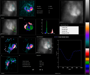
Figure 4.5 is an image of a MUGA (multigated acquisition) scan. The MUGA scan is used to assess the heart to determine how well it functions and, more specifically, how well it pumps blood to the organs in the body (Alberta Health Services, 2022b).
Examples of diagnostic test orders:
- Keep O2 level greater than or equal to 90%
- Add Mg, Na, and P to labs tomorrow
Explanation of diagnostic test orders:
- Keep oxygen level greater than or equal to 90 percent.
- Add magnesium, sodium, and phosphorus to laboratory tests tomorrow.
| ABBREVIATION | MEANING |
| PFT | pulmonary function test |
| PSA | prostate-specific antigen |
| PT/INR | prothrombin time/international normalized ratio |
| PTT | partial thromboplastin time |
| rbc | red blood cell |
| RBC | red blood count |
| spec | specimen |
| T | temperature |
| TENS | transcutaneous electrical nerve stimulation |
Key Concepts
A PT (prothrombin time) blood test is done to determine how long it takes blood to clot. This test might be ordered to determine whether a person has a bleeding disorder or if medications, such as coumadin, are working correctly. This test is also called INR (international normalized ratio) and is used to ensure that no matter how the test is completed, the results will all always be standardized (Alberta Health Services, 2022c).
PTT (partial thromboplastin time) is another test used to determine how long it takes blood to clot, and it is often done at the same time as a PT blood test. This test is also used to assess whether a person has a bleeding disorder such as hemophilia or if certain medications, such as heparin, are working effectively (Alberta Health Services, 2022c).
Examples of diagnostic test orders:
- Patient to have PFT to assess treatment effectiveness
- PT level 0800 call for coumadin orders after results
Explanation of diagnostic test orders:
- Patient to have pulmonary function test to assess treatment effectiveness.
- Prothrombin time level at 8 o’clock in the morning, call for coumadin orders after results.
Table 4.10. Diagnostic Tests
| ABBREVIATION | MEANING |
| U/A | urinalysis |
| U/O | urine output |
| UGI | upper gastrointestinal |
| US | ultrasound |
| VQ scan | ventilation-perfusion scan of the lungs |
| wbc | white blood cell |
| WBC | white blood count |
Key Concept
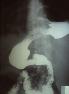
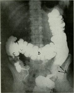
Figure 4.6 is an image of a UGI (upper gastrointestinal) series, and Fig 4.7 is an image of a BaE (barium enema) lower gastrointestinal series. Both of these diagnostic procedures involve the introduction of the contrast medium barium. For a UGI series, the barium would be swallowed, and for the lower gastrointestinal series, it would be given as an enema. X-rays are then taken of the upper and lower gastrointestinal system structures (Johns Hopkins, 2020).
Examples of diagnostic test orders:
- Throat swab for C&S
- 24 hr urine spec for Ca
- CBC, diff, Hgb & ESR
Explanation of diagnostic test orders:
- Take a throat swab for culture and sensitivity testing.
- Collect all the urine voided in a 24-hour period to be tested for calcium.
- Complete blood count, differential test for white blood cells, hemoglobin, and erythrocyte sedimentation rate.
Exercises
Attribution
Unless otherwise indicated, material on this page has been adapted from the following resource:
Carter, K., & Rutherford, M. (2020). Building a medical terminology foundation. eCampusOntario. https://ecampusontario.pressbooks.pub/medicalterminology/ licensed under CC BY 4.0
References
Alberta Health Services. (2022a). Blood glucose test. https://myhealth.alberta.ca/Health/Pages/conditions.aspx?hwid=hw8252
Alberta Health Services. (2022b). MUGA scan: About this test. https://myhealth.alberta.ca/health/AfterCareInformation/pages/conditions.aspx?HwId=abs1885
Alberta Health Services. (2022c). Partial thromboplastin time (PTT) test. https://myhealth.alberta.ca/health/tests-treatments/pages/conditions.aspx?Hwid=hw203152
Johns Hopkins. (2020). Kidney, ureter, and bladder X-ray. https://www.hopkinsmedicine.org/health/treatment-tests-and-therapies/kidney-ureter-and-bladder-xray#:~:text=A%20kidney%2C%20ureter%2C%20and%20bladder%20(KUB)%20X%2D,to%20assess%20the%20urinary%20system
LeClair, R. J. (2021). Cell biology, genetics, and biochemistry for pre-clinical students. Virginia Tech Carilion School of Medicine in association with Virginia Tech Publishing. https://pressbooks.lib.vt.edu/cellbio/ licensed under CC BY-NC-SA 4.0
Whitten, C. (2021). What is a differential blood count? WebMD. https://www.webmd.com/a-to-z-guides/what-is-differential-blood-count#:~:text=A%20differential%20blood%20count%20is,for%20infections%20and%20other%20problems
Image Credits (images are listed in order of appearance)
Stethoscope A by U.S. Air Force, Public domain
Antibiotic sensitivity and resistance by Dr. Graham Beards, CC BY-SA 4.0
Echocardiogram by BruceBlaus, CC BY-SA 4.0
American X-ray journal (1903) (14754979231) by Internet Archive Book Images, Public domain
Normal MUGA scan 1 by Jmarchn, CC BY-SA 3.0
Radiology 0032 Nevit by Nevit Dilmen, CC BY-SA 3.0
Image from page 525 of “The American journal of roentgenology, radium therapy and nuclear medicine” (1906) by Internet Archive Book Images, Public domain