
21.4: The Stomach
- Page ID
- 61628

\( \newcommand{\vecs}[1]{\overset { \scriptstyle \rightharpoonup} {\mathbf{#1}} } \)
\( \newcommand{\vecd}[1]{\overset{-\!-\!\rightharpoonup}{\vphantom{a}\smash {#1}}} \)
\( \newcommand{\dsum}{\displaystyle\sum\limits} \)
\( \newcommand{\dint}{\displaystyle\int\limits} \)
\( \newcommand{\dlim}{\displaystyle\lim\limits} \)
\( \newcommand{\id}{\mathrm{id}}\) \( \newcommand{\Span}{\mathrm{span}}\)
( \newcommand{\kernel}{\mathrm{null}\,}\) \( \newcommand{\range}{\mathrm{range}\,}\)
\( \newcommand{\RealPart}{\mathrm{Re}}\) \( \newcommand{\ImaginaryPart}{\mathrm{Im}}\)
\( \newcommand{\Argument}{\mathrm{Arg}}\) \( \newcommand{\norm}[1]{\| #1 \|}\)
\( \newcommand{\inner}[2]{\langle #1, #2 \rangle}\)
\( \newcommand{\Span}{\mathrm{span}}\)
\( \newcommand{\id}{\mathrm{id}}\)
\( \newcommand{\Span}{\mathrm{span}}\)
\( \newcommand{\kernel}{\mathrm{null}\,}\)
\( \newcommand{\range}{\mathrm{range}\,}\)
\( \newcommand{\RealPart}{\mathrm{Re}}\)
\( \newcommand{\ImaginaryPart}{\mathrm{Im}}\)
\( \newcommand{\Argument}{\mathrm{Arg}}\)
\( \newcommand{\norm}[1]{\| #1 \|}\)
\( \newcommand{\inner}[2]{\langle #1, #2 \rangle}\)
\( \newcommand{\Span}{\mathrm{span}}\) \( \newcommand{\AA}{\unicode[.8,0]{x212B}}\)
\( \newcommand{\vectorA}[1]{\vec{#1}} % arrow\)
\( \newcommand{\vectorAt}[1]{\vec{\text{#1}}} % arrow\)
\( \newcommand{\vectorB}[1]{\overset { \scriptstyle \rightharpoonup} {\mathbf{#1}} } \)
\( \newcommand{\vectorC}[1]{\textbf{#1}} \)
\( \newcommand{\vectorD}[1]{\overrightarrow{#1}} \)
\( \newcommand{\vectorDt}[1]{\overrightarrow{\text{#1}}} \)
\( \newcommand{\vectE}[1]{\overset{-\!-\!\rightharpoonup}{\vphantom{a}\smash{\mathbf {#1}}}} \)
\( \newcommand{\vecs}[1]{\overset { \scriptstyle \rightharpoonup} {\mathbf{#1}} } \)
\(\newcommand{\longvect}{\overrightarrow}\)
\( \newcommand{\vecd}[1]{\overset{-\!-\!\rightharpoonup}{\vphantom{a}\smash {#1}}} \)
\(\newcommand{\avec}{\mathbf a}\) \(\newcommand{\bvec}{\mathbf b}\) \(\newcommand{\cvec}{\mathbf c}\) \(\newcommand{\dvec}{\mathbf d}\) \(\newcommand{\dtil}{\widetilde{\mathbf d}}\) \(\newcommand{\evec}{\mathbf e}\) \(\newcommand{\fvec}{\mathbf f}\) \(\newcommand{\nvec}{\mathbf n}\) \(\newcommand{\pvec}{\mathbf p}\) \(\newcommand{\qvec}{\mathbf q}\) \(\newcommand{\svec}{\mathbf s}\) \(\newcommand{\tvec}{\mathbf t}\) \(\newcommand{\uvec}{\mathbf u}\) \(\newcommand{\vvec}{\mathbf v}\) \(\newcommand{\wvec}{\mathbf w}\) \(\newcommand{\xvec}{\mathbf x}\) \(\newcommand{\yvec}{\mathbf y}\) \(\newcommand{\zvec}{\mathbf z}\) \(\newcommand{\rvec}{\mathbf r}\) \(\newcommand{\mvec}{\mathbf m}\) \(\newcommand{\zerovec}{\mathbf 0}\) \(\newcommand{\onevec}{\mathbf 1}\) \(\newcommand{\real}{\mathbb R}\) \(\newcommand{\twovec}[2]{\left[\begin{array}{r}#1 \\ #2 \end{array}\right]}\) \(\newcommand{\ctwovec}[2]{\left[\begin{array}{c}#1 \\ #2 \end{array}\right]}\) \(\newcommand{\threevec}[3]{\left[\begin{array}{r}#1 \\ #2 \\ #3 \end{array}\right]}\) \(\newcommand{\cthreevec}[3]{\left[\begin{array}{c}#1 \\ #2 \\ #3 \end{array}\right]}\) \(\newcommand{\fourvec}[4]{\left[\begin{array}{r}#1 \\ #2 \\ #3 \\ #4 \end{array}\right]}\) \(\newcommand{\cfourvec}[4]{\left[\begin{array}{c}#1 \\ #2 \\ #3 \\ #4 \end{array}\right]}\) \(\newcommand{\fivevec}[5]{\left[\begin{array}{r}#1 \\ #2 \\ #3 \\ #4 \\ #5 \\ \end{array}\right]}\) \(\newcommand{\cfivevec}[5]{\left[\begin{array}{c}#1 \\ #2 \\ #3 \\ #4 \\ #5 \\ \end{array}\right]}\) \(\newcommand{\mattwo}[4]{\left[\begin{array}{rr}#1 \amp #2 \\ #3 \amp #4 \\ \end{array}\right]}\) \(\newcommand{\laspan}[1]{\text{Span}\{#1\}}\) \(\newcommand{\bcal}{\cal B}\) \(\newcommand{\ccal}{\cal C}\) \(\newcommand{\scal}{\cal S}\) \(\newcommand{\wcal}{\cal W}\) \(\newcommand{\ecal}{\cal E}\) \(\newcommand{\coords}[2]{\left\{#1\right\}_{#2}}\) \(\newcommand{\gray}[1]{\color{gray}{#1}}\) \(\newcommand{\lgray}[1]{\color{lightgray}{#1}}\) \(\newcommand{\rank}{\operatorname{rank}}\) \(\newcommand{\row}{\text{Row}}\) \(\newcommand{\col}{\text{Col}}\) \(\renewcommand{\row}{\text{Row}}\) \(\newcommand{\nul}{\text{Nul}}\) \(\newcommand{\var}{\text{Var}}\) \(\newcommand{\corr}{\text{corr}}\) \(\newcommand{\len}[1]{\left|#1\right|}\) \(\newcommand{\bbar}{\overline{\bvec}}\) \(\newcommand{\bhat}{\widehat{\bvec}}\) \(\newcommand{\bperp}{\bvec^\perp}\) \(\newcommand{\xhat}{\widehat{\xvec}}\) \(\newcommand{\vhat}{\widehat{\vvec}}\) \(\newcommand{\uhat}{\widehat{\uvec}}\) \(\newcommand{\what}{\widehat{\wvec}}\) \(\newcommand{\Sighat}{\widehat{\Sigma}}\) \(\newcommand{\lt}{<}\) \(\newcommand{\gt}{>}\) \(\newcommand{\amp}{&}\) \(\definecolor{fillinmathshade}{gray}{0.9}\)- Label on a diagram the four main regions of the stomach, its curvatures, and its sphincter
- Identify the four main types of secreting cells in gastric glands, and their important products
- Explain why the stomach does not digest itself
- Describe the mechanical and chemical digestion of food entering the stomach
Although a minimal amount of carbohydrate digestion occurs in the mouth, chemical digestion really gets underway in the stomach. An expansion of the alimentary canal that lies immediately inferior to the esophagus, the stomach links the esophagus to the first part of the small intestine (the duodenum) and is relatively fixed in place at its esophageal and duodenal ends. In between, however, it can be a highly active structure, contracting and continually changing position and size. These contractions provide mechanical assistance to digestion. The empty stomach is only about the size of your fist, but can stretch to hold as much as 4 liters of food and fluid, or more than 75 times its empty volume, and then return to its resting size when empty. Although you might think that the size of a person’s stomach is related to how much food that individual consumes, body weight does not correlate with stomach size. Rather, when you eat greater quantities of food—such as at holiday dinner—you stretch the stomach more than when you eat less.
Popular culture tends to refer to the stomach as the location where all digestion takes place. Of course, this is not true. An important function of the stomach is to serve as a temporary holding chamber. You can ingest a meal far more quickly than it can be digested and absorbed by the small intestine. Thus, the stomach holds food and parses only small amounts into the small intestine at a time. Foods are not processed in the order they are eaten; rather, they are mixed together with digestive juices in the stomach until they are converted into chyme, which is released into the small intestine.
As you will see in the sections that follow, the stomach plays several important roles in chemical digestion, including the continued digestion of carbohydrates and the initial digestion of proteins and triglycerides. Little if any nutrient absorption occurs in the stomach, with the exception of the negligible amount of nutrients in alcohol.
Structure
There are four main regions in the stomach: the cardia, fundus, body, and pylorus (Figure \(\PageIndex{1}\)). The cardia (or cardiac region) is the point where the esophagus connects to the stomach and through which food passes into the stomach. Located inferior to the diaphragm, above and to the left of the cardia, is the dome-shaped fundus. Below the fundus is the body, the main part of the stomach. The funnel-shaped pylorus connects the stomach to the duodenum. The wider end of the funnel, the pyloric antrum, connects to the body of the stomach. The narrower end is called the pyloric canal, which connects to the duodenum. The smooth muscle pyloric sphincter is located at this latter point of connection and controls stomach emptying. In the absence of food, the stomach deflates inward, and its mucosa and submucosa fall into a large fold called a ruga.
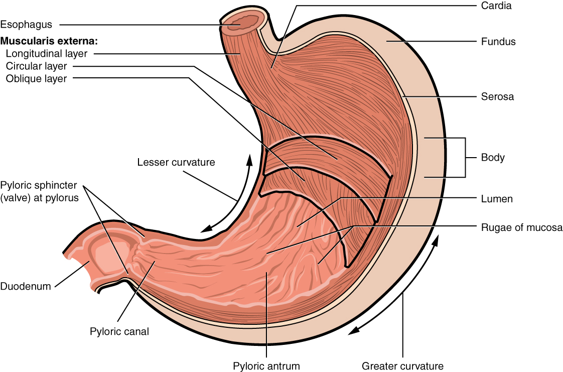
The convex lateral surface of the stomach is called the greater curvature; the concave medial border is the lesser curvature. The stomach is held in place by the lesser omentum, which extends from the liver to the lesser curvature, and the greater omentum, which runs from the greater curvature to the posterior abdominal wall.
Histology
The wall of the stomach is made of the same four layers as most of the rest of the alimentary canal, but with adaptations to the mucosa and muscularis for the unique functions of this organ. In addition to the typical circular and longitudinal smooth muscle layers, the muscularis has an inner oblique smooth muscle layer (Figure \(\PageIndex{2}\)). As a result, in addition to moving food through the canal, the stomach can vigorously churn food, mechanically breaking it down into smaller particles.
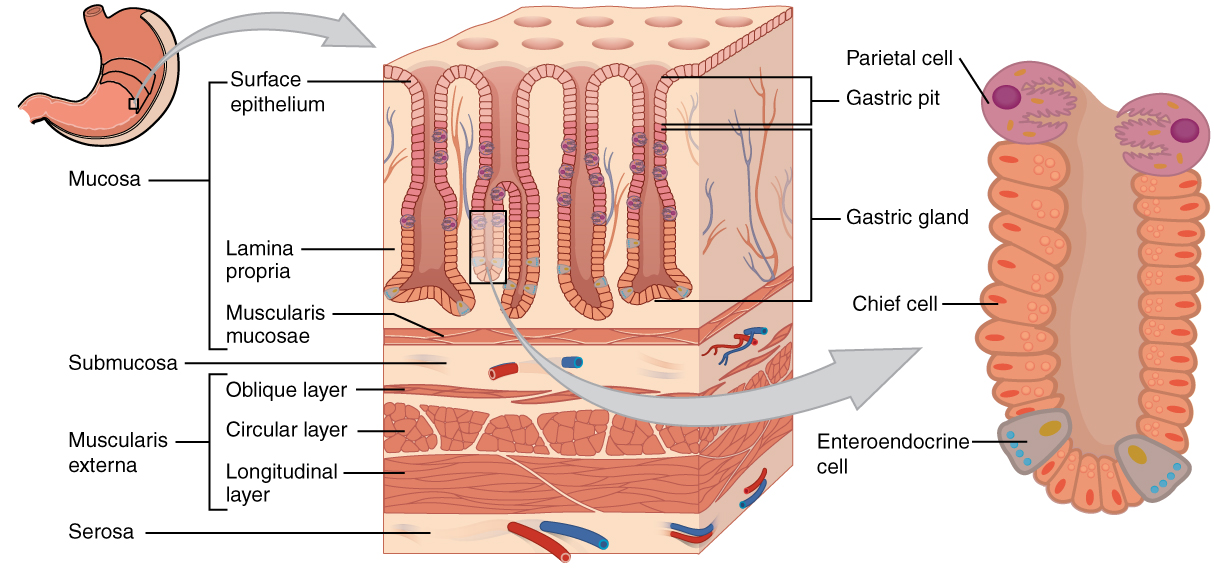
The stomach mucosa’s epithelial lining consists only of surface mucus cells, which secrete a protective coat of alkaline mucus. A vast number of gastric pits dot the surface of the epithelium, giving it the appearance of a well-used pincushion, and mark the entry to each gastric gland, which secretes a complex digestive fluid referred to as gastric juice.
Although the walls of the gastric pits are made up primarily of mucus cells, the gastric glands are made up of different types of cells. The glands of the cardia and pylorus are composed primarily of mucus-secreting cells. Cells that make up the pyloric antrum secrete mucus and a number of hormones, including the majority of the stimulatory hormone, gastrin. The much larger glands of the fundus and body of the stomach, the site of most chemical digestion, produce most of the gastric secretions. These glands are made up of a variety of secretory cells. These include parietal cells, chief cells, mucous neck cells, and enteroendocrine cells.
Parietal cells—Located primarily in the middle region of the gastric glands are parietal cells, which are among the most highly differentiated of the body’s epithelial cells. These relatively large cells produce both hydrochloric acid (HCl) and intrinsic factor. HCl is responsible for the high acidity (pH 1.5 to 3.5) of the stomach contents and is needed to activate the protein-digesting enzyme, pepsin. The acidity also kills much of the bacteria you ingest with food and helps to denature proteins, making them more available for enzymatic digestion. Intrinsic factor is a glycoprotein necessary for the absorption of vitamin B12 in the small intestine.
Chief cells—Located primarily in the basal regions of gastric glands are chief cells, which secrete pepsinogen, the inactive proenzyme form of pepsin. HCl is necessary for the conversion of pepsinogen to pepsin.
Mucous neck cells—Gastric glands in the upper part of the stomach contain mucous neck cells that secrete thin, acidic mucus that is much different from the mucus secreted by the goblet cells of the surface epithelium. The role of this mucus is not currently known.
Enteroendocrine cells—Finally, enteroendocrine cells found in the gastric glands secrete various hormones into the interstitial fluid of the lamina propria. These include gastrin, which is released mainly by enteroendocrine G cells.
Table \(\PageIndex{1}\) describes the digestive functions of important hormones secreted by the stomach.

Watch this animation that depicts the structure of the stomach and how this structure functions in the initiation of protein digestion. This view of the stomach shows the characteristic rugae. What is the function of these rugae?
| Hormone | Production site | Production stimulus | Target organ | Action |
|---|---|---|---|---|
| Gastrin | Stomach mucosa, mainly G cells of the pyloric antrum | Presence of peptides and amino acids in stomach | Stomach | Increases secretion by gastric glands; promotes gastric emptying |
| Gastrin | Stomach mucosa, mainly G cells of the pyloric antrum | Presence of peptides and amino acids in stomach | Small intestine | Promotes intestinal muscle contraction |
| Gastrin | Stomach mucosa, mainly G cells of the pyloric antrum | Presence of peptides and amino acids in stomach | Ileocecal valve | Relaxes valve |
| Gastrin | Stomach mucosa, mainly G cells of the pyloric antrum | Presence of peptides and amino acids in stomach | Large intestine | Triggers mass movements |
| Ghrelin | Stomach mucosa, mainly fundus | Fasting state (levels increase just prior to meals) | Hypothalamus | Regulates food intake, primarily by stimulating hunger and satiety |
| Histamine | Stomach mucosa | Presence of food in the stomach | Stomach | Stimulates parietal cells to release HCl |
| Serotonin | Stomach mucosa | Presence of food in the stomach | Stomach | Contracts stomach muscle |
| Somatostatin | Mucosa of stomach, especially pyloric antrum; also duodenum | Presence of food in the stomach; sympathetic axon stimulation | Stomach | Restricts all gastric secretions, gastric motility, and emptying |
| Somatostatin | Mucosa of stomach, especially pyloric antrum; also duodenum | Presence of food in the stomach; sympathetic axon stimulation | Pancreas | Restricts pancreatic secretions |
| Somatostatin | Mucosa of stomach, especially pyloric antrum; also duodenum | Presence of food in the stomach; sympathetic axon stimulation | Small intestine | Reduces intestinal absorption by reducing blood flow |
Chapter Review
The stomach participates in all digestive activities except ingestion and defecation. It vigorously churns food. It secretes gastric juices that break down food and absorbs certain drugs, including aspirin and some alcohol. The stomach begins the digestion of protein and continues the digestion of carbohydrates and fats. It stores food as an acidic liquid called chyme, and releases it gradually into the small intestine through the pyloric sphincter.
Interactive Link Questions
Watch this animation that depicts the structure of the stomach and how this structure functions in the initiation of protein digestion. This view of the stomach shows the characteristic rugae. What is the function of these rugae?
Answer: Answers may vary.
Review Questions
Q. Which of these cells secrete hormones?
A. parietal cells
B. mucous neck cells
C. enteroendocrine cells
D. chief cells
Answer: C
Q. Parietal cells secrete ________.
A. gastrin
B. hydrochloric acid
C. pepsin
D. pepsinogen
Answer: B
Critical Thinking Questions
Q. Describe unique anatomical features that enable the stomach to perform digestive functions.
A. The stomach has an additional inner oblique smooth muscle layer that helps the muscularis churn and mix food. The epithelium includes gastric glands that secrete gastric fluid. The gastric fluid consists mainly of mucous, HCl, and the enzyme pepsin released as pepsinogen.
Glossary
- body
- mid-portion of the stomach
- cardia
- (also, cardiac region) part of the stomach surrounding the cardiac orifice (esophageal hiatus)
- cephalic phase
- (also, reflex phase) initial phase of gastric secretion that occurs before food enters the stomach
- chief cell
- gastric gland cell that secretes pepsinogen
- enteroendocrine cell
- gastric gland cell that releases hormones
- fundus
- dome-shaped region of the stomach above and to the left of the cardia
- G cell
- gastrin-secreting enteroendocrine cell
- gastric emptying
- process by which mixing waves gradually cause the release of chyme into the duodenum
- gastric gland
- gland in the stomach mucosal epithelium that produces gastric juice
- gastric phase
- phase of gastric secretion that begins when food enters the stomach
- gastric pit
- narrow channel formed by the epithelial lining of the stomach mucosa
- gastrin
- peptide hormone that stimulates secretion of hydrochloric acid and gut motility
- hydrochloric acid (HCl)
- digestive acid secreted by parietal cells in the stomach
- intrinsic factor
- glycoprotein required for vitamin B12 absorption in the small intestine
- intestinal phase
- phase of gastric secretion that begins when chyme enters the intestine
- mixing wave
- unique type of peristalsis that occurs in the stomach
- mucosal barrier
- protective barrier that prevents gastric juice from destroying the stomach itself
- mucous neck cell
- gastric gland cell that secretes a uniquely acidic mucus
- parietal cell
- gastric gland cell that secretes hydrochloric acid and intrinsic factor
- pepsinogen
- inactive form of pepsin
- pyloric antrum
- wider, more superior part of the pylorus
- pyloric canal
- narrow, more inferior part of the pylorus
- pyloric sphincter
- sphincter that controls stomach emptying
- pylorus
- lower, funnel-shaped part of the stomach that is continuous with the duodenum
- ruga
- fold of alimentary canal mucosa and submucosa in the empty stomach and other organs
- stomach
- alimentary canal organ that contributes to chemical and mechanical digestion of food from the esophagus before releasing it, as chyme, to the small intestine
Contributors and Attributions
OpenStax Anatomy & Physiology (CC BY 4.0). Access for free at https://openstax.org/books/anatomy-and-physiology