
2.5: Lab Exercise 6- The Skeletal System—Microscopic and Gross Anatomy of Bones
- Page ID
- 72635
\( \newcommand{\vecs}[1]{\overset { \scriptstyle \rightharpoonup} {\mathbf{#1}} } \)
\( \newcommand{\vecd}[1]{\overset{-\!-\!\rightharpoonup}{\vphantom{a}\smash {#1}}} \)
\( \newcommand{\id}{\mathrm{id}}\) \( \newcommand{\Span}{\mathrm{span}}\)
( \newcommand{\kernel}{\mathrm{null}\,}\) \( \newcommand{\range}{\mathrm{range}\,}\)
\( \newcommand{\RealPart}{\mathrm{Re}}\) \( \newcommand{\ImaginaryPart}{\mathrm{Im}}\)
\( \newcommand{\Argument}{\mathrm{Arg}}\) \( \newcommand{\norm}[1]{\| #1 \|}\)
\( \newcommand{\inner}[2]{\langle #1, #2 \rangle}\)
\( \newcommand{\Span}{\mathrm{span}}\)
\( \newcommand{\id}{\mathrm{id}}\)
\( \newcommand{\Span}{\mathrm{span}}\)
\( \newcommand{\kernel}{\mathrm{null}\,}\)
\( \newcommand{\range}{\mathrm{range}\,}\)
\( \newcommand{\RealPart}{\mathrm{Re}}\)
\( \newcommand{\ImaginaryPart}{\mathrm{Im}}\)
\( \newcommand{\Argument}{\mathrm{Arg}}\)
\( \newcommand{\norm}[1]{\| #1 \|}\)
\( \newcommand{\inner}[2]{\langle #1, #2 \rangle}\)
\( \newcommand{\Span}{\mathrm{span}}\) \( \newcommand{\AA}{\unicode[.8,0]{x212B}}\)
\( \newcommand{\vectorA}[1]{\vec{#1}} % arrow\)
\( \newcommand{\vectorAt}[1]{\vec{\text{#1}}} % arrow\)
\( \newcommand{\vectorB}[1]{\overset { \scriptstyle \rightharpoonup} {\mathbf{#1}} } \)
\( \newcommand{\vectorC}[1]{\textbf{#1}} \)
\( \newcommand{\vectorD}[1]{\overrightarrow{#1}} \)
\( \newcommand{\vectorDt}[1]{\overrightarrow{\text{#1}}} \)
\( \newcommand{\vectE}[1]{\overset{-\!-\!\rightharpoonup}{\vphantom{a}\smash{\mathbf {#1}}}} \)
\( \newcommand{\vecs}[1]{\overset { \scriptstyle \rightharpoonup} {\mathbf{#1}} } \)
\( \newcommand{\vecd}[1]{\overset{-\!-\!\rightharpoonup}{\vphantom{a}\smash {#1}}} \)
Lab Summary: In this lab, you will explore the macroscopic and microscopic anatomy and physiology of bones and their tissues. You will also review functions of some key accessory structures of the skeletal system, including the locations and functions of ligaments, tendons, hyaline cartilage, elastic cartilage, and fibrocartilage.
Your objectives for this lab are:
- Differentiate between the appendicular and axial skeleton in terms of function and bones included in each division
- List examples of long, short, sesamoid, irregular, and flat bones
- In a real bone and in models of bone structure, locate and define the functions of the following macroscopic features
- Compact/cancellous bone
- Spongy bone/trabecular bone o Epiphysis
- Diaphysis
- Medullary cavity
- Red bone marrow
- Yellow bone marrow
- Articular cartilage
- Epiphyseal line
- Periosteum
- Endosteum
- In models of microscopic bone structure, locate and define the functions of o Central canal
- Lacuna(e)
- Canaliculi
- Osteocytes
- Lamella(e)
- Trabeculae
- Sharpey’s fibers o Osteon
- Compact/cancellous bone
- Spongy bone/trabecular bone o Medullary cavity
- Red bone marrow
- Periosteum
- Endosteum
- In a slide of compact bone, identify and define the functions of o Central canal
- Lacuna(e)
- Canaliculi
- Osteocytes
- Lamella(e)
- Explain bone.
the effects of acid and heat on the molecular composition and macroscopic structure of a long
- Identify the locations and functions of hyaline cartilage, fibrocartilage, and elastic cartilage
- Identify the tissues that composes tendons and ligaments
- Identify the locations and functions of tendons and ligaments
Background Information
Bone, or osseous tissue, is a hard, dense connective tissue that forms most of the adult skeleton. In the areas of the skeleton where bones move (for example, the ribcage and joints), cartilage, a semi-rigid form of connective tissue, provides flexibility and smooth surfaces for movement. The skeletal system is composed of bones, ligaments, and cartilages that enable it to perform several critical functions for the human body including supporting the body, facilitating movement, protecting internal organs, producing blood cells, and storing and releasing minerals and fat.
Activity 6.1: Major Divisions of the Skeleton & Classifications of Bones
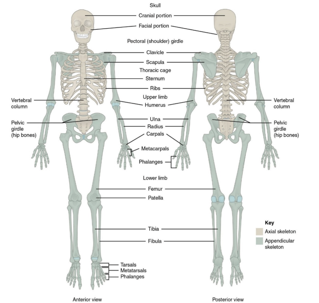
For adults, there are 206 bones in the skeleton. Younger individuals have higher numbers of bones because some bones fuse together during childhood and adolescence to form an adult bone. The skeleton is subdivided into two major divisions—the axial and appendicular.
The axial skeleton, consisting of the 80 bones of the skull vertebral column, and thoracic cage, forms the vertical, central axis of the body (Figure 6.1). It serves to protect the brain, spinal cord, heart, and lungs. It also serves as the attachment site for muscles that move the head, neck, and back, and for muscles that act across the shoulder and hip joints to move their corresponding limbs.
The appendicular skeleton includes all 126 bones of the upper and lower limbs, plus the bones that attach each limb to the axial skeleton (Figure \(\PageIndex{1}\)). These bones are divided into two groups: the bones that are located within the limbs themselves, and the girdle bones that attach the limbs to the axial skeleton. The bones of the shoulder region form the pectoral girdle, which anchors the upper limb to the thoracic cage of the axial skeleton. The lower limb is attached to the vertebral column by the pelvic girdle.
Classification of Bone Shapes
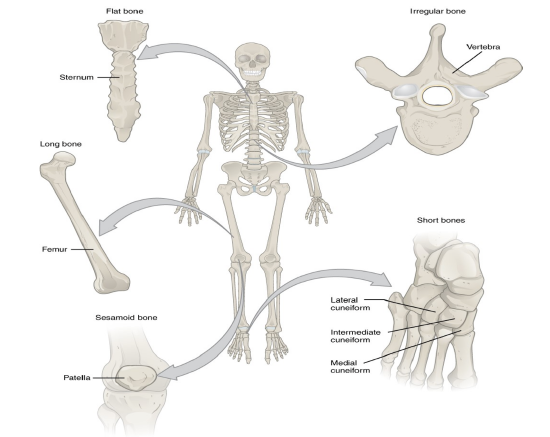
The 206 bones that compose the adult skeleton are divided into five categories based on their shapes (Figure \(\PageIndex{2}\)). Their shapes and their functions are related such that each categorical shape of bone has a distinct function.
Long Bones A long bone is one that is cylindrical and is longer than it is wide. Long bones are found in the arms (humerus, ulna, radius) and legs (femur, tibia, fibula), as well as in the fingers (metacarpals, phalanges) and toes (metatarsals, phalanges). Long bones function as levers; they move when muscles contract.
Short Bones A short bone is one that is cube-like in shape, being approximately equal in length, width, and thickness. The only short bones in the human skeleton are in the carpals of the wrists and the tarsals of the ankles. Short bones provide stability and support as well as some limited motion.
Flat Bones The term “flat bone” is somewhat of a misnomer because, although a flat bone is typically thin, it is also often curved. Examples include the cranial (skull) bones, the scapulae (shoulder blades), the sternum (breastbone), and the ribs. Flat bones serve as points of attachment for muscles and often protect internal organs.
Irregular Bones An irregular bone is one that does not have any easily characterized shape and therefore does not fit any other classification. These bones tend to have more complex shapes, like the vertebrae that support the spinal cord and protect it from compressive forces. Many facial bones, particularly the ones containing sinuses, are classified as irregular bones.
Sesamoid Bones A sesamoid bone is a small, round bone that, as the name suggests, is shaped like a sesame seed. These bones form in tendons (the sheaths of tissue that connect bones to muscles) where a great deal of pressure is generated in a joint. Sesamoid bones protect tendons by helping them overcome compressive forces. Sesamoid bones vary in number and placement from person to person but are typically found in tendons associated with the feet, hands, and knees. The patellae (singular = patella) are the only sesamoid bones found in common with every person.
Procedure for Activity 6.1: Classifications of Bones
1. Obtain a skeleton from your lab kit or the classroom lab supplies. Use the information above and your skeleton to fill in the “Classification of Bone Shape” and “Is this an axial or appendicular bone?” columns for each bone listed on the left in the table below.
Note if you are doing this lab remotely,
- you can use Complete Anatomy (if you have a student subscription; you are not required to purchase a subscription if you are doing this lab with RVCC), or
- you can a picture, such as this one: https://s3-us-west-2.amazonaws.com/courses-images-archive-read-only/wp content/uploads/sites/18/2014/07/19181404/801_Appendicular_Skeleton.jpg
|
Bone Name |
Classification of Bone Shape |
Is this an axial or appendicular bone? |
|
Skull bones of the face |
|
|
|
Scapula (“shoulder” bone) |
|
|
|
Radius (lateral forearm bone) |
|
|
|
Tarsal bones (7 bones of the ankle) |
|
|
|
Phalanges (“finger and toe” bones) |
|
|
|
Ribs |
|
|
|
Patella (“knee” bone) |
|
|
Activity 6.2: Gross Anatomy of Long Bones
The structure of a long bone allows for the best visualization of all of the parts of a bone (Figure \(\PageIndex{3}\)). A long bone has two major parts: the diaphysis and the epiphysis. The diaphysis is the tubular shaft that runs between the proximal and distal ends of the bone which are called epiphyses. The hollow region in the diaphysis is called the medullary cavity, which is filled with yellow marrow in adults. The walls of the diaphysis are composed of dense and hard compact bone.
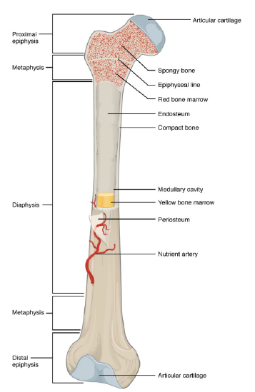
The medullary cavity has a delicate membranous lining called the endosteum (end- = “inside”; oste- = “bone”), where bone growth, repair, and remodeling occur. The outer surface of the bone is covered with a fibrous membrane called the periosteum (peri- = “around” or “surrounding”). The periosteum contains blood vessels, nerves, and lymphatic vessels that nourish compact bone. Tendons and ligaments also attach to bones at the periosteum. The periosteum covers the entire outer surface except where the epiphyses meet other bones to form joints (Figure \(\PageIndex{3}\)). In this region, the epiphyses are covered with articular cartilage, a thin layer of cartilage that reduces friction and acts as a shock absorber.
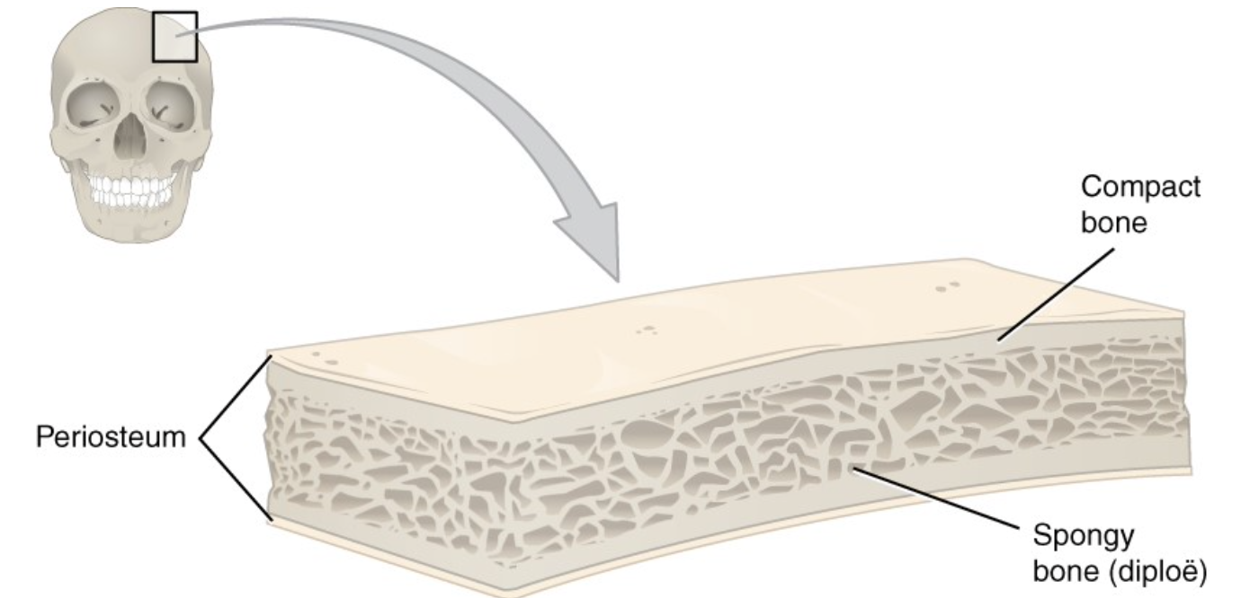
Flat bones, like those of the cranium, consist of a layer of diploë (spongy bone), lined on either side by a layer of compact bone (Figure \(\PageIndex{4}\)). The two layers of compact bone and the interior spongy bone work together to protect the internal organs. If the outer layer of a cranial bone fractures, the brain is still protected by the intact inner layer.
Procedure for Activity 6.2: Gross Anatomy of Long Bones
- Obtain a model or preserved specimen* of a long bone that has been cut longitudinally.
- In the bone(s), locate Compact/cancellous bone, Spongy bone/trabecular bone, Epiphysis, Diaphysis, Medullary cavity/canal, Red bone marrow, Yellow bone marrow, Articular cartilage, Epiphyseal line, Periosteum, Endosteum.
- *Alternately, you can use cracked poultry bones that have been softened by during the bone broth making process.
- Review the function of each part listed in Step 2 of this procedure.
Activity 6.3: Microscopic Anatomy of Long Bones
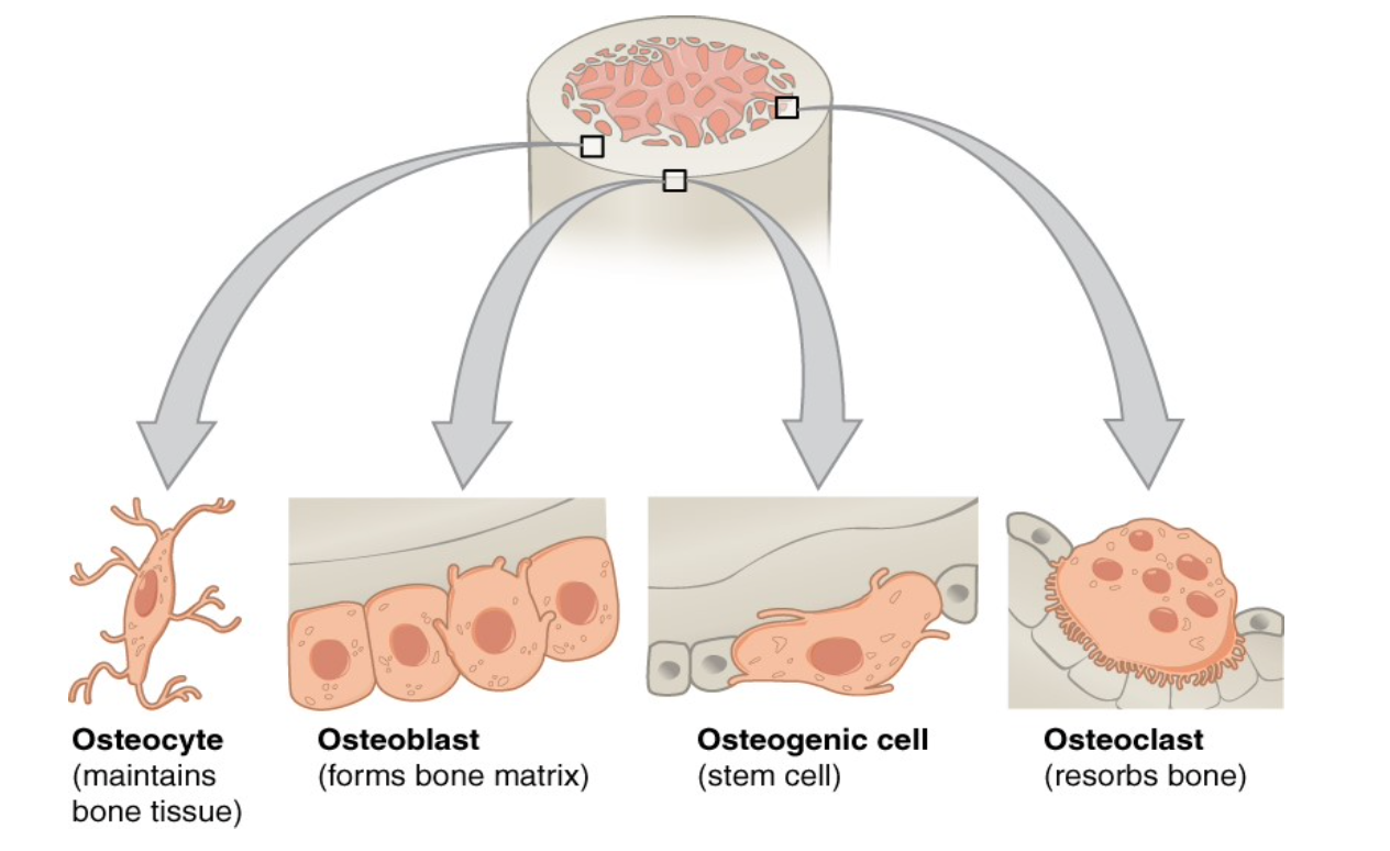
Bone contains a relatively small number of cells entrenched in a crystallized matrix of collagen fibers and inorganic salt crystals. These salt crystals form when calcium phosphate and calcium carbonate combine to create hydroxyapatite, which incorporates other inorganic salts like magnesium hydroxide, fluoride, and sulfate as it crystallizes, or calcifies, on the collagen fibers. The hydroxyapatite crystals give bones their hardness and strength, while the collagen fibers give them flexibility so that they are not brittle. Although bone cells compose a small amount of the bone volume, they are crucial to the function of bones. Four types of cells are found within bone tissue: osteoblasts, osteocytes, osteogenic cells, and osteoclasts (Figure \(\PageIndex{5}\)).
Bone Cells
Osteogenic cells are undifferentiated with high mitotic activity; they are the only bone cells that divide. Immature osteogenic cells are found in the deep layers of the periosteum and the marrow. They differentiate and develop into osteoblasts. Osteoblasts are responsible for forming new bone matrix and are found in the growing portions of bone, including the periosteum and endosteum. Moreover, the activation of these cells requires several vitamins include vitamins K2, C, and D to name a few. Osteoblasts, which do not divide, synthesize and secrete the collagen matrix and calcium salts. As the secreted matrix surrounding the osteoblast calcifies under the influence of alkaline phosphatase enzyme, the osteoblast becomes trapped within it; as a result, it changes in structure and becomes an osteocyte, the primary cell of mature bone and the most common type of bone cell. Each osteocyte is located in a space called a lacuna and is surrounded by bone tissue. Osteocytes maintain the mineral concentration of the matrix via the secretion of enzymes. Like osteoblasts, osteocytes lack mitotic activity. They can communicate with each other and receive nutrients via long cytoplasmic processes that extend through canaliculi (singular = canaliculus), channels within the bone matrix.
The dynamic nature of bone means that new tissue is constantly formed, and old, injured, or unnecessary bone is dissolved for repair or for calcium release. The cell responsible for bone resorption, or breakdown, is the osteoclast. They are found on bone surfaces, are multinucleated, and originate from monocytes and macrophages, two types of white blood cells, not from osteogenic cells. Osteoclasts are continually breaking down old bone, while osteoblasts are continually forming new bone. The ongoing balance between osteoblasts and osteoclasts is responsible for the constant but subtle reshaping of bone.
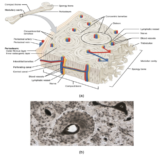
Compact/ Cancellous Bone
Compact bone is the denser, stronger of the two types of bone tissue (Figure \(\PageIndex{6}\)). It is constructed to withstand compressive forces. It can be found under the periosteum and in the diaphysis of long bones, where it provides support and protection. The microscopic structural unit of compact bone is called an osteon, or Haversian system. Each osteon is composed of concentric rings of calcified matrix called lamellae (singular = lamella). Running down the center of each osteon is the central canal which contains blood vessels, nerves, and lymphatic vessels. These vessels and nerves branch off at right angles through a perforating canal to extend to the periosteum and endosteum. Osteocytes are located inside spaces called lacunae (singular = lacuna), found at the borders of adjacent lamellae. As described above, canaliculi connect with the canaliculi of other lacunae and eventually with the central canal. This system allows nutrients to be transported to osteocytes and wastes to be removed from them.
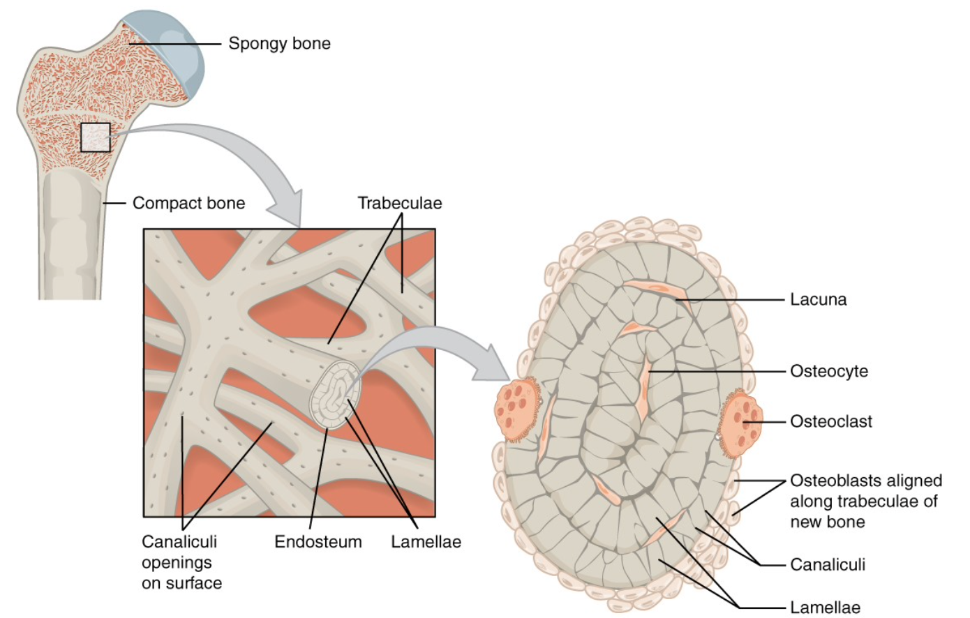
Spongy/Trabecular/Lamellar Bone
Like compact bone, spongy bone, also known as trabecular or lamellar bone, contains osteocytes housed in lacunae, but they are not usually arranged in concentric circles. Instead, the lacunae and osteocytes are found in a latticelike network of matrix spikes called trabeculae (singular = trabecula) (Figure \(\PageIndex{7}\)). The trabeculae may appear to be a random network, but each trabecula forms along lines of stress to provide strength, by enabling support for shifts in weight distribution or dissipation of forces applied to the bone. to the bone. The spaces of the trabeculated network provide balance to the dense and heavy compact bone by making bones lighter so that muscles can move them more easily. In addition, the spaces in some spongy bones contain red marrow, protected by the trabeculae, where blood cell production (hematopoiesis) occurs.
Procedure for Activity 6.3: Microscopic Anatomy of Long Bones in Models
Part 1:
- Obtain a model showing the microscopic anatomy of a long bone.
- In the bone(s), locate Central canal, Lacuna(e), Canaliculi, Osteocytes, Lamella(e), Trabeculae, Sharpey’s fibers, Osteon, Compact/cancellous bone, Spongy /trabecular bone, Medullary cavity, Red bone marrow, Periosteum, Endosteum.
- Review the function of each part listed in Step 2 of this procedure.
Part 2:
- Obtain a slide of osseous tissue. You are going to revisit this slide (originally seen in lab exercise 5) with the new knowledge you have learned in this lab.
- In the bone(s), locate Central canal, Lacuna(e), Canaliculi, Osteocytes, Lamella(e).
- Review the function of each part listed in Step 2 of this procedure.
Part 3: (You are welcomed to touch the bones. You must wear goggles and use a fresh pair of gloves for each group of bones that you touch.)
- Go to the designated area in the lab to observe raw bones, baked bones and bones soaked in acid.
- Observe the raw bones as a baseline for comparison to the baked bones and bones soaked in acid.
- Compare the visual and physical characteristics of the three sets of bones. Make notes about the differences below. Then answer questions a and b below.
a. Rickets disease is a childhood disease in which bones do not properly mineralize (do not form calcium phosphate properly). This is often due to deficiencies in vitamin D. The vitamin D deficiency will also lead to deficiencies in calcium. The lack of mineralization can often lead to the bowing or bending of bones, especially those in the legs when the child starts to stand and bear weight. Given this information and your observations of the three sets of bones, explain which set of bones you think most closely represents what happens to bones in Rickets disease.
b. Osteoporosis is a disease characterized by osteopenia and lower than normal bone density, often due to a lack of calcium or vitamin D. This results in bones that are more likely to fracture. Given this information and your observations of the three sets of bones, explain which set of bones you think most closely represents what happens to bones in osteoporosis.
Additional Learning Resources:
- Watch this video to learn about key structures found in a long bone for Activities 1 and 2: https://www.youtube.com/watch?v=vwN22f6AI6g&feature=youtu.be
- Watch this video to learn about microscopic structures found in compact bone for Activity 3: https://www.youtube.com/watch?v=ZUvZ_XHsi50&feature=youtu.be
- Practice identifying parts of the microscopic bone model here: