
2.8: Lab Exercise 10- Major Muscles of the Body
- Page ID
- 72638
\( \newcommand{\vecs}[1]{\overset { \scriptstyle \rightharpoonup} {\mathbf{#1}} } \)
\( \newcommand{\vecd}[1]{\overset{-\!-\!\rightharpoonup}{\vphantom{a}\smash {#1}}} \)
\( \newcommand{\id}{\mathrm{id}}\) \( \newcommand{\Span}{\mathrm{span}}\)
( \newcommand{\kernel}{\mathrm{null}\,}\) \( \newcommand{\range}{\mathrm{range}\,}\)
\( \newcommand{\RealPart}{\mathrm{Re}}\) \( \newcommand{\ImaginaryPart}{\mathrm{Im}}\)
\( \newcommand{\Argument}{\mathrm{Arg}}\) \( \newcommand{\norm}[1]{\| #1 \|}\)
\( \newcommand{\inner}[2]{\langle #1, #2 \rangle}\)
\( \newcommand{\Span}{\mathrm{span}}\)
\( \newcommand{\id}{\mathrm{id}}\)
\( \newcommand{\Span}{\mathrm{span}}\)
\( \newcommand{\kernel}{\mathrm{null}\,}\)
\( \newcommand{\range}{\mathrm{range}\,}\)
\( \newcommand{\RealPart}{\mathrm{Re}}\)
\( \newcommand{\ImaginaryPart}{\mathrm{Im}}\)
\( \newcommand{\Argument}{\mathrm{Arg}}\)
\( \newcommand{\norm}[1]{\| #1 \|}\)
\( \newcommand{\inner}[2]{\langle #1, #2 \rangle}\)
\( \newcommand{\Span}{\mathrm{span}}\) \( \newcommand{\AA}{\unicode[.8,0]{x212B}}\)
\( \newcommand{\vectorA}[1]{\vec{#1}} % arrow\)
\( \newcommand{\vectorAt}[1]{\vec{\text{#1}}} % arrow\)
\( \newcommand{\vectorB}[1]{\overset { \scriptstyle \rightharpoonup} {\mathbf{#1}} } \)
\( \newcommand{\vectorC}[1]{\textbf{#1}} \)
\( \newcommand{\vectorD}[1]{\overrightarrow{#1}} \)
\( \newcommand{\vectorDt}[1]{\overrightarrow{\text{#1}}} \)
\( \newcommand{\vectE}[1]{\overset{-\!-\!\rightharpoonup}{\vphantom{a}\smash{\mathbf {#1}}}} \)
\( \newcommand{\vecs}[1]{\overset { \scriptstyle \rightharpoonup} {\mathbf{#1}} } \)
\( \newcommand{\vecd}[1]{\overset{-\!-\!\rightharpoonup}{\vphantom{a}\smash {#1}}} \)
Lab Summary: In this lab, you will review and practice terminology used to describe the movement of joints and limbs, which is produced by contraction of skeletal muscles. You will learn the locations, origins, insertions, and actions of a select group of muscles, most of which are superficial muscles. This is a challenging topic, but the more time you spend reviewing, the better you will become at visualizing
muscles. For muscle actions, knowing the origin, insertion, and fascicle arrangement can help you determine an action without memorizing it. As you work with models in the lab and with videos online, make sure to palpate your own muscles as you move joints to engage in kinesthetic learning. Remember that actions should always include a verb and the joint or structure moved (ex. “biceps brachii flexes the elbow” or “shoulder flexion is caused by biceps brachii” NOT simply “biceps flexes” or “flexion”). Identifying a movement without referencing the joint does not provide enough information. In some fields of practice, it is conventional to identify the joint first and the action second.
Your objectives for this lab are:
- Identify the three functional joint classifications.
- Define the following terms of motion and be able to define them
- Flexion, Extension, Hyperextension
- Abduction, Adduction
- Plantar flexion, Dorsiflexion
- Rotation, Circumduction, Internal rotation, External Rotation o Inversion, Eversion
- Pronation, Supination
- Protraction, retraction
- Using proper terminology, list the types of movements enabled by the structure of the hip, knee, shoulder, elbow, ankle, and temporomandibular joints.
- Define the terms agonist, antagonist, synergist, and fixator, and
- Provide examples of agonists, antagonists, synergists, and fixators
- Define the following terms related to movement: Tendon, Aponeurosis, Ligament
- Describe criteria used to name muscle and include examples (such as biceps brachii is named for its
- location and the number of heads), including
- Direction of muscle fibers o Relative size of muscle
- Location of muscle
- Number of origins
- Location of origin and insertion o Shape of muscle
- Action of muscle
- Fiber arrangement
- Analyze a muscle’s movement and determine its antagonists and synergists
- Identify the functions of tendons, aponeuroses, ligaments
- Locate, identify, and give the origins, insertions and actions the muscles listed below (in models, videos, and pictures):
Muscles of the Head and Face
- Orbicularis oculi, Orbicularis oris,
- Frontalis, Occipitalis, Temporalis,
- Masseter, Buccinator
Muscles of the Neck, Thorax, and Back
- Scalenes group, Sternocleidomastoideus (SCM), Levator scapulae
- Latissimus dorsi, Trapezius
- Pectoralis major, Pectoralis minor,
- External intercostals, Internal intercostals, Diaphragm
- Erector spinae
Muscles of the Abdomen
- Quadratus lumborum
- Rectus abdominis, External oblique, Internal oblique, Transversus abdominis
Muscles of the Upper Limb
- Deltoid, Triceps brachii, Biceps brachii
- Teres major, Supraspinatus*, Infraspinatus*, Teres minor*, Subscapularis*
- *Be able to list these four muscles as part of the rotator cuff group; they are also known as the SITS muscles
Muscles of the Lower Limb
- Gluteus maximus, Gluteus medius, Piriformis
- Tensor fascia lata
- Iliopsoas
- Sartorius
- Gracilis, Adductor magnus, Adductor longus
- Rectus femoris**, Vastus lateralis**, Vastus medialis**, Vastus intermedius**
- ** Be able to list these four muscles as part of the quadriceps group
- Biceps femoris***, Semitendinosus***, Semimembranosus***
- *** Be able to list these as part of the hamstrings group
- Gastrocnemius, Soleus, Tibialis anterior
Activity 10.1: Demonstrating Movements at Synovial Joints
Synovial joints allow the body a tremendous range of movements. Each movement at a synovial joint results from the contraction or relaxation of the muscles that are attached to the bones on either side of the articulation. The type of movement that can be produced at a synovial joint is determined by its structural type. While the ball-and-socket joint gives the greatest range of movement at an individual joint, in other regions of the body, several joints may work together to produce a particular movement. Movement types are generally paired, with one being the opposite of the other. Body movements are always described in relation to the anatomical position of the body: upright stance, upper limbs to the side of the body, and palms facing anteriorly. Refer to Figures 7.5 and 7.6 as you work through this section.
Interactions of Muscles
To pull on a bone, that is, to change the angle at its joint, which essentially moves the skeleton, a skeletal muscle must also be attached to a fixed part of the skeleton. The moveable end of the muscle that attaches to the bone being pulled is called the muscle’s insertion, and the end of the muscle attached to a fixed (stabilized) bone is called the origin. During elbow flexion—bending the elbow—the biceps brachii brings the forearm closer to the arm, and the brachialis and brachioradialis assists in this motion.
For now, here are some brief explanations of other terms related to muscle actions:
- Prime mover, or agonist: the principal muscle involved in an action. For example, the biceps brachii is the prime mover of elbow flexion.
- Synergist: a muscle with similar action that assists the agonist. For example, the brachialis is a synergist of elbow flexion.
- Fixator: a muscle that stabilizes the bone that is the attachment for the prime mover’s origin. For example, the coracobrachialis muscle is a fixator for biceps brachii.
- Antagonist: a muscle with the opposite action of the prime mover. For example, the triceps brachii serves as an antagonist to elbow flexion because it causes elbow extension.
Before you move on, stop and write down definitions of agonist, synergist, fixator, and antagonist in your own words.
Muscle Action Terms
Flexion and Extension
Flexion and extension are movements that take place within the sagittal plane and involve anterior or posterior movements of the body or limbs. For the vertebral column, flexion (anterior flexion) is an anterior (forward) bending of the neck or body, while extension involves a posterior-directed motion, such as straightening from a flexed position or bending backward. Lateral flexion is the bending of the neck or body toward the right or left side. In the limbs, flexion decreases the angle between the bones (bending of the joint), while extension increases the angle and straightens the joint. For the upper limb, all anterior- going motions are flexion, and all posterior-going motions are extension. These include anterior-posterior movements of the arm at the shoulder, the forearm at the elbow, the hand at the wrist, and the fingers at the metacarpophalangeal and interphalangeal joints. For the thumb (with the upper extremity in anatomical position), extension moves the thumb away from the palm of the hand, within the same plane as the palm, while flexion brings the thumb back against the index finger or into the palm. In the lower limb, bringing the thigh forward and upward is flexion at the hip joint, while any posterior-going motion of the thigh is extension. Note that extension of the thigh beyond the anatomical (standing) position is greatly limited by the ligaments that support the hip joint; this is referred to as hyperextension and is discussed later. Knee flexion is the bending of the knee to bring the foot toward the posterior thigh, and extension is the straightening of the knee.
Hyperextension is movement of the joint beyond its normal anatomical position. Sometimes, this action can cause injury or damage when the joint is hyperextended beyond the natural limits of the bony and soft tissue structures’ capacity. Other times, this action is simply part of the joint’s normal range of motion (ex. in the hip, shoulder, neck, and vertebral column). Similarly, hyperflexion is excessive flexion at a joint. Hyperextension injuries are common at hinge joints such as the knee or elbow. In cases of “whiplash” when the head is suddenly moved backward and then forward, a person may experience both hyperextension and hyperflexion of the cervical region.
Abduction and Adduction
Abduction and adduction motions occur within the coronal plane and involve medial-lateral motions of the limbs, fingers, toes, or thumb. Abduction moves the limb laterally away from the midline of the body, while adduction is the opposing movement that brings the limb toward the body or across the midline. For example, abduction is raising the arm at the shoulder joint, moving it laterally away from the body, while adduction brings the arm down to the side of the body. Similarly, abduction and adduction at the wrist moves the hand away from or toward the midline of the body. Spreading the fingers or toes apart is also abduction, while bringing the fingers or toes together is adduction. For the thumb (with the upper extremity in anatomical position), abduction is the anterior movement that brings the thumb to a 90° perpendicular position, pointing straight out from the palm. Adduction moves the thumb back to the anatomical position, next to the index finger.
Circumduction
Circumduction is the movement of a body region in a circular manner, in which one end of the body region being moved stays relatively stationary while the other end describes a circle. It involves the sequential combination of flexion, adduction, extension, and abduction at a joint. This is typically seen in the upper and lower limbs when the proximal joint (hip or shoulder) doesn’t move, but the rest of the limb moves through all planes of motion.
Rotation
Rotation can occur within the vertebral column, at a pivot joint, or at a ball-and-socket joint. Rotation of the neck or body is the twisting movement produced by the summation of the small rotational movements available between adjacent vertebrae. At a pivot joint, one bone rotates in relation to another bone. This is a uniaxial joint, and thus rotation is the only motion allowed at a pivot joint. For example, at the atlantoaxial joint, the first cervical (C1) vertebra (atlas) rotates around the dens, the upward projection from the second cervical (C2) vertebra (axis). This allows the head to rotate from side to side as when shaking the head “no.” The proximal radioulnar joint is a pivot joint formed by the head of the radius and its articulation with the ulna. This joint allows for the radius to rotate along its length during pronation and supination movements of the forearm. Rotation can also occur at the ball-and-socket joints of the shoulder and hip. Here, the humerus and femur rotate around their long axis, which moves the anterior surface of the arm or thigh either toward or away from the midline of the body. Movement that brings the anterior surface of the limb toward the midline of the body is called medial (internal) rotation. Conversely, rotation of the limb so that the anterior surface moves away from the midline is lateral (external) rotation. Be sure to distinguish medial and lateral rotation, which can only occur at the shoulder and hip joints, from circumduction.
Supination and Pronation
Supination and pronation are movements of the forearm. In the anatomical position, the upper limb is held next to the body with the palm facing forward. This is the supinated position of the forearm. In this position, the radius and ulna are parallel to each other. When the palm of the hand faces backward, the forearm is in the pronated position, and the radius and ulna form an X-shape. Supination and pronation are the movements of the forearm that go between these two positions. Pronation is the motion that moves the forearm from the supinated (anatomical) position to the pronated (palm backward) position. Supination is the opposite motion, in which rotation of the radius returns the bones to their parallel positions and moves the palm to the anterior facing (supinated) position. It helps to remember that supination is the motion you use when scooping up soup with a spoon.
Dorsiflexion and Plantar Flexion
Dorsiflexion and plantar flexion are movements at the ankle joint, which is a hinge joint. Lifting the front of the foot, so that the top of the foot moves toward the anterior leg is dorsiflexion, while lifting the heel of the foot from the ground or pointing the toes downward is plantar flexion.
Inversion and Eversion
Inversion and eversion are complex movements that involve multiple plane joints among the tarsal bones of the posterior foot and thus are not motions that take place at the ankle joint. This motion begins in the most posterior tarsal bone (the calcaneus), then the other tarsal and metatarsal bones move as a result of their connections to the calcaneus. Inversion is the turning of the foot to angle the bottom of the foot toward the midline, while eversion turns the bottom of the foot away from the midline. The foot has a greater range of inversion than eversion motion. These are important motions that help to stabilize the foot when walking or running on an uneven surface and aid in the quick side-to-side changes in direction used during active sports such as basketball, tennis, parkour, or soccer.
Protraction and Retraction
Protraction and retraction are anterior-posterior movements of the scapula or mandible. Protraction of the scapula occurs when the shoulder is moved forward, as when pushing against something or throwing a ball. Retraction is the opposite motion, with the scapula being pulled posteriorly and medially, toward the vertebral column. For the mandible, protraction occurs when the lower jaw is pushed forward, to stick out the chin, while retraction pulls the lower jaw backward.
Depression and Elevation
Depression and elevation are downward and upward movements of the scapula or mandible. The upward movement of the scapula and shoulder is elevation, while a downward movement is depression. These movements are used to shrug your shoulders. Similarly, elevation of the mandible is the upward movement of the lower jaw used to close the mouth or bite on something, and depression is the downward movement that produces opening of the mouth.
Opposition and Reposition
Opposition is the thumb movement that brings the tip of the thumb in contact with the tip of a finger. Thumb opposition is produced by a combination of flexion and abduction of the thumb at this joint. Returning the thumb to its anatomical position next to the index finger is called reposition.
You can watch this video to see these joint motions alone and in combination with other movements. Please note that at minute 7:03, The Funky Professor refers to extension and flexion of the ankle. In the US, we refer to these as dorsiflexion and plantar flexion, respectively; these are the terms you will use for our course. https://www.youtube.com/watch?v=X5RUFXZZBH4


Procedure for Activity 10.1: Demonstrating Movements at Synovial Joints: After reading the information above, work with a partner to describe and demonstrate some important movement at selected synovial joints. This means that as you perform each movement, you will also describe it verbally to reinforce the definition and applications of the vital terms. You can verify with your instructor that you are performing the movements correctly.
- Hip Joint: Demonstrate and define abduction, adduction, flexion, extension, circumduction, external rotation, and internal rotation
- Shoulder Joint: Demonstrate and define abduction, adduction, flexion, extension, circumduction, internal rotation, external rotation, elevation, depression, protraction, and retraction
- Elbow Joint: Demonstrate and define supination, pronation, flexion, and extension
- Temporomandibular Joint: Demonstrate and define elevation, depression, protraction, and retraction
- Knee Joint: Demonstrate and define flexion, extension, hyperextension (be careful), and gentle rotation (be careful)
- Vertebral Column (as a whole, includes joints of the thoracic and lumbar spine): Demonstrate and define flexion, extension, hyperextension, and lateral flexion
- Neck (includes the joints of C0 to C7): Demonstrate and define flexion, extension, hyperextension, lateral flexion, rotation
- Hand: Demonstrate and define flexion and extension of the metacarpophalangeal joints, opposition of the thumb; adduction and abduction of digit 5 at the metacarpophalangeal joint
- Ankle Joint: Demonstrate and define dorsiflexion, plantar flexion, inversion, and eversion. (Recall that inversion and eversion mostly occur in the joint of the foot, but what we see externally is movement of the ankle.)
- As you’ve demonstrated and defined, the shoulder joint is highly mobile, whereas the knee joint allows for many fewer movements. What does it mean when we say that a joint sacrifices stability in favor of mobility? How does the shape of the joint surfaces influence either stability or mobility?
Activity 10.2: Criteria for Naming Muscles
Anatomists name the skeletal muscles according to a number of criteria, each of which describes the muscle in some way. Muscles are most commonly named for
- their shape (often derived from Latin)
- size compared to other muscles in the area
- location in the body or the location of their attachments to the skeleton
- how many origins they have
- their action(s)
- arrangement of the fascicles between the muscle’s origin and insertion
The skeletal muscle’s anatomical location or its relationship to a particular bone often determines its name. For example, the frontalis muscle is located on top of the frontal bone of the skull. Similarly, the shapes of some muscles are very distinctive and the names, such as orbicularis, reflect the shape. For the buttocks, the size of the muscles influences the names: gluteus maximus (largest), gluteus medius (medium), and the gluteus minimus (smallest). Some names were given to indicate length—brevis (short), longus (long)—and to identify position relative to the midline: lateralis (to the outside away from the midline), and medialis (toward the midline). The direction of the muscle fibers and fascicles are used to describe muscles relative to the midline, such as the rectus (straight) abdominis, or the oblique (at an angle) muscles of the abdomen.
Some muscle names indicate the number of muscles in a group. One example of this is the quadriceps, a group of four muscles located on the anterior (front) thigh. Other muscle names can provide information as to how many origins a particular muscle has, such as the biceps brachii. The prefix bi indicates that the muscle has two origins and tri indicates three origins.
The location of a muscle’s attachment can also appear in its name. When the name of a muscle is based on the attachments, the origin is always named first. For instance, the sternocleidomastoid muscle of the neck has a dual origin on the sternum (sterno-) and clavicle (-cleido-), and it inserts on the mastoid process of the temporal bone (-mastoideus). The last feature by which to name a muscle is its action. When muscles are named for the movement they produce, one can find action words in their name. Some examples
are flexor (decreases the angle at the joint), extensor (increases the angle at the joint), abductor (moves the bone away from the midline), adductor (moves the bone toward the midline), and levator (elevates the bone).
Procedure for Activity 10.2: As you work through the rest of the activities, make notes about how the muscles are named and be able to provide examples of each criteria listed above.
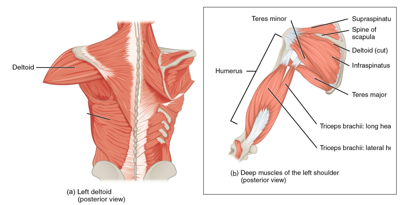
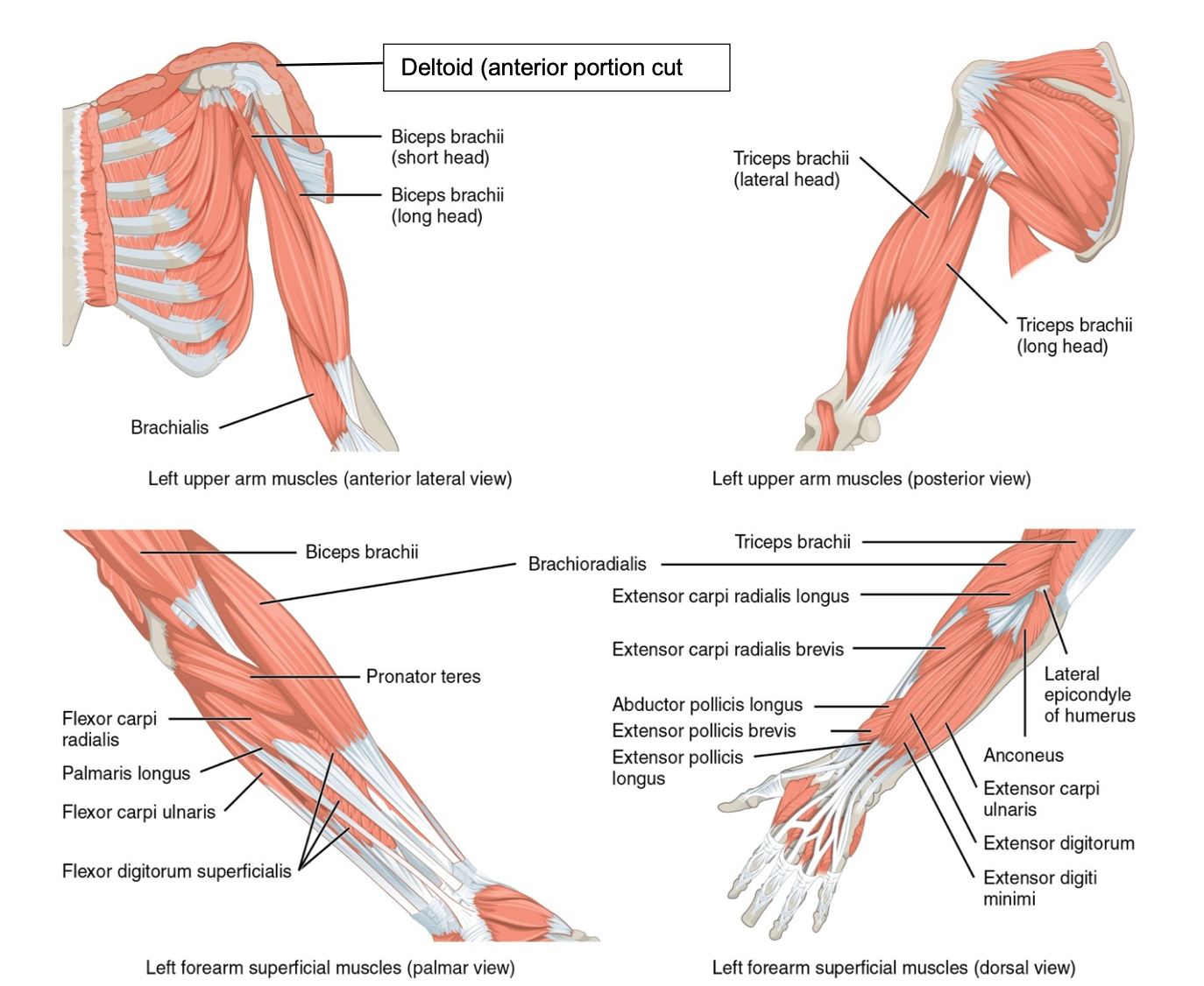
Activity 10.3: Selected Muscles of the Upper Limb
The muscles of the upper limb work through a much wider range of motion than other parts of the body, which allows a different set of functions compared to the lower limb. Muscles of the shoulder and upper limb can be divided into four groups: muscles that stabilize and position the pectoral girdle, muscles that move the arm, muscles that move the forearm, and muscles that move the wrists, hands, and fingers.
Procedure for Activity 10.3: Use the pictures and charts below to locate required muscles on the models. As you work, palpate the muscles and make notes about their appearance. You also need to know their origins, insertions, and actions.
|
Name |
Primary Actions |
Origin |
Insertion |
|
Deltoid |
Abduct shoulder; flex and extend shoulder; medial and lateral rotation of shoulder |
Clavicle; acromion process and scapular spine |
Deltoid tuberosity |
|
Supraspinatus |
Abduct shoulder |
Supraspinous fossa |
Greater tubercle |
|
Infraspinatus |
Extend and adduct shoulder |
Infraspinous fossa |
Greater tubercle |
|
Subscapularis |
Medially rotate shoulder |
Subscapular fossa |
Lesser tubercle |
|
Teres major |
Extend and adduct shoulder |
Inferior angle of scapula, posterior surface of scapula |
Intertubercular sulcus of humerus |
|
Teres minor |
Extend and adduct shoulder |
Lateral border of scapula |
Greater tubercle |
|
Triceps brachii |
Extend elbow |
Infraglenoid tubercle; posterior diaphysis and distal radial groove of humerus |
Olecranon process |
|
Biceps brachii |
Flex elbow; supinate forearm |
Coracoid process and supraglenoid tubercle of scapula |
Radial tuberosity, bicipital aponeurosis |

Activity 10.4: Selected Muscles of the Lower Limb
The muscles of the leg position and stabilize the pelvic girdle and work with the bones of the leg to allow you to stand, walk, and run.
Procedure for Activity 10.4: Use the pictures and charts below to locate required muscles on the models. As you work, palpate the muscles and make notes about their appearance. You also need to know their origins, insertions, and actions.
|
Muscle Name |
Primary Action |
Origin |
Insertion |
|
Iliopsoas |
Flex and laterally rotate hip; flex torso and vertebral column |
(Psoas major) T12 and L1-L5 (Iliacus) Iliac fossa; iliac crest; lateral sacrum |
Lesser trochanter |
|
Gluteus maximus |
Extend hip |
Posterolateral ilium; sacrum; coccyx |
Gluteal tuberosity of femur; IT tract (iliotibial tract) |
|
Gluteus medius |
Abduct hip |
Superolateral surface of ilium |
Greater trochanter of femur |
|
Piriformis |
Laterally rotate hip; adduction of hip |
Anterolateral surface of the sacrum |
Greater trochanter of femur |
|
Gracilis |
Adduct hip and flex knee |
Inferior ramus and body of pubis; ischial ramus |
Medial surface of tibia |
|
Sartorius |
Flex, abduct and laterally rotate hip; flex knee |
Anterior superior iliac spine (ASIS) |
Medial aspect of proximal tibia |
|
Adductor magnus |
Adduct hip |
Pubic ramus; ischial tuberosity |
Linea aspera and adductor tubercle of femur |
|
Adductor longus |
Adduct hip |
Pubic bone |
Linea aspera of femur |
|
Tensor Fascia lata |
Extend knee; laterally rotate hip; aids in maintaining balance at pelvis |
Iliac crest; anterior superior iliac spine
|
IT tract (iliotibial tract) |
|
Rectus femoris |
Extend knee; assists with flexing hip |
Anterior inferior iliac spine; superior margin of acetabulum |
Patella; tibial tuberosity |
|
Vastus lateralis |
Extend knee |
Greater trochanter; linea aspera |
Patella; tibial tuberosity |
|
Vastus medialis |
Extend knee |
Linea aspera |
Patella; tibial tuberosity |
|
Vastus intermedius |
Extend knee |
Proximal femur shaft |
Patella; tibial tuberosity |
|
Biceps femoris |
Flex knee; extend and laterally rotate hip |
Ischial tuberosity; linea aspera; distal femur |
Proximal fibula; lateral condyle of tibia |
|
Semitendinosus |
Flex knee; extend and medially rotate hip |
Ischial tuberosity |
Medial, proximal tibia |
|
Semimembranosus |
Flex knee; extend and medially rotate hip |
Ischial tuberosity |
Medial condyle of tibia |
| Gastrocnemius | Plantar flex ankle; some flexion of knee | Medial and lateral condyles of femur | Calcaneal/ Achille’s tendon |
| Soleus | Plantar flex ankle | Proximal tibia; fibula | Calcaneal/ Achille’s tendon |
| Tibialis anterior | Dorsiflex ankle; invert foot at the ankle | Lateral condyle and proximal portion of tibia | Medial cuneiform; metatarsal #1 |


Activity 10.5: Selected Muscles of the Axial Skeleton
The muscles of the head, neck, and trunk provide strength and stability to the trunk of the body while also allowing some special functions such as creating facial expressions and breathing. In this lesson, students will identify select muscles of the head, neck, and trunk and work to understand their function via muscle attachments, actions, and innervation.
Procedure for Activity 10.5: Use the pictures and charts below to locate required muscles on the models. As you work, palpate the muscles and make notes about their appearance. You also need to know their origins, insertions, and actions.
|
Muscle Name |
Primary Action |
Origin |
Insertion |
|
Frontalis |
Raise eyebrows, draw scalp anteriorly |
Epicranial aponeurosis |
Subcutaneous tissue of eyebrows |
|
Occipitalis |
Moves the scalp |
Superior nuchal line; |
Epicranial aponeurosis |
|
Temporalis |
Elevate mandible to close mouth |
Temporal fossa |
Coronoid process and ramus of mandible |
|
Masseter |
Elevate mandible to close mouth |
Zygomatic arch; zygomatic bone |
Angle and ramus of mandible |
|
Buccinator |
Compress cheek |
Maxilla and mandible near molar teeth |
Orbicularis oris |
|
Orbicularis oculi |
Close eyelid; wink eye; blink eyes |
Frontal bone; maxilla |
Skin of eyelid |
|
Orbicularis oris |
Close mouth; puckering of lips |
Skin of the mouth |
Skin near lips
|

|
Muscle Name |
Primary Actions |
Origin |
Insertion |
|
Levator scapulae |
Elevate scapula, abduct and rotate neck |
Vertebrae C1-4 |
Medial border of scapula |
|
Scalenes group (anterior, middle, posterior) |
Flex and rotate neck at cervical spine, elevate ribs 1 and 2 |
Transverse process of cervical vertebrae |
Ribs 1 and 2 |
|
Sternocleidomastoideus (SCM) |
Rotate and flex head at cervical spine |
Manubrium of sternum, medial clavicle |
Mastoid process of temporal bone |
|
Pectoralis major |
Flex, adduct, and medially rotate humerus |
Clavicle, sternum, cartilages of ribs 1-7 |
Crest of greater tubercle of humerus |
|
Pectoralis minor |
Depress shoulder |
Ribs 3-5 |
Coracoid process of scapula |
|
External intercostals |
Elevate ribs (increase volume in thorax during inhalation) |
Inferior border of ribs 1-11 |
Superior border of next inferior rib |
|
Internal intercostals |
Depress ribs (decrease volume in thorax during forced exhalation) |
Superior border of ribs 2-12 |
Inferior border of next superior rib |
|
Diaphragm |
Increases volume in thorax during inhalation |
Xiphoid process of sternum, ribs 7-12, superior lumbar vertebrae |
Central tendon of diaphragm |
|
Erector spinae group (iliocostalis, longissimus, spinalis) |
Extend and rotate vertebral column and head |
Nuchal ligament, thoracic and lumbar vertebrae, sacrum, ilium, ribs 3-12 |
All ribs, all thoracic vertebrae, temporal bones |
|
Trapezius |
Extend and abduct head; rotate and adduct scapula; fixator of scapula |
Posterior occipital bone, nuchal ligament, vertebrae C7-T4 |
Clavicle, acromion, scapular spine |
|
Latissimus dorsi |
Extend, adduct, and medially rotate humerus; depress shoulder |
T7-12, L1-5, ribs 9-12, iliac crest
|
Intertubercular sulcus of humerus |


|
Muscle Name |
Primary Actions |
Origin |
Insertion |
|
Rectus abdominis |
Flex vertebral column (when both are used), laterally flex vertebral column (when one side is used); compress abdominal wall |
Crest of pubis, pubic symphysis |
Costal cartilages 5-7, xiphoid process |
|
External abdominal oblique |
Flex vertebral column (when both are used), laterally flex vertebral column (when one side is used), compress abdominal wall, lateral rotation of trunk |
Ribs 5-12 |
Linea alba, pubis, Iliac crest, ASIS |
|
Internal abdominal oblique |
Compress abdominal wall, lateral rotation of vertebral column (trunk) |
Inguinal ligament, iliac crest |
Pubis, ribs 10-12 |
|
Transverse abdominal |
Compress abdominal wall, laterally rotate trunk at waist |
Inguinal ligament, iliac crest, costal cartilages 7-12 |
Linea alba, pubis |
|
Quadratus lumborum |
Laterally flex vertebral column; extend lumbar vertebrae |
Iliac crest |
Transverse processes of L1-4, rib 12 |
Activity 10.6: Analyze Muscles as Agonists, Antagonist, Synergists, and Fixators
To pull on a bone, that is, to change the angle at its joint, which essentially moves the skeleton, a skeletal muscle must also be attached to a fixed part of the skeleton. The moveable end of the muscle that attaches to the bone being pulled is called the muscle’s insertion, and the end of the muscle attached to a fixed (stabilized) bone is called the origin. During elbow flexion—bending the elbow—the biceps brachii brings the forearm closer to the arm, and the brachialis and brachioradialis assists in this motion.
Although a number of muscles may be involved in an action, the principal muscle involved is called the prime mover, or agonist. To lift a cup, the biceps brachii is actually the prime mover; however, because it can be assisted by the brachialis and brachioradialis muscles, the brachialis and brachioradialis are called synergists in this action (Figure \(\PageIndex{21}\)). A synergist can also be a fixator that stabilizes the bone that is the attachment for the prime mover’s origin.

A muscle with the opposite action of the prime mover is called an antagonist. Antagonists play two important roles in muscle function: (1) they maintain body or limb position, such as holding the arm out or standing erect; and (2) they control rapid movement, as in shadow boxing without landing a punch or the ability to check the motion of a limb.
For example, to extend the knee, a group of four muscles called the quadriceps femoris in the anterior compartment of the thigh are activated (and would be called the agonists of knee extension). However, to flex the knee joint, an opposite or antagonistic set of muscles called the hamstrings is activated.
As you can see, these terms would also be reversed for the opposing action. If you consider the first action as the knee bending, the hamstrings would be called the agonists and the quadriceps femoris would then be called the antagonists. See Table 11.1 in the OpenStax A&P textbook for a list of more agonists and antagonists.
Procedure for Activity 10.6: Using the names of muscles you’ve studied in this lab, provide examples of agonists, antagonists, synergists, and fixators as directed below.
- Name an antagonist of the brachialis muscle: ______________________________________________
- Name an agonist of knee flexion: _______________________________________________________
- Name a synergist of the triceps brachii: __________________________________________________
- Name a fixator of the hip joint: _________________________________________________________
- Name an antagonist of rectus femoris: ___________________________________________________
- Name a synergist of the soleus muscle: __________________________________________________
- Name a prime mover of dorsiflexion of the foot: ____________________________________________
Additional Learning Resources:
- Watch these videos to review learn about the synovial joints listed above and the terminology used to describe joint movements:
- https://www.youtube.com/watch?v=-I9Ufqpcc30&feature=youtu.be
- This video has a memory trick for the definitions of joint movement terminology: https://www.youtube.com/watch?v=oA6HiaV1RlU
- Watch these videos to learn about the muscles listed above in a human cadaver (with Amanda Couitt, DPT of RVCC’s PTA department) and to learn how to identify muscles in models
- Activity 1 (Head and Neck Muscles): https://www.youtube.com/watch?v=Gv-TDM3oIjI&feature=youtu.be
- Activity 2 (Muscles of the Trunk): https://www.youtube.com/watch?v=7hjw_5MI-g0&feature=youtu.be
- Activity 3 (Muscles of the Upper Limb): https://www.youtube.com/watch?v=o_3epUCoqeA&feature=youtu.be
- Activity 4 (Muscles of the Lower Limb): https://www.youtube.com/watch?v=Fm4SSPYLkBM&feature=youtu.be
- Leg Model: https://www.youtube.com/watch?v=q0WDD704P1M
- Arm Model: https://www.youtube.com/watch?v=ToNL6jMEjuE
- Large Muscled Torso: https://www.youtube.com/watch?v=F_O0Rj3IWn8
- Muscle Fiber Model: https://www.youtube.com/watch?v=v-9VegCKF6U&t=3s (up to min 6:58)
- Mini Man 1: https://www.youtube.com/watch?v=G7QvRKMO78Y
- Mini Man 2: https://www.youtube.com/watch?v=Oq4CXI470ds
- Practice identifying muscles in models and identifying structure in a muscle fiber with this document: Muscle Model Labeling Practice.pdf (found in the Canvas Module containing the electronic copy of the SLM)
- Practice identifying muscles in pictures and models, as well as origin, insertion, and action practice (This site is very helpful but includes word banks as drop down menus; your lab practicals do not have a word banks): https://webanatomy.umn.edu/muscular-system