
14.6: Nervous System Worksheet
- Page ID
- 2840

\( \newcommand{\vecs}[1]{\overset { \scriptstyle \rightharpoonup} {\mathbf{#1}} } \)
\( \newcommand{\vecd}[1]{\overset{-\!-\!\rightharpoonup}{\vphantom{a}\smash {#1}}} \)
\( \newcommand{\dsum}{\displaystyle\sum\limits} \)
\( \newcommand{\dint}{\displaystyle\int\limits} \)
\( \newcommand{\dlim}{\displaystyle\lim\limits} \)
\( \newcommand{\id}{\mathrm{id}}\) \( \newcommand{\Span}{\mathrm{span}}\)
( \newcommand{\kernel}{\mathrm{null}\,}\) \( \newcommand{\range}{\mathrm{range}\,}\)
\( \newcommand{\RealPart}{\mathrm{Re}}\) \( \newcommand{\ImaginaryPart}{\mathrm{Im}}\)
\( \newcommand{\Argument}{\mathrm{Arg}}\) \( \newcommand{\norm}[1]{\| #1 \|}\)
\( \newcommand{\inner}[2]{\langle #1, #2 \rangle}\)
\( \newcommand{\Span}{\mathrm{span}}\)
\( \newcommand{\id}{\mathrm{id}}\)
\( \newcommand{\Span}{\mathrm{span}}\)
\( \newcommand{\kernel}{\mathrm{null}\,}\)
\( \newcommand{\range}{\mathrm{range}\,}\)
\( \newcommand{\RealPart}{\mathrm{Re}}\)
\( \newcommand{\ImaginaryPart}{\mathrm{Im}}\)
\( \newcommand{\Argument}{\mathrm{Arg}}\)
\( \newcommand{\norm}[1]{\| #1 \|}\)
\( \newcommand{\inner}[2]{\langle #1, #2 \rangle}\)
\( \newcommand{\Span}{\mathrm{span}}\) \( \newcommand{\AA}{\unicode[.8,0]{x212B}}\)
\( \newcommand{\vectorA}[1]{\vec{#1}} % arrow\)
\( \newcommand{\vectorAt}[1]{\vec{\text{#1}}} % arrow\)
\( \newcommand{\vectorB}[1]{\overset { \scriptstyle \rightharpoonup} {\mathbf{#1}} } \)
\( \newcommand{\vectorC}[1]{\textbf{#1}} \)
\( \newcommand{\vectorD}[1]{\overrightarrow{#1}} \)
\( \newcommand{\vectorDt}[1]{\overrightarrow{\text{#1}}} \)
\( \newcommand{\vectE}[1]{\overset{-\!-\!\rightharpoonup}{\vphantom{a}\smash{\mathbf {#1}}}} \)
\( \newcommand{\vecs}[1]{\overset { \scriptstyle \rightharpoonup} {\mathbf{#1}} } \)
\(\newcommand{\longvect}{\overrightarrow}\)
\( \newcommand{\vecd}[1]{\overset{-\!-\!\rightharpoonup}{\vphantom{a}\smash {#1}}} \)
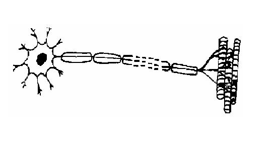
\(\newcommand{\avec}{\mathbf a}\) \(\newcommand{\bvec}{\mathbf b}\) \(\newcommand{\cvec}{\mathbf c}\) \(\newcommand{\dvec}{\mathbf d}\) \(\newcommand{\dtil}{\widetilde{\mathbf d}}\) \(\newcommand{\evec}{\mathbf e}\) \(\newcommand{\fvec}{\mathbf f}\) \(\newcommand{\nvec}{\mathbf n}\) \(\newcommand{\pvec}{\mathbf p}\) \(\newcommand{\qvec}{\mathbf q}\) \(\newcommand{\svec}{\mathbf s}\) \(\newcommand{\tvec}{\mathbf t}\) \(\newcommand{\uvec}{\mathbf u}\) \(\newcommand{\vvec}{\mathbf v}\) \(\newcommand{\wvec}{\mathbf w}\) \(\newcommand{\xvec}{\mathbf x}\) \(\newcommand{\yvec}{\mathbf y}\) \(\newcommand{\zvec}{\mathbf z}\) \(\newcommand{\rvec}{\mathbf r}\) \(\newcommand{\mvec}{\mathbf m}\) \(\newcommand{\zerovec}{\mathbf 0}\) \(\newcommand{\onevec}{\mathbf 1}\) \(\newcommand{\real}{\mathbb R}\) \(\newcommand{\twovec}[2]{\left[\begin{array}{r}#1 \\ #2 \end{array}\right]}\) \(\newcommand{\ctwovec}[2]{\left[\begin{array}{c}#1 \\ #2 \end{array}\right]}\) \(\newcommand{\threevec}[3]{\left[\begin{array}{r}#1 \\ #2 \\ #3 \end{array}\right]}\) \(\newcommand{\cthreevec}[3]{\left[\begin{array}{c}#1 \\ #2 \\ #3 \end{array}\right]}\) \(\newcommand{\fourvec}[4]{\left[\begin{array}{r}#1 \\ #2 \\ #3 \\ #4 \end{array}\right]}\) \(\newcommand{\cfourvec}[4]{\left[\begin{array}{c}#1 \\ #2 \\ #3 \\ #4 \end{array}\right]}\) \(\newcommand{\fivevec}[5]{\left[\begin{array}{r}#1 \\ #2 \\ #3 \\ #4 \\ #5 \\ \end{array}\right]}\) \(\newcommand{\cfivevec}[5]{\left[\begin{array}{c}#1 \\ #2 \\ #3 \\ #4 \\ #5 \\ \end{array}\right]}\) \(\newcommand{\mattwo}[4]{\left[\begin{array}{rr}#1 \amp #2 \\ #3 \amp #4 \\ \end{array}\right]}\) \(\newcommand{\laspan}[1]{\text{Span}\{#1\}}\) \(\newcommand{\bcal}{\cal B}\) \(\newcommand{\ccal}{\cal C}\) \(\newcommand{\scal}{\cal S}\) \(\newcommand{\wcal}{\cal W}\) \(\newcommand{\ecal}{\cal E}\) \(\newcommand{\coords}[2]{\left\{#1\right\}_{#2}}\) \(\newcommand{\gray}[1]{\color{gray}{#1}}\) \(\newcommand{\lgray}[1]{\color{lightgray}{#1}}\) \(\newcommand{\rank}{\operatorname{rank}}\) \(\newcommand{\row}{\text{Row}}\) \(\newcommand{\col}{\text{Col}}\) \(\renewcommand{\row}{\text{Row}}\) \(\newcommand{\nul}{\text{Nul}}\) \(\newcommand{\var}{\text{Var}}\) \(\newcommand{\corr}{\text{corr}}\) \(\newcommand{\len}[1]{\left|#1\right|}\) \(\newcommand{\bbar}{\overline{\bvec}}\) \(\newcommand{\bhat}{\widehat{\bvec}}\) \(\newcommand{\bperp}{\bvec^\perp}\) \(\newcommand{\xhat}{\widehat{\xvec}}\) \(\newcommand{\vhat}{\widehat{\vvec}}\) \(\newcommand{\uhat}{\widehat{\uvec}}\) \(\newcommand{\what}{\widehat{\wvec}}\) \(\newcommand{\Sighat}{\widehat{\Sigma}}\) \(\newcommand{\lt}{<}\) \(\newcommand{\gt}{>}\) \(\newcommand{\amp}{&}\) \(\definecolor{fillinmathshade}{gray}{0.9}\)1. The diagram below is of a nerve cell or neurone.
- i. Add the following labels to the diagram.
- Axon; Myelin sheath; Cell body; Dendrites; Muscle fibres;
- ii. If you like, colour in the diagram as suggested below.
- Axon - purple;
- Myelin sheath - yellow;
- Cell body - blue;
- Dendrites - green;
- Muscle fibres – red;
- iii. Now indicate the direction that the nerve impulse travels.
2. There are three different kinds of neurone or nerve cell. Match each kind with its function.
- A. Motor neuron; B. Sensory neuron; C. Relay neuron;
| Kind of neurone | Function |
|---|---|
| ................................... |
The nerve cell that carries impulses from a sense receptor to the brain or spinal cord. |
| .................................... | The nerve cell that connects sensory and motor neurons |
| ..................................... |
The nerve cell that transmits impulses from the brain or spinal cord to a muscle or gland |
3. Match the descriptions in the table below with the terms in the list.
- A. Synapse; B. Axon; C. Myelin sheath; D. Nerve impulse; E. Sense receptor; F. Response;
- G.Reflex; H. Cell body; I. Dendrite; J. Nerve; K. Neurotransmitter; L. Axon terminal
| Term | Function |
|---|---|
| .............................. | 1. The long fibre that carries the nerve impulses. |
| .............................. | 2. A bundle of axons. |
| .............................. | 3. The connection between adjacent neurons. |
| ............................... |
4. The chemical secreted into the gap between neurons at a synapse. |
| ............................... | 5. A rapid automatic response to a stimulus. |
| ............................... |
6. The covering of fatty material that speeds up the passage of nerve impulses. |
| ................................... |
7. The structure at the end of an axon that produces neurotransmitters to transmit the nerve impulse across the synapse. |
| ................................ | 8. The high speed signals that pass along the axons of nerve cells. |
| ................................ |
9. The branching filaments that conduct nerve impulses towards the cell. |
| ..................................... |
10. The sense organ or cells that receive stimuli from within and outside the body. |
| ..................................... | 11. The reaction to a stimulus by a muscle or gland. |
| ..................................... | 12.The part of the nerve cell containing the nucleus. |
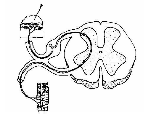
4. The diagram below shows a cross-section of the spinal cord. Add the following labels to the diagram.
- Central canal; White matter; Dorsal root; Grey matter; Ventral root; Skin;
- Muscle; Sensory neuron; Relay neuron; Motor neuron; Pain receptors in skin
5.
- a) List in order the 3 different neurons involved in a reflex arc from the stimulus to the response.
| Stimulus | ........................... | ........................... | ........................... | Response |
|---|
- b) Name 3 different reflexes found in animals.
Reflex 1..............................
Reflex 2..............................
Reflex 3..............................
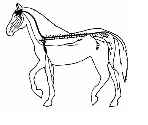
6. The diagram below shows the nervous system of a horse. Add the following labels.
- Brain; Spinal cord; Cranial nerves; Spinal nerves; Sciatic nerve; Nerves of the autonomic nervous system; Vagus nerve; Network of nerves to forelimb.
7. Indicate whether the following parts of the nervous system are part of the Central Nervous System CNS) or the Peripheral Nervous System (PNS).
| Part of nervous system | CNS or PNS? |
|---|---|
| Brain | ........................ |
| Autonomic nervous system | ........................ |
| Spinal nerves | ........................ |
| Spinal cord | ........................ |
| Cranial nerves | ......................... |
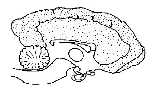
8. The diagram below shows a section of a dog’s brain. Add the labels in the list below and, if you like, colour in the diagram as suggested.
- Cerebellum - blue;
- Spinal cord - green;
- Medulla oblongata - orange;
- Hypothalamus - purple;
- Pituitary gland - red;
- Cerebral hemispheres – yellow.
9. Match the descriptions below with the terms in the list. You may need to use some terms more than once.
- A. Cerebral hemispheres; B. White matter; C. Cerebellum; D.Medulla oblongata; E. Hypothalamus; F. Pituitary; G. Grey matter; H. Meninges; I. Ventricles; J. Cerebrospinal fluid; K. Sulcus; L. Carotid artery
| Term | Description |
|---|---|
| ............................. | 1. Controls water balance and body temperature. |
| .............................. | 2. Where the respiratory rate is controlled. |
| ............................... |
3. Where posture, balance and voluntary muscle movements are controlled. |
| ............................... |
4. Contains centres governing mental activity, including intelligence, memory, and learning. |
| ............................... |
5. The tough fibrous envelope enclosing the brain and spinal cord. |
| ............................... | 6. The “master” gland of the endocrine system. |
| ............................... | 7. Responsible for instigating voluntary movements. |
| ............................... | 8. The fluid that surrounds the brain and spinal cord. |
| .............................. | 9. Composed of cell bodies and nuclei. |
| .............................. | 10. Composed of axons. |
| ............................... |
11. Where the sensations of sight, sound, taste etc. are interpreted. |
| ................................ | 12. Spaces in the brain filled with cerebral spinal fluid. |
| ................................ | 13. A fold in the cerebral cortex. |
| ................................. | 14. The artery that supplies the brain with oxygenated blood. |
10. Match the descriptions below with the parts of the nervous system in the list. You may need to use some terms more than once.
- A. Autonomic nervous system; B. Central nervous system; C. Peripheral nervous system;
- D. Parasympathetic nervous system; E. Sympathetic nervous system
| Description | Part of the nervous system |
|---|---|
|
1. Part of the nervous system that is composed of the brain and the spinal cord. |
........................ |
|
2. Part of the nervous system that is composed of the cranial and spinal nerves. |
.......................... |
|
3. The part of the peripheral nervous system that regulates the activity of the heart and smooth muscle. |
........................... |
|
4. The part of the autonomic nervous system that increases heart and respiratory rates, increases blood flow to the skeletal muscles and dilates the pupils of the eye. |
............................ |
|
5. The part of the autonomic nervous system that increases gut activity and decreases heart and respiratory rates. |
............................ |
11. Name the nerves described below using the choices in the list.
- Olfactory nerve; Sciatic nerve; Vagus nerve; Optic nerve; Vestibular nerve
| Nerve | Description |
|---|---|
| .......................... |
1. The 8th cranial nerve that carries impulses from the organs of balance and hearing to the brain. |
| ........................... |
2. The 2nd cranial nerve that carries nervous impulses from the retina of the eye to the brain. |
| ............................ | 3. The largest nerve in the body serving the muscles of the leg. |
| ............................ |
4. The 1st cranial nerve that carries impulses from the organ of smell in the nose to the brain. |
| .............................. |
5. The 10th cranial nerve that supplies the pharynx, lungs, heart, stomach and most of the abdominal organs. |