
2.5: Airway management
- Page ID
- 56787
\( \newcommand{\vecs}[1]{\overset { \scriptstyle \rightharpoonup} {\mathbf{#1}} } \)
\( \newcommand{\vecd}[1]{\overset{-\!-\!\rightharpoonup}{\vphantom{a}\smash {#1}}} \)
\( \newcommand{\dsum}{\displaystyle\sum\limits} \)
\( \newcommand{\dint}{\displaystyle\int\limits} \)
\( \newcommand{\dlim}{\displaystyle\lim\limits} \)
\( \newcommand{\id}{\mathrm{id}}\) \( \newcommand{\Span}{\mathrm{span}}\)
( \newcommand{\kernel}{\mathrm{null}\,}\) \( \newcommand{\range}{\mathrm{range}\,}\)
\( \newcommand{\RealPart}{\mathrm{Re}}\) \( \newcommand{\ImaginaryPart}{\mathrm{Im}}\)
\( \newcommand{\Argument}{\mathrm{Arg}}\) \( \newcommand{\norm}[1]{\| #1 \|}\)
\( \newcommand{\inner}[2]{\langle #1, #2 \rangle}\)
\( \newcommand{\Span}{\mathrm{span}}\)
\( \newcommand{\id}{\mathrm{id}}\)
\( \newcommand{\Span}{\mathrm{span}}\)
\( \newcommand{\kernel}{\mathrm{null}\,}\)
\( \newcommand{\range}{\mathrm{range}\,}\)
\( \newcommand{\RealPart}{\mathrm{Re}}\)
\( \newcommand{\ImaginaryPart}{\mathrm{Im}}\)
\( \newcommand{\Argument}{\mathrm{Arg}}\)
\( \newcommand{\norm}[1]{\| #1 \|}\)
\( \newcommand{\inner}[2]{\langle #1, #2 \rangle}\)
\( \newcommand{\Span}{\mathrm{span}}\) \( \newcommand{\AA}{\unicode[.8,0]{x212B}}\)
\( \newcommand{\vectorA}[1]{\vec{#1}} % arrow\)
\( \newcommand{\vectorAt}[1]{\vec{\text{#1}}} % arrow\)
\( \newcommand{\vectorB}[1]{\overset { \scriptstyle \rightharpoonup} {\mathbf{#1}} } \)
\( \newcommand{\vectorC}[1]{\textbf{#1}} \)
\( \newcommand{\vectorD}[1]{\overrightarrow{#1}} \)
\( \newcommand{\vectorDt}[1]{\overrightarrow{\text{#1}}} \)
\( \newcommand{\vectE}[1]{\overset{-\!-\!\rightharpoonup}{\vphantom{a}\smash{\mathbf {#1}}}} \)
\( \newcommand{\vecs}[1]{\overset { \scriptstyle \rightharpoonup} {\mathbf{#1}} } \)
\(\newcommand{\longvect}{\overrightarrow}\)
\( \newcommand{\vecd}[1]{\overset{-\!-\!\rightharpoonup}{\vphantom{a}\smash {#1}}} \)
\(\newcommand{\avec}{\mathbf a}\) \(\newcommand{\bvec}{\mathbf b}\) \(\newcommand{\cvec}{\mathbf c}\) \(\newcommand{\dvec}{\mathbf d}\) \(\newcommand{\dtil}{\widetilde{\mathbf d}}\) \(\newcommand{\evec}{\mathbf e}\) \(\newcommand{\fvec}{\mathbf f}\) \(\newcommand{\nvec}{\mathbf n}\) \(\newcommand{\pvec}{\mathbf p}\) \(\newcommand{\qvec}{\mathbf q}\) \(\newcommand{\svec}{\mathbf s}\) \(\newcommand{\tvec}{\mathbf t}\) \(\newcommand{\uvec}{\mathbf u}\) \(\newcommand{\vvec}{\mathbf v}\) \(\newcommand{\wvec}{\mathbf w}\) \(\newcommand{\xvec}{\mathbf x}\) \(\newcommand{\yvec}{\mathbf y}\) \(\newcommand{\zvec}{\mathbf z}\) \(\newcommand{\rvec}{\mathbf r}\) \(\newcommand{\mvec}{\mathbf m}\) \(\newcommand{\zerovec}{\mathbf 0}\) \(\newcommand{\onevec}{\mathbf 1}\) \(\newcommand{\real}{\mathbb R}\) \(\newcommand{\twovec}[2]{\left[\begin{array}{r}#1 \\ #2 \end{array}\right]}\) \(\newcommand{\ctwovec}[2]{\left[\begin{array}{c}#1 \\ #2 \end{array}\right]}\) \(\newcommand{\threevec}[3]{\left[\begin{array}{r}#1 \\ #2 \\ #3 \end{array}\right]}\) \(\newcommand{\cthreevec}[3]{\left[\begin{array}{c}#1 \\ #2 \\ #3 \end{array}\right]}\) \(\newcommand{\fourvec}[4]{\left[\begin{array}{r}#1 \\ #2 \\ #3 \\ #4 \end{array}\right]}\) \(\newcommand{\cfourvec}[4]{\left[\begin{array}{c}#1 \\ #2 \\ #3 \\ #4 \end{array}\right]}\) \(\newcommand{\fivevec}[5]{\left[\begin{array}{r}#1 \\ #2 \\ #3 \\ #4 \\ #5 \\ \end{array}\right]}\) \(\newcommand{\cfivevec}[5]{\left[\begin{array}{c}#1 \\ #2 \\ #3 \\ #4 \\ #5 \\ \end{array}\right]}\) \(\newcommand{\mattwo}[4]{\left[\begin{array}{rr}#1 \amp #2 \\ #3 \amp #4 \\ \end{array}\right]}\) \(\newcommand{\laspan}[1]{\text{Span}\{#1\}}\) \(\newcommand{\bcal}{\cal B}\) \(\newcommand{\ccal}{\cal C}\) \(\newcommand{\scal}{\cal S}\) \(\newcommand{\wcal}{\cal W}\) \(\newcommand{\ecal}{\cal E}\) \(\newcommand{\coords}[2]{\left\{#1\right\}_{#2}}\) \(\newcommand{\gray}[1]{\color{gray}{#1}}\) \(\newcommand{\lgray}[1]{\color{lightgray}{#1}}\) \(\newcommand{\rank}{\operatorname{rank}}\) \(\newcommand{\row}{\text{Row}}\) \(\newcommand{\col}{\text{Col}}\) \(\renewcommand{\row}{\text{Row}}\) \(\newcommand{\nul}{\text{Nul}}\) \(\newcommand{\var}{\text{Var}}\) \(\newcommand{\corr}{\text{corr}}\) \(\newcommand{\len}[1]{\left|#1\right|}\) \(\newcommand{\bbar}{\overline{\bvec}}\) \(\newcommand{\bhat}{\widehat{\bvec}}\) \(\newcommand{\bperp}{\bvec^\perp}\) \(\newcommand{\xhat}{\widehat{\xvec}}\) \(\newcommand{\vhat}{\widehat{\vvec}}\) \(\newcommand{\uhat}{\widehat{\uvec}}\) \(\newcommand{\what}{\widehat{\wvec}}\) \(\newcommand{\Sighat}{\widehat{\Sigma}}\) \(\newcommand{\lt}{<}\) \(\newcommand{\gt}{>}\) \(\newcommand{\amp}{&}\) \(\definecolor{fillinmathshade}{gray}{0.9}\)The priorities of basic life support are airway (A), breathing (B) and circulation (C)
Anesthetists must be very skilled at airway management.
All patients must have a careful preoperative assessment of their airway. The anesthetist will then decide on a plan to manage the patient’s airway. The anesthetist must also decide what alternative action they will take if the airway is difficult to manage. Finally, the anesthetist must be prepared and skilled in managing a patient who cannot be intubated or ventilated.
28% of deaths related to anaesthesia occur because the anesthetist was unable to mask ventilate or intubate.
Careful preoperative evaluation and planning will prevent morbidity and mortality.
The Unconscious Patient
The unconscious patient or any severely ill patient lying on their back may have an obstructed airway. The first step in basic life support is to keep the airway clear. Look,listen and feel. Look to see if the chest is rising and falling with respiration. Partial or complete obstruction makes the diaphragm muscle work harder. The abdomen will continue to move but there will be less rise of the chest (paradoxical movement) and there will be indrawing of the spaces between the ribs and above the collar bones during inspiration. Listen for airway noises (stridor). A partially obstructed airway may have noises on inspiration or expiration. A completely obstructed airway may be silent. Feel for breaths at the mouth and nose.
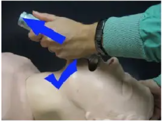
Clear the airway. Do not try to clear the airway without looking. Sweeping a finger “blindly” in the airway may push the obstruction further in.Turn the patient on their side and check the airway is clear. In the anesthestized patient or patients who cannot be turned onto their side, the airway is kept open by extending the neck and pulling the jaw forwards (jaw thrust). The tongue will be lifted forward by the genioglossus muscle that is attached to the base of the point of the jaw.
If the airway remains obstructed place an artificial airway (e.g. oropharyngeal airway,nasopharyngeal airway, laryngeal mask or endotracheal tube).
Continually monitor the patient. Look, listen and feel. Use a stethoscope to check there is air entry and that it is bilateral. If available use pulse oximetry. Cyanosis may be a nunreliable sign of hypoxaemia. Skin pigmentation, room lighting and the experience of the observer can affect it. Cyanosis occurs when there is 5 g/dl of unoxygenated blood.An anaemic patient may be severely hypoxic without showing cyanosis. A patient with haemoglobin of 15 g/dl would become cyanotic at a SaO2 of 78% (PaO2 44 mmHg). A patient with haemoglobin of 9 g/dl would show cyanosis at a SaO2 of 63% (PaO2 33mmHg). A patient with haemoglobin less than 9 g/dl would be likely to die before showing cyanosis.
Changes in heart rate and blood pressure are late signs of hypoxia.
Artificial Airways
Oropharyngeal airways (Guedel) are hollow curved plastic devices with a rigid flange.When correctly placed the curved portion holds the tongue clear of the posterior oropharyngeal wall and the flange sits against the lips. The correct sized oropharyngeal airway will reach from the angle of the mouth to the ear. The wrong size oral airway may worsen obstruction. If the airway is too short it may push the tongue down, if the airway is too long it may lie against the epiglottis. The airway is inserted “upside down”(with the concave surface facing up) until the tip is beyond the end of the tongue. It is then rotated 180 degrees. The anesthetist may need to continue to extend the neck and pull the jaw forwards to maintain a clear airway. A cuffed oropharyngeal airway with a 15 mm connector for attachment to a breathing system is available. They may not be tolerated if the patient has an intact gag reflex.
Nasopharyngeal airways are smooth non-cuffed tubes with a flange to prevent pushing them completely into the nose. Nasopharyngeal airways avoid damage to the teeth and can be inserted if the mouth cannot be opened, but they may cause the nose to bleed which may cause further obstruction. They are well tolerated by awake or sedated patients with an intact gag reflex. An un-cuffed endotracheal tube with a safety pin though one end may be used as a nasopharyngeal airway. The correct size nasopharyngeal airway will reach from the tip of the nose to the tragus of the ear. They must be lubricated before insertion. Gently insert along the floor of the nostril, perpendicular to the face (never upwards towards the cribriform plate). If there is resistance to insertion, gently rotate the nasal airway, try the other nostril or use a smaller tube.
The laryngeal mask is a spoon-shaped mask attached at 30 degrees to a connecting tube. When correctly placed it forms a low pressure seal around the laryngeal inlet. The laryngeal mask provides a more secure airway than mask ventilation, allows the anesthetist to attend to other tasks and avoids many of the complications of endotracheal intubation. It is simple to use. Though aspiration is uncommon, the laryngeal mask does not protect against aspiration. An air leak will occur with positive pressure ventilation greater than 15 to 20 cm H-20. The laryngeal mask is available in sizes 1 (less than 5 kg), 1.5 (5 to 10 kg), 2 (10 to 20 kg), 2.5 (20 to 30 kg), 3 (30 to 50 kg), 4 (small adult), 5 (large adult).The laryngeal mask is best used for routine general anaesthesia without muscle relaxation or as an emergency airway device when unable to intubate or ventilate. There are modified laryngeal masks for positive pressure ventilation (Proseal ®) and intubation (Fastrach ®).
The Proseal has a larger wedge shaped mask that creates a better seal, allowing the proseal to be used for positive pressure ventilation. The Proseal also has a drainage tube,which will direct regurgitated contents away from the laryngeal inlet.The Fastrach is a rigid laryngeal mask, which will direct an endotracheal tube centrally and anteriorly towards the laryngeal inlet. An endotracheal tube can be passed though a laryngeal mask but the success is much greater using a Fastrach.
[Laryngeal mask size/endotracheal size: 1/3.5, 2/4.5, 3/5.0, 4/6.0, and 5/7.5]
The laryngeal mask is inserted blindly into the pharynx. There are several ways of inserting a laryngeal mask. The first technique is based on how food is swallowed. The patient is placed in the sniffing position (head extended on the neck and neck flexed on the chest) and the mouth opened. The tip of a deflated and lubricated laryngeal mask is placed against the hard palate. The index finger is placed at the join of the cuff and connecting tube, and the laryngeal mask is pushed around the curve of the hard palate until resistance is felt. The cuff should be inflated without holding the tube. If correctly placed, the laryngeal mask will rise about 1.5 cm. The longitudinal black line along the tube should be in the midline against the upper lip. Other methods include placing the laryngeal mask in “upside down” (inlet facing up) and rotating the tube 180 degrees after passing passed the tongue, like an oropharyngeal airway.
A bite-block, usually folded gauze, is inserted in the mouth to protect the laryngeal mask from being bitten.
Mask ventilation is used for pre oxygenation, short operations (when there is no risk of aspiration) and for resuscitation. This is the most important skill an anesthetist has.Patients do not die because they can’t be intubated; they die because they can’t be ventilated.
The correct sized mask will fit around the bridge of the nose and over the cheeks and mouth. The mask is usually held with the left hand but with difficult mask ventilation the anesthetist may need to hold the mask with both hands while an assistant ventilates the patient. An oropharyngeal or nasopharyngeal airway may help maintain a clear airway.
The mask may be placed over the bridge of the nose and rolled forward into place. The thumb and index fingers are on the neck of the mask. The third and fourth fingers are placed along the mandible. These fingers should not push into the soft tissues of the neck. The little finger is placed at the angle of the jaw. The jaw should be pulled forward into the mask. The mask should not be pushed down onto the jaw. This would cause the neck to flex at the head and obstruct the airway. If there is a leak from one side of the mask the anesthetist can roll the mask slightly to one side or ask the assistant to push the cheek up into the mask.
A mask does not protect against regurgitation and aspiration.


Insertion of a Laryngeal Mask: The index finger is placed at the junction of the mask and the shaft. The lubricated laryngeal mask is pushed back against the roof of the mouth and down into the oropharynx. Once placed,the mask is inflated. (Copyright; Pescod)

Blind Intubation With a Fastrach: Once a lubricated fastrach laryngeal mask is inserted, a well-lubricated fastrach endotracheal tube can be blindly passed though the laryngeal inlet. (Copyright; Pescod)
ENDOTRACHEAL INTUBATION
The first attempt at intubation is usually the best attempt. The anesthetist must be prepared before attempting intubation. Drugs and equipment are checked. The assistant is ready. The patient is carefully positioned. The anesthetist must be ready to deal with a difficult intubation at any time.
The trachea may be intubated with the patient awake, anesthetized and breathing spontaneously or anesthetized and paralyzed. Maintaining spontaneous respiration is safer if the anesthetist believes that airway management may be difficult. Endotracheal intubation needs to be learnt and practiced. In an emergency the airway must be clear and the patient must be ventilated. Consider alternative airway management (e.g. laryngeal mask,oropharyngeal airway and mask ventilation) if not skilled in intubation.
Indications
Endotracheal intubation may be required in several conditions including respiratory arrest, respiratory failure, airway obstruction, reduced conscious state (Glasgow comas core less than 8), protection from aspiration, suctioning of the trachea and bronchi, drug administration, prolonged ventilation, inhalation injury, unstable mid-face fracture, large flail segment and anaesthesia.
Preoperative Assessment
All patients for anaesthesia must have careful airway assessment regardless of the planned anaesthetic technique. Patients receiving local anaesthesia may have complications that require the anesthetist to intubate. Mask or laryngeal mask ventilation may be inadequate and the patient may need intubation. Surgery may become more complicated/extensive requiring intubation. The anesthetist must have a plan for the management of the patient’s airway and a secondary plan to manage problems with ventilation. The anesthetist must always be prepared to establish an emergency airway.Patients must never be given a muscle relaxant unless the anesthetist is certain of being able to ventilate them.
Equipment
Correct equipment includes two working laryngoscopes and a selection of blades, a variety of endotracheal tubes, introducers for endotracheal tubes (rigid stylets and flexible bougies), Magill forceps, oral and nasal airways, face masks, suction, alternative airway (e.g. laryngeal mask) and an emergency airway (e.g. cricothyroid puncture kit).
Positioning
Position the patient in the sniffing position. Successful direct laryngoscopy requires alignment of the oral, pharyngeal and laryngeal axes. The neck should be flexed 25 to 35 degrees at the chest and extended at the head (atlanto-occiptal joint). This can be achieved by elevating the head about 10 cm with a firm pillow or pads beneath the occiput with the shoulders remaining on the table.The occiput of children less than 2 years of age naturally extends the head. They may not need a pillow.
Apply monitoring to the patient if available.
Endotracheal tube
Select the endotracheal tube and laryngoscope . Modern endotracheal tubes are available in sizes from 2.5 mm internal diameter (I.D.) to 9.0 mm I.D. in 0.5 mm increments. Adult tubes have a high volume low-pressure cuff to seal the trachea. The cuff should not be inflated to a pressure of more than 25 mmHg. This is the perfusion pressure in the tracheal mucosa. Inflation of the cuff to more than 25 mmHg may cause tracheal mucosa ischaemia. The pressure in a red rubber endotracheal tube often exceeds 25 mmHg. The narrowest part of the trachea is the cricoid cartilage in children. Un-cuffed tubes should be used before puberty.
The larger the endotracheal tube the lower the airway resistance and the less chance that there will be herniation of the cuff from over inflation but the smaller the tube the less chance of sore throat and the easier the intubation. Airway resistance does not increase significantly unless the tube is less than a size 6.0 mm I.D.
The endotracheal tube should have a hole cut in the wall opposite the bevelled end(Murphy eye). This allows gas to flow even if the bevelled end is obstructed.
Laryngoscopes
There are many different types of laryngoscopes (including McCoy, Bell, Miller and Polio). The standard rigid laryngoscope consists of a detachable blade with a removable bulb (or fibreoptic light) that attaches to a battery-containing handle. The blade has a flange on the left side for displacing the tongue. The blade may be curved (e.g.Macintosh) or straight (e.g. Miller). The curved blade may present more room in the mouth as the blade matches the curve of the oropharynx. The straight blade may be better when mouth opening is vertically limited or the larynx is anterior. The anesthetist should be trained in the use of both blades. When laryngoscopy is difficult with one blade, the other blade may be useful. Laryngoscope blades are available in different lengths. Adults usually need a size #3 or #4 Macintosh blade, children less than 8 years of age a size #2 Macintosh blade and term infants a size #1 Miller blade and premature infants a size #0 Miller blade.
When choosing the size of the endotracheal tube and length of the laryngoscope blade remember that it is easier to intubate with a small tube and long blade than a large tube and a short blade.
All patients should be pre-oxygenated for at least thee minutes.


Laryngoscopy
Laryngoscopy is performed with the laryngoscope held in the left hand. The blade is inserted into the right side of the mouth. Be careful not to pinch the lips or knock the incisor teeth. The assistant can help by pulling the lower lip out of the way. Be careful that the assistant does not flex the head while pulling the lip. At the tonsillar pillars sweep the tongue to the left and identify the uvula. Advance the laryngoscope blade slowly down the midline over the base of the tongue until the epiglottis is seen. A common mistake is to insert the blade too far down and into the oesophagus. If unsure,withdraw the laryngoscope slowly and the epiglottis may fall into view. A curved blade should have its tip in the vallecula. The tip of a straight blade is placed over the epiglottis. Exposure of the laryngeal inlet is improved by lifting the laryngoscope in the direction of the handle. Do not use the blade as a lever on the teeth and gums. The laryngoscope should only be moved in the direction of the handle, not back towards the anesthetist.
Insert the endotracheal tube from the right corner of the mouth. Rotating the tube 90 degrees clockwise may improve the view of the larynx. The tip of the endotracheal tube may be difficult to push pass the arytenoids and the base of the laryngeal inlet. Rotating the tube 90 degrees anti-clockwise may help pass the tube though the laryngeal inlet.
If only the base of the larynx can be seen or the endotracheal tube cannot be positioned anteriorly to pass though the larynx, the assistant can help by pressing on the cricoid.The assistant should push the cricoid backwards, upwards and to the right (BURP).Alternatively the anesthetist can push the cricoid until there is a good view of the larynx then have the assistant hold the cricoid in that position.
The rigid stylet and flexible bougie are excellent intubation aids. The rigid stylet is placed in the endotracheal tube and the tube is bent into a more useful shape. Usually the curve at the distal end is increased. The stylet should not extend beyond the end of the endotracheal tube. It could cause tracheal trauma.
The flexible bougie (gum elastic bougie) should be 60 cm long with a J shape at the distal tip. It should be soft and flexible to prevent trauma to the trachea. The flexible bougie is used as a guide for the endotracheal tube. Perform laryngoscopy. At least the tip of the epiglottis must be seen but ideally the arytenoids should also be seen. BURP may help improve the view. Pass the flexible bougie behind the mid point of the epiglottis with the J tip facing anteriorly. Gently push the flexible bougie in an anterior direction. If successful, “clicks” may be heard as the tip passes over the tracheal rings. A tracheal ring or the carina will stop the flexible bougie. If there are no clicks and the passage of the flexible bougie is not stopped, then the bougie may be in the oesophagus.If in doubt remove the bougie, ventilate the patient with 100% oxygen and try again.Hold the bougie firmly at the level of the mouth. Pass the endotracheal tube over the bougie until the proximal end of the bougie emerges. Have the assistant hold the proximal end of the bougie firmly. Maintain laryngoscopy. Gently push the endotracheal tube down the bougie. The tip of the tube may be stopped by the arytenoids. Turning the tube 90 degrees anticlockwise may help.

The patient’s head is placed in the “sniffing position” with the neck flexed on the trunk and the head extended on the neck. The laryngoscope is gently inserted with the left hand into the right side of the mouth. The tongue is swept to the right side of the mouth as the laryngoscope is pushed to the centre. The laryngoscope is gently inserted. The uvula is seen first then the epiglottis. A common error with laryngoscopy is to insert the laryngoscope too far without identifying oropharyngeal structures. This usually results with the tip of the laryngoscope in the oesophagus. If the laryngoscope is slowly removed the epiglottis will fall into view.


The gum elastic bougie is placed in the trachea with direct laryngoscopy. The anesthetist may feel clicks as the tip of the bogie passes over the tracheal rings. It is not possible to fully insert the gum elastic bougie in the trachea. If the bougie can be inserted fully,it is probably in the oesophagus.
Once the bougie is positioned in the trachea the endotracheal tube is slid along the bougie and into the trachea. The anesthetist should maintain laryngoscopy. It may be necessary to rotate the endotracheal tube to help it pass though the cords.
The gum elastic bougie is then removed and tracheal placement of the endotracheal tube confirmed.
Nasotracheal Intubation
Nasotracheal intubation may be required for intraoral surgery. It is contraindicated for patients with a fractured base of skull, fractured nose and coagulopathy. The patient will require a smaller endotracheal tube (6.0 to 7.0 mm). Spraying the nose with a vasoconstrictor and warming the tube in hot water will reduce the incidence of bleeding.The anesthetist should check which nostril is patent. The right nostril is preferred, as the bevel of the endotracheal tube will face the flat septum. This is less likely to cause trauma. Once in the pharynx, a Magill forceps may be needed to direct the tube though the laryngeal inlet.
Oesophageal Intubation
Always confirm that the endotracheal tube is in the trachea. If in doubt pull it out and mask ventilate with 100% oxygen before trying again.The cuff should be placed just below the cords. In an adult the marking on the tube at the lip is usually between 20 and 24 cm.
The best way to assess the tube position is to see the endotracheal tube pass though the cords and check for the presence of end-tidal carbon dioxide. Look for symmetrical chest movement and listen for breath sounds on both sides of the chest. Check that there are no breath sounds over the stomach. Look for vapor condensation on the inside of the tube with exhalation.
Oesophageal Detector
Unrecognized oesophageal intubation will cause gastric dilatation, aspiration, hypoxia and death. When capnography is not available an oesophageal detector device is a simple way of detecting oesophageal intubation. Oesophageal detector devices work by aspirating air. If the endotracheal tube is in the trachea, air is easily aspirated. (The trachea is a rigid structure and will not collapse). It is difficult to aspirate air if the endotracheal tube is in the oesophagus, as it will collapse.
A 60 ml catheter tip syringe can be connected to an endotracheal tube connector by a short length of rubber tubing. If the tube is in the oesophagus there will be resistance with aspiration and the plunger will return to its original position when released.Aspiration of 30 ml of air is a good sign that the tube is in the trachea. It is not 100%accurate. Some false results can occur. If there is distension of the oesophagus or stomach with air, or if the joins of the oesophageal detector device are not airtight, there may be aspiration of air. Bronchial intubation, bronchospasm, tracheal compression,obesity and chronic obstructive airways disease may cause resistance to aspiration of air.
Failed Intubation
Always have a plan for a failed intubation. Don’t waste time trying to intubate, instead mask ventilate, re-evaluate and try again. If pulse oximetry is available stop attempting intubation if the SaO2 falls below 90% and do not try again until the SaO2 is greater than 95%.
No attempt at intubation should be longer than 30 seconds.
If the patient is at risk of regurgitation and aspiration, maintain the cricoid pressure .Ideally the patient at risk of aspiration should have been given suxamethonium and will begin to breathe within 3 to 5 minutes. If the patient becomes hypoxic before the suxamethonium stops working,The anesthetist should be give gentle mask ventilation whilst maintaining the cricoid pressure. If the patient at risk of aspiration has unfortunately been given a long-acting muscle relaxant, they will need cricoid pressure and ventilation by mask until the muscle relaxant can be safely reversed.
If the first attempt at intubation (less than 30 seconds) fails, mask ventilate the patient.(Always make sure the patient is oxygenated). Reassess the patient’s position and the equipment. Is the patient in the sniffing position? Is it the correct size laryngoscope?Would BURP help? Would an intubation aid help? Can you call for help?Try to intubate again. If intubation fails thee times consider abandoning endotracheal intubation.
If endotracheal intubation is abandoned the anesthetist must decide how to manage the airway and decide if the surgery should continue. Alternative airway management to endotracheal intubation includes mask ventilation, laryngeal mask, Proseal and Fastrach. Neither will protect against regurgitation (though the Proseal laryngeal mask offers some protection). Laryngeal masks have the advantage that the anesthetist can attempt to pass an endotracheal tube blindly though the laryngeal mask.The anesthetist must also be aware that because of repeated attempts at intubation the airway may become more difficult to manage. Airway bleeding and oedema may make mask ventilation more difficult. If there are surgical complications it may be difficult for the anesthetist to manage both an unsecured airway and the surgical complication. Most surgery can be delayed at least a few hours while the patient is allowed to wake up and an alternative plan made. Reassess the need for intubation.
Regional anaesthesia may be possible for some surgery. However the anesthetist must be aware that rare complications from regional anaesthesia (e.g. convulsion, high spinal) may require intubation.
Awake Intubation
If the surgery cannot be done under regional anaesthesia the anesthetist must decide on a plan for airway management. With more airway aids and the help of a more experienced anesthetist it may be possible to attempt endotracheal intubation again.Alternative airway management includes awake intubation or awake surgical or percutaneous tracheostomy.
The aim of awake intubation is to anesthetize the upper airway using local anesthesia in order to pass an endotracheal tube.An awake intubation should be performed as an elective procedure. It is not a good choice of emergency airway management when intubation fails. It is a good choice when intubation is assessed preoperatively as being difficult.
Awake intubation can be performed with a flexible fibreoptic bronchoscope or using direct laryngoscopy. The endotracheal tube may be placed though the nose or the mouth.The nasal route may be less stimulating. The patient must be carefully prepared. The patient must be co-operative. They will need a full explanation of the procedure.Premedication with intramuscular atropine will dry up oral secretions and improve visibility but may be uncomfortable for the patient. The patient may require minimal amounts of sedation but the anesthetist must be aware that an awake intubation is performed because the airway may be difficult. If the anesthetist gives too much sedation the airway may become obstructed and intubation may be impossible. It is safer to take more time than give more sedation.
The local anaesthetic can be given topically or by nerve blocks. Remember not to exceed the recommended maximum dose. Lignocaine can be “sprayed as you go”.Laryngoscopy must be performed very slowly. Lignocaine is sprayed over the exposed mucosa and time is allowed for it to work before advancing. The patient will cough when lignocaine is sprayed on the cords and down the trachea. Alternatively lignocaine may be nebulized (5 ml of 4%). The patient will often also need some extra sprays.
The nose may be anesthetized with sprays of lignocaine with adrenaline or with cocaine(4% up to 2 mg/kg). It is important to use a vasoconstrictor to reduce the chance of nasal haemorrhage. The endotracheal tube must be well lubricated.
The airway may be anaesthetised by gargling 4% lignocaine (or a glossopharyngeal nerve block ) and then performing superior laryngeal nerve blocks and a trans-tracheal injection of local anaesthetic.
A superior laryngeal nerve block will anesthetise the supra-glottic structures: 2 to 3 ml of lignocaine is injected between the greater cornu of the hyoid bone and the thyroidcartilage.A trans-tracheal injection of 3 ml of 2 % lignocaine with a 23-gauge needle though the cricothyroid membrane will anesthetize the trachea. The needle must be quickly removed because most patients will cough vigorously.
The glossopharyngeal nerves may be blocked by 5 ml of 1% lignocaine injected into the area where the base of the tongue touches the palatoglossal fold. The needle must be aspirated to avoid intravascular injection. Gargling with lignocaine followed by a spray of 10% lignocaine is equally effective.
If the laryngeal structures are easily seen during awake laryngoscopy it may be safe to induce general anaesthesia and intubate the patient. Remember that some patients may depend on the normal muscle tone in the upper airway to maintain a patent airway and with induction of anaesthesia the structure of the upper airway may change making intubation difficult. For example it may be possible to view the larynx with awake laryngoscopy in a patient with a large intra-oral tumour but with induction of anesthesia and loss of airway muscle tone the tumour may move to completely obstruct the larynx.
Awake blind nasal intubation can be attempted after adequate local anaesthesia of the airway. A well-lubricated endotracheal tube is passed though the nose into the hypopharynx. The anesthetist listens for breath sounds at the end of the tube. The tubeis advanced slowly towards the maximum breath sounds. If breath sounds disappear thetube has passed into the oesophagus. The tube is passed into the larynx duringinspiration or the patient is asked to pant which will maintain the cords in an open position.
Extubation
Extubation of patients that were difficult to intubate must be done with care because they may need to be re-intubated. The patient should be fully awake, reversed and receive 100% oxygen before removing the tube. Emergency airway management equipment should be prepared. If in doubt, insert a bougie or a guide wire though the endotracheal tube and extubate the patient. The endotracheal tube may then be re-inserted over the bougie if the patient needs re-intubation.
CAN’T INTUBATE – CAN’T VENTILATE!
Good preparation and careful assessment should prevent the anesthetist ever having this nightmare. The anesthetist must rapidly establish an emergency airway
(cricothyroidotomy).
Cricothyroidotomy
With a cricothyroidotomy the patient can only be oxygenated. Carbon dioxide cannot be removed and respiratory acidosis will occur. A cricothyroidotomy should be converted to a tracheostomy as soon as possible. It is not possible to breathe spontaneously though a cricothyroidotomy.
Use a large intravenous cannula attached to a high-pressure oxygen source to ventilate the patient. A 12 or 14 gauge intravenous cannula is connected to a 10 ml syringe half full with liquid (e.g. saline) and introduced though the cricothyroid membrane until air is aspirated. Placing saline in the syringe makes it easier to see the air being aspirated. One hand can hold the trachea between the thumb and index finger. Once air is aspirated the cannula is advanced off the needle. The syringe with saline should be re-connected tothe cannula and aspirated to confirm the cannula has entered the trachea.
The cannula can then be connected to an oxygen source by several methods. It is wise to have a cricothyroidotomy kit prepared for each operating room. Time may be wasted trying to remember how to attach the oxygen. A 3 mm endotracheal tube adaptor will connect directly to the cannula or a 7.5 mm endotracheal tube connector will attach to the barrel of a 3 ml syringe that will attach to the intravenous cannula. Another excellent alternative is to attach a thee-way tap to the intravenous cannula. The correct size oxygen tubing will connect to the opposite side of the thee-way tap. The tap is turned toopen all thee channels. The oxygen supply is turned up to 12 to 15 litre/minute. Theanaesthetist can then intermittently obstruct the third channel of the thee-way tap with afinger. This will direct oxygen into the lungs. This method of connection ensures the anesthetist’s hand remains in contact with the cannula and patient. There is less chance of the emergency airway being lost. This method also delivers high volumes of oxygen.
The anesthetists should also attempt to prevent obstruction of the patient’s airway (jaw thrust, neck extension, artificial airway). The gas delivered to the patient will not be able to be expired if the patient’s airway is totally obstructed. This will cause barotrauma.


The endotracheal connector of a 7.5 endotracheal tube will fit into the barrel of a3 ml syringe which can be connected to the intravenous catheter.
The endotracheal connector of a 3.5 mm endotracheal tube will fitdirectly into the intravenous catheter.
Another excellent alternative is to attach a thee-way tap to the intravenous cannula. The correct size oxygen tubing will connect to the opposite side of the thee-way tap. The tap is turned to open all thee channels. The oxygen supply is turned up to 12 to 15 litres/minute. The anaesthetist can then intermittently obstruct the third channel of the thee-way tap with a finger.This will direct oxygen into the lungs. This method of connection ensures the anesthetist's hand remains in contact with the cannula and patient. There is less chance of the emergency airway being lost. This method also delivers high volumes of oxygen.
The anesthetist must have a plan of action if there is abnormal ventilation or inadequate oxygenation in an intubated patient.
Detach the patient from the ventilator and manually ventilate.
If the patient improves there is a problem with the anaesthetic machine. If ventilation or oxygenation remains abnormal then there is a problem with the airway or patient.
Check the position of the endotracheal tube.
Ensure that it is in the trachea. Ensure that it is not endobronchial. Listen to both sides of the chest. Make sure it is not kinked. Inspect it for obstructions. Pass a suction catheter down the tube. If a suction catheter passes easily it is unlikely to be obstructed. If the catheter does not pass easily deflate the cuff. The cuff may have herniated over the end of the tube. If in doubt, pull it out and mask ventilate. Replace the endotracheal tube.
Check the patient.
There are many patient causes of abnormal ventilation or inadequate oxygenation including bronchospasm (asthma, anaphylaxis), pneumothorax, aspiration and cardiac failure.