
10.4: Monocytes
- Page ID
- 38822

\( \newcommand{\vecs}[1]{\overset { \scriptstyle \rightharpoonup} {\mathbf{#1}} } \) \( \newcommand{\vecd}[1]{\overset{-\!-\!\rightharpoonup}{\vphantom{a}\smash {#1}}} \)\(\newcommand{\id}{\mathrm{id}}\) \( \newcommand{\Span}{\mathrm{span}}\) \( \newcommand{\kernel}{\mathrm{null}\,}\) \( \newcommand{\range}{\mathrm{range}\,}\) \( \newcommand{\RealPart}{\mathrm{Re}}\) \( \newcommand{\ImaginaryPart}{\mathrm{Im}}\) \( \newcommand{\Argument}{\mathrm{Arg}}\) \( \newcommand{\norm}[1]{\| #1 \|}\) \( \newcommand{\inner}[2]{\langle #1, #2 \rangle}\) \( \newcommand{\Span}{\mathrm{span}}\) \(\newcommand{\id}{\mathrm{id}}\) \( \newcommand{\Span}{\mathrm{span}}\) \( \newcommand{\kernel}{\mathrm{null}\,}\) \( \newcommand{\range}{\mathrm{range}\,}\) \( \newcommand{\RealPart}{\mathrm{Re}}\) \( \newcommand{\ImaginaryPart}{\mathrm{Im}}\) \( \newcommand{\Argument}{\mathrm{Arg}}\) \( \newcommand{\norm}[1]{\| #1 \|}\) \( \newcommand{\inner}[2]{\langle #1, #2 \rangle}\) \( \newcommand{\Span}{\mathrm{span}}\)\(\newcommand{\AA}{\unicode[.8,0]{x212B}}\)
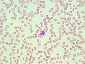
- An image from a peripheral blood smear showing a monocyte in the center. 50x oil immersion.From MLS Collection, University of Alberta, https://doi.org/10.7939/R3Q52FV0F
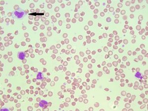
- An image from a peripheral blood smear showing a monocyte in the top left corner (indicated with an arrow). 50x oil immersion. From MLS Collection, University of Alberta, https://doi.org/10.7939/R3FN1172B
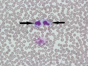
- An image from a peripheral blood smear showing two monocytes (indicated by arrows) with prominent vacuoles. 60x oil immersion. From MLS Collection, University of Alberta, https://doi.org/10.7939/R3MG7GB06
Notes: Monocyte nuclear and cytoplasmic morphology can be highly variable.1
Nucleus-to-Cytoplasm Ratio: Variable 2
Nucleoli: Not visible 2
Nucleus:1,2
Variable shapes (Folds, kidney shaped)
Loose, lacy, violet chromatin
Cytoplasm:1,2
Blue-gray cytoplasm (Ground glass appearance due to fine, diffuse granules)
May have pseudopods
May have vacuoles
Normal % in Bone Marrow: 2%2
Normal % in Peripheral Blood: 3-11%2
References:
1. Landis-Piwowar K. Granulocytes and Monocytes. In: Clinical laboratory hematology. 3rd ed. New Jersey: Pearson; 2015. p. 97-121.
2. Rodak BF, Carr JH. Monocyte maturation. In: Clinical hematology atlas. 5th ed. St. Louis, Missouri: Elsevier Inc.; 2017. p. 55-64.