
4.21: Hip Pain
- Page ID
- 59246
Hip-related pain is common in young to middle aged active adults (usually aged 18–50 years) and has a significant impact on physical activity and quality of life (Kemp et al., 2020).
Pathophysiology
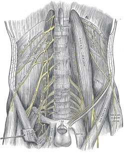
The presentation of hip pain does not always mean that the joint is the primary contributor to pain. Another peripheral generator that is often overlooked is peripheral nerve irritation, namely, sciatic, pudendal, obturator, femoral, and lateral femoral cutaneous (Martin et al., 2017). There are also twenty-one muscles that cross the hip providing both movement and stability between the femur and acetabulum, all of this contributes to the complex clinical picture of hip pain.
Classification of hip-related pain
Osteoarthritis Related Hip Pain
Osteoarthritis of the hip is a common finding in the general population, and in a majority of cases these degenerative changes are asymptomatic. However, in some cases this condition involves sensitization of nociceptive pathways, which may result in patients with osteoarthritis perceiving relatively low level stimuli as being overtly painful (Hunter & Bierma-Zeinstra, 2019).
Gluteal Tendinopathy
Tendinopathy of the gluteus medius and gluteus minimus tendons is now recognized as a primary local source of lateral hip pain. Many cases of hip “bursitis” should be more correctly classified as a non-inflammatory insertional tendinopathy of the gluteus medius or gluteus minimus tendons, that attach just deep to the greater trochanteric bursa. This condition interferes with sleep (side lying) and common weight-bearing tasks. The cardinal sign for this diagnosis is pain on palpation of the soft tissues over the greater trochanter.
Greater Trochanteric Pain Syndrome
An umbrella term used to encompass trochanteric bursitis, snapping hip syndrome, and abductor tendinopathy.
Femoroacetabular Impingement (FAI) Syndrome
The diagnosis of FAI syndrome currently includes bony morphological changes in the hip which may cause aberrant joint forces during hip movements and possible damage to the intra-articular structures of the joint.
Ischiofemoral Impingement
Refers to the painful entrapment of the quadratus femoris muscle between the lesser trochanter and the ischial tuberosity. The quadratus femoris acts synergistically with the other short external rotators but also serves as a secondary adductor of the hip.
Snapping Hip Syndrome
(iliopsoas tendinitis, or dancer’s hip) is characterized by a snapping sensation felt when the hip is flexed and extended. This may be accompanied by an audible snapping or popping noise and pain or discomfort. Pain often decreases with rest and diminished activity. Snapping hip syndrome is classified by location of the snapping, either extra-articular or intra-articular.
- Intra-articular
Because the iliopsoas or hip flexor crosses directly over the anterior superior labrum of the hip, an intra-articular hip derangement (i.e., labral tears, hip impingement, loose bodies) can lead to an effusion that subsequently produces internal snapping hip symptoms. - Extra-articular
- Lateral extra-articular (More common) Occurs when the iliotibial band, tensor fasciae latae, or gluteus medius tendon slides back and forth across the greater trochanter. This normal action becomes a snapping hip syndrome when one of these connective tissue bands thickens and catches with motion. The underlying bursa may also become inflamed, causing a painful external snapping hip syndrome.
- Medial extra-articular (Less common) The iliopsoas tendon catches on the anterior inferior iliac spine, the lesser trochanter, or the iliopectineal ridge during hip extension, as the tendon moves from an anterolateral to a posterior medial position. With overuse, the resultant friction may eventually cause painful symptoms, resulting in muscle trauma, bursitis, or inflammation in the area.
Examination
A thorough health history intake can be done to gather information about patients’ limitations, course of pain, and prognostic factors for delayed recovery (e.g., low self-efficacy, fear of movement, ineffective coping strategies, fear-avoidance, pain catastrophizing) and answers to health-related questions. Screen patients to identify those with a higher likelihood of serious pathology/red flag conditions. Then undertake a physical examination: neurological screening test, assess mobility and/or muscle strength.
Outcome Measurements
Incorporate one or more of the following outcome measurements when assessing and monitoring patient progress:
- Self-Rated Recovery Question
- Patient Specific Functional Scale
- Brief Pain Inventory (BPI)
- Visual Analog Scale (VAS)
- The Western Ontario and McMaster Universities Arthritis Index (WOMAC)
- Lower Extremity Functional Scale (LEFS)
Physical Examination
Incorporate one or more of the following physical examination tools and interpret examination results in the context of all clinical exam findings.
- FABER (Flexion-Abduction-External Rotation) Test (Patrick’s Test)
- FADDIR (Flexion-Adduction-Internal Rotation) Test
- Resisted External Derotation Test
- Trendelenburg Sign
- Thomas Test
- Ely’s Test
- Ober’s Test
- 90-90 Straight Leg Test
- Piriformis Strength Test (Pace Maneuver)
Clinical Signs of Osteoarthritis
According to a recent systematic review the most useful clinical finding to identify patients most likely to have osteoarthritis of the hip are (Metcalfe et al., 2019):
- Posterior Pain with squatting
- Groin pain with passive abduction or adduction
- Hip abductor weakness
- Decreased passive hip adduction or internal rotation as measured by a goniometer or compared with the contralateral leg.
Treatment
Education
Provide reassurance and patient education on condition and management options and encourage the use of active approaches (lifestyle, physical activity) to help manage symptoms.
Manual Therapy
The use of massage therapy has been shown to improve outcomes in post-operative hip patients. One recent randomized controlled trial published in the journal PM&R, looked at the use of manual therapy following total hip arthroplasty (Busato et al., 2016). In this study two treatment sessions were able to significantly improve functional outcomes in patients when used in addition to usual treatment.
Structures to be Aware of When Treating Hip Pain
A massage therapy treatment plan should be implemented based on patient-specific assessment findings and patient tolerance. Structures to keep in mind while assessing and treating patients suffering from hip pain may include neurovascular structures and investing fascia of:
- Iliopsoas (iliacus and psoas major)
- Hip Adductors (adductor brevis, adductor longus, adductor magnus, pectineus, gracilis)
- External Rotators of The Hip (piriformis, gemellus superior, externus and internus obturators, gemellus inferior, and quadratus femoris)
- Quadricep Muscles (rectus femoris, vastus lateralis, vastus mediali, vastus intermedius)
- Hamstring Muscles (semimembranosus, semitendinosus and biceps femoris)
- Gluteal Muscles (gluteus maximus, gluteus medius, gluteus minimus, and tensor fasciae latae)
- Erector Spinae (iliocostalis, longissimus, spinalis) & Multifidus
- Quadratus Lumborum
- Thoracolumbar Fascia & Latissimus Dorsi
Rehabilitation Program
Massage therapists not only provide hands-on treatment they can also develop self-management programs to help patients manage symptoms. Simple home-care recommendations such as patient education to remove or reduce loads that exacerbate symptoms, this may be sitting or standing with crossed legs, standing out onto one hip, and side lying (without pillows between the knees) may be useful for people with hip pain.
Prognosis
Prognosis is good, manual therapy is supported by clinical practice guidelines for the management of hip pain and mobility deficits (Ceballos-Laita et al. 2019; Cibulka et al., 2017).
Massage Tutorial: Hip Pain
Contemporary multimodal massage therapists are uniquely suited to incorporate a number of rehabilitation strategies for acute and chronic hip pain based on patient-specific assessment findings including, but not limited to:
- Manual Therapy (soft tissue massage, neural mobilization, joint mobilization)
- Education that is Person-Centered (e.g., biopsychosocial model of health and disease, self-efficacy beliefs, active coping strategies)
- Stretching & Loading Programs (e.g., concentric, eccentric, isometric exercises)
- Hydrotherapy (hot & cold)
- Self-Management Strategies (e.g., engaging in physical activity and exercise, social activities, and healthy sleep habits)
References and Sources
Alentorn-Geli, E., Samuelsson, K., Musahl, V., Green, C. L., Bhandari, M., & Karlsson, J. (2017). The Association of Recreational and Competitive Running With Hip and Knee Osteoarthritis: A Systematic Review and Meta-analysis. The Journal of orthopaedic and sports physical therapy, 47(6), 373–390. doi:10.2519/jospt.2017.7137
Battaglia, P. J., D’Angelo, K., & Kettner, N. W. (2016). Posterior, Lateral, and Anterior Hip Pain Due to Musculoskeletal Origin: A Narrative Literature Review of History, Physical Examination, and Diagnostic Imaging. Journal of chiropractic medicine, 15(4), 281–293. doi:10.1016/j.jcm.2016.08.004
Busato, M., Quagliati, C., Magri, L., Filippi, A., Sanna, A., Branchini, M., … Stecco, A. (2016). Fascial Manipulation Associated With Standard Care Compared to Only Standard Postsurgical Care for Total Hip Arthroplasty: A Randomized Controlled Trial. PM & R: the journal of injury, function, and rehabilitation, 8(12), 1142–1150. doi:10.1016/j.pmrj.2016.04.007
Ceballos-Laita, L., Estébanez-de-Miguel, E., Martín-Nieto, G., Bueno-Gracia, E., Fortún-Agúd, M., & Jiménez-Del-Barrio, S. (2019). Effects of non-pharmacological conservative treatment on pain, range of motion and physical function in patients with mild to moderate hip osteoarthritis. A systematic review. Complementary therapies in medicine, 42, 214–222. https://doi.org/10.1016/j.ctim.2018.11.021
Ceballos-Laita, L., Jiménez-Del-Barrio, S., Marín-Zurdo, J., Moreno-Calvo, A., Marín-Boné, J., Albarova-Corral, M. I., & Estébanez-de-Miguel, E. (2019). Effects of dry needling in HIP muscles in patients with HIP osteoarthritis: A randomized controlled trial. Musculoskeletal science & practice, 43, 76–82. doi:10.1016/j.msksp.2019.07.006
Cheatham, S. W., Kolber, M. J., & Salamh, P. A. (2013). Meralgia paresthetica: a review of the literature. International journal of sports physical therapy, 8(6), 883–893.
Cibulka, M. T., Bloom, N. J., Enseki, K. R., Macdonald, C. W., Woehrle, J., & McDonough, C. M. (2017). Hip Pain and Mobility Deficits-Hip Osteoarthritis: Revision 2017. The Journal of orthopaedic and sports physical therapy, 47(6), A1–A37. doi:10.2519/jospt.2017.0301
Cowan, R. M., Semciw, A. I., Pizzari, T., Cook, J., Rixon, M. K., Gupta, G., Plass, L. M., & Ganderton, C. L. (2020). Muscle Size and Quality of the Gluteal Muscles and Tensor Fasciae Latae in Women with Greater Trochanteric Pain Syndrome. Clinical anatomy (New York, N.Y.), 33(7), 1082–1090. https://doi.org/10.1002/ca.23510
Czuppon, S., Prather, H., Hunt, D. M., Steger-May, K., Bloom, N. J., Clohisy, J. C., … Harris-Hayes, M. (2017). Gender-Dependent Differences in Hip Range of Motion and Impingement Testing in Asymptomatic College Freshman Athletes. PM & R: the journal of injury, function, and rehabilitation, 9(7), 660–667. doi:10.1016/j.pmrj.2016.10.022
Ferguson, R. J., Palmer, A. J., Taylor, A., Porter, M. L., Malchau, H., & Glyn-Jones, S. (2018). Hip replacement. Lancet (London, England), 392(10158), 1662–1671. doi:10.1016/S0140-6736(18)31777-X
Grimaldi, A., Mellor, R., Hodges, P., Bennell, K., Wajswelner, H., & Vicenzino, B. (2015). Gluteal Tendinopathy: A Review of Mechanisms, Assessment and Management. Sports medicine (Auckland, N.Z.), 45(8), 1107–1119. doi:10.1007/s40279-015-0336-5
Ganderton, C., Semciw, A., Cook, J., & Pizzari, T. (2017). Demystifying the Clinical Diagnosis of Greater Trochanteric Pain Syndrome in Women. Journal of women’s health (2002), 26(6), 633–643. doi:10.1089/jwh.2016.5889
Hunter, D. J., & Bierma-Zeinstra, S. (2019). Osteoarthritis. Lancet (London, England), 393(10182), 1745–1759. doi:10.1016/S0140-6736(19)30417-9
Kemp, J. L., Risberg, M. A., Mosler, A., Harris-Hayes, M., Serner, A., Moksnes, H., … Bizzini, M. (2020). Physiotherapist-led treatment for young to middle-aged active adults with hip-related pain: consensus recommendations from the International Hip-related Pain Research Network, Zurich 2018. British journal of sports medicine, 54(9), 504–511. https://doi.org/10.1136/bjsports-2019-101458
Kemp, J. L., Mosler, A. B., Hart, H., Bizzini, M., Chang, S., Scholes, M. J., Semciw, A. I., & Crossley, K. M. (2020). Improving function in people with hip-related pain: a systematic review and meta-analysis of physiotherapist-led interventions for hip-related pain. British journal of sports medicine, bjsports-2019-101690. Advance online publication. https://doi.org/10.1136/bjsports-2019-101690
Kolasinski, S. L., Neogi, T., Hochberg, M. C., Oatis, C., Guyatt, G., Block, J., … Reston, J. (2020). 2019 American College of Rheumatology/Arthritis Foundation Guideline for the Management of Osteoarthritis of the Hand, Hip, and Knee. Arthritis & rheumatology (Hoboken, N.J.), 72(2), 220–233. https://doi.org/10.1002/art.41142
Kompel, A. J., Roemer, F. W., Murakami, A. M., Diaz, L. E., Crema, M. D., & Guermazi, A. (2019). Intra-articular Corticosteroid Injections in the Hip and Knee: Perhaps Not as Safe as We Thought?. Radiology, 293(3), 656–663. doi:10.1148/radiol.2019190341
Martin, R., Martin, H. D., & Kivlan, B. R. (2017). Nerve Entrapment in the Hip region: Current Concepts Review. International journal of sports physical therapy, 12(7), 1163–1173. https://doi.org/10.26603/ijspt20171163
Mellor, R., Bennell, K., Grimaldi, A., Nicolson, P., Kasza, J., Hodges, P., … Vicenzino, B. (2018). Education plus exercise versus corticosteroid injection use versus a wait and see approach on global outcome and pain from gluteal tendinopathy: prospective, single blinded, randomised clinical trial. BMJ (Clinical research ed.), 361, k1662. doi:10.1136/bmj.k1662
Metcalfe, D., Perry, D. C., Claireaux, H. A., Simel, D. L., Zogg, C. K., & Costa, M. L. (2019). Does This Patient Have Hip Osteoarthritis?: The Rational Clinical Examination Systematic Review. JAMA, 322(23), 2323–2333. doi:10.1001/jama.2019.19413
Neumann, D. A. (2010). Kinesiology of the hip: a focus on muscular actions. The Journal of orthopaedic and sports physical therapy, 40(2), 82–94. doi:10.2519/jospt.2010.3025
Reiman, M. P., Mather, R. C., 3rd, & Cook, C. E. (2015). Physical examination tests for hip dysfunction and injury. British journal of sports medicine, 49(6), 357–361. doi:10.1136/bjsports-2012-091929
Reiman, M. P., Agricola, R., Kemp, J. L., Heerey, J. J., Weir, A., van Klij, P., Kassarjian, A., Mosler, A. B., Ageberg, E., Hölmich, P., Warholm, K. M., Griffin, D., Mayes, S., Khan, K. M., Crossley, K. M., Bizzini, M., Bloom, N., Casartelli, N. C., Diamond, L. E., Di Stasi, S., … Dijkstra, H. P. (2020). Consensus recommendations on the classification, definition and diagnostic criteria of hip-related pain in young and middle-aged active adults from the International Hip-related Pain Research Network, Zurich 2018. British journal of sports medicine, 54(11), 631–641. https://doi.org/10.1136/bjsports-2019-101453