
1.1: Cell Biology Review
- Page ID
- 41537

\( \newcommand{\vecs}[1]{\overset { \scriptstyle \rightharpoonup} {\mathbf{#1}} } \)
\( \newcommand{\vecd}[1]{\overset{-\!-\!\rightharpoonup}{\vphantom{a}\smash {#1}}} \)
\( \newcommand{\id}{\mathrm{id}}\) \( \newcommand{\Span}{\mathrm{span}}\)
( \newcommand{\kernel}{\mathrm{null}\,}\) \( \newcommand{\range}{\mathrm{range}\,}\)
\( \newcommand{\RealPart}{\mathrm{Re}}\) \( \newcommand{\ImaginaryPart}{\mathrm{Im}}\)
\( \newcommand{\Argument}{\mathrm{Arg}}\) \( \newcommand{\norm}[1]{\| #1 \|}\)
\( \newcommand{\inner}[2]{\langle #1, #2 \rangle}\)
\( \newcommand{\Span}{\mathrm{span}}\)
\( \newcommand{\id}{\mathrm{id}}\)
\( \newcommand{\Span}{\mathrm{span}}\)
\( \newcommand{\kernel}{\mathrm{null}\,}\)
\( \newcommand{\range}{\mathrm{range}\,}\)
\( \newcommand{\RealPart}{\mathrm{Re}}\)
\( \newcommand{\ImaginaryPart}{\mathrm{Im}}\)
\( \newcommand{\Argument}{\mathrm{Arg}}\)
\( \newcommand{\norm}[1]{\| #1 \|}\)
\( \newcommand{\inner}[2]{\langle #1, #2 \rangle}\)
\( \newcommand{\Span}{\mathrm{span}}\) \( \newcommand{\AA}{\unicode[.8,0]{x212B}}\)
\( \newcommand{\vectorA}[1]{\vec{#1}} % arrow\)
\( \newcommand{\vectorAt}[1]{\vec{\text{#1}}} % arrow\)
\( \newcommand{\vectorB}[1]{\overset { \scriptstyle \rightharpoonup} {\mathbf{#1}} } \)
\( \newcommand{\vectorC}[1]{\textbf{#1}} \)
\( \newcommand{\vectorD}[1]{\overrightarrow{#1}} \)
\( \newcommand{\vectorDt}[1]{\overrightarrow{\text{#1}}} \)
\( \newcommand{\vectE}[1]{\overset{-\!-\!\rightharpoonup}{\vphantom{a}\smash{\mathbf {#1}}}} \)
\( \newcommand{\vecs}[1]{\overset { \scriptstyle \rightharpoonup} {\mathbf{#1}} } \)
\( \newcommand{\vecd}[1]{\overset{-\!-\!\rightharpoonup}{\vphantom{a}\smash {#1}}} \)
\(\newcommand{\avec}{\mathbf a}\) \(\newcommand{\bvec}{\mathbf b}\) \(\newcommand{\cvec}{\mathbf c}\) \(\newcommand{\dvec}{\mathbf d}\) \(\newcommand{\dtil}{\widetilde{\mathbf d}}\) \(\newcommand{\evec}{\mathbf e}\) \(\newcommand{\fvec}{\mathbf f}\) \(\newcommand{\nvec}{\mathbf n}\) \(\newcommand{\pvec}{\mathbf p}\) \(\newcommand{\qvec}{\mathbf q}\) \(\newcommand{\svec}{\mathbf s}\) \(\newcommand{\tvec}{\mathbf t}\) \(\newcommand{\uvec}{\mathbf u}\) \(\newcommand{\vvec}{\mathbf v}\) \(\newcommand{\wvec}{\mathbf w}\) \(\newcommand{\xvec}{\mathbf x}\) \(\newcommand{\yvec}{\mathbf y}\) \(\newcommand{\zvec}{\mathbf z}\) \(\newcommand{\rvec}{\mathbf r}\) \(\newcommand{\mvec}{\mathbf m}\) \(\newcommand{\zerovec}{\mathbf 0}\) \(\newcommand{\onevec}{\mathbf 1}\) \(\newcommand{\real}{\mathbb R}\) \(\newcommand{\twovec}[2]{\left[\begin{array}{r}#1 \\ #2 \end{array}\right]}\) \(\newcommand{\ctwovec}[2]{\left[\begin{array}{c}#1 \\ #2 \end{array}\right]}\) \(\newcommand{\threevec}[3]{\left[\begin{array}{r}#1 \\ #2 \\ #3 \end{array}\right]}\) \(\newcommand{\cthreevec}[3]{\left[\begin{array}{c}#1 \\ #2 \\ #3 \end{array}\right]}\) \(\newcommand{\fourvec}[4]{\left[\begin{array}{r}#1 \\ #2 \\ #3 \\ #4 \end{array}\right]}\) \(\newcommand{\cfourvec}[4]{\left[\begin{array}{c}#1 \\ #2 \\ #3 \\ #4 \end{array}\right]}\) \(\newcommand{\fivevec}[5]{\left[\begin{array}{r}#1 \\ #2 \\ #3 \\ #4 \\ #5 \\ \end{array}\right]}\) \(\newcommand{\cfivevec}[5]{\left[\begin{array}{c}#1 \\ #2 \\ #3 \\ #4 \\ #5 \\ \end{array}\right]}\) \(\newcommand{\mattwo}[4]{\left[\begin{array}{rr}#1 \amp #2 \\ #3 \amp #4 \\ \end{array}\right]}\) \(\newcommand{\laspan}[1]{\text{Span}\{#1\}}\) \(\newcommand{\bcal}{\cal B}\) \(\newcommand{\ccal}{\cal C}\) \(\newcommand{\scal}{\cal S}\) \(\newcommand{\wcal}{\cal W}\) \(\newcommand{\ecal}{\cal E}\) \(\newcommand{\coords}[2]{\left\{#1\right\}_{#2}}\) \(\newcommand{\gray}[1]{\color{gray}{#1}}\) \(\newcommand{\lgray}[1]{\color{lightgray}{#1}}\) \(\newcommand{\rank}{\operatorname{rank}}\) \(\newcommand{\row}{\text{Row}}\) \(\newcommand{\col}{\text{Col}}\) \(\renewcommand{\row}{\text{Row}}\) \(\newcommand{\nul}{\text{Nul}}\) \(\newcommand{\var}{\text{Var}}\) \(\newcommand{\corr}{\text{corr}}\) \(\newcommand{\len}[1]{\left|#1\right|}\) \(\newcommand{\bbar}{\overline{\bvec}}\) \(\newcommand{\bhat}{\widehat{\bvec}}\) \(\newcommand{\bperp}{\bvec^\perp}\) \(\newcommand{\xhat}{\widehat{\xvec}}\) \(\newcommand{\vhat}{\widehat{\vvec}}\) \(\newcommand{\uhat}{\widehat{\uvec}}\) \(\newcommand{\what}{\widehat{\wvec}}\) \(\newcommand{\Sighat}{\widehat{\Sigma}}\) \(\newcommand{\lt}{<}\) \(\newcommand{\gt}{>}\) \(\newcommand{\amp}{&}\) \(\definecolor{fillinmathshade}{gray}{0.9}\)- Overview of cell biology
- Inside the cell:
- Cell membrane, cytoplasm
- Organelles
- Outside the cell:
- Extra-Cellular Matrix (ECM)
- Extra-Cellular Fluid (ECF)
- Neither in nor out:
- Cell division
- Cell death
- Cell junctions

Overview of cell biology
The cell is the basic unit of life. It is the smallest thing that we call living (without arguing about viruses), and the human body is made of 10 trillion of them. We started off as a single cell, and the purpose of this class to learn a little bit about how that one cell developed into the trillion-celled organism you are today. Nearly all of the instructions for making trillions of cells– how to make them, when to make them, where to make them– are found within that single cell. Different cells have different functions, even though almost every cell in one person has the exact same DNA. To become different from one another, cells express different DNA. This is an important process called differentiation, which is really no more complicated in concept than cells going from looking like boring generic cells to looking and behaving differently from other cells. We give different names to cells as they make these changes. For instance, a stem cell that gives rise to, oh let’s say, a smurf, would be named a Smurfal Stem Cell. As the Smurfal Stem Cells divide and differentiate into cells that make a smurf, the cells actually making the smurf would be named Smurfoblasts, and when the Smurf was finished, the cells inside that Smurf would be named Smurfocytes. The stem cells found in a tissue called mesenchyme are named mesenchymal stem cells, the cells that make dentin are named odontoblasts, the cells within mature cementum are named cementocytes. Note that differentiation is not a single step, but a series of steps along a spectrum, from the least differentiated, to more differentiated, until we reach a terminally differentiated cell. The aim of this chapter is to review aspects of a cell biology class that come up in this textbook, and not much more. We assume you have covered cell biology in a per-requisite to this class. We need to review the parts of a cell that help us to explain difficult concepts like differentiation and development. If you find you need more than a quick refresher, here are some link from the NIH, who have a number of very useful publications and videos (for free), such as:
- 3D animations of the human cell
- Learn genetics (interactive website)
- Inside the Cell (pdf and epub downloads)
Two useful (and free) eBooks you may wish to download are from OpenStax:

Inside the cell
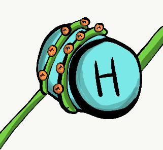
Cell membrane and cytoplasm
Every human cell is surrounded by the plasma membrane. The plasma membrane separates the cell from its environment, and allows certain materials to enter and leave the cell. Phospholipids and cholesterol form a barrier that separates the cell from the external environment, which in turn allows us to point at that cell and call it a thing. Trans-membrane proteins span the plasma membrane, and regulate what goes in or out. Other trans-membrane proteins receive signals from other cells or the environment, and relay that information to the inside of the cell. Still others mediate attachment to structures outside of the cell. Other integral membrane proteins are located on the outer or inner surface of the plasma membrane, but do not span the entire width of the phospholipid bilayer. Many biology classes focus heavily on the nucleus, a trend that was admonished by one of the first African Americans to earn a Ph.D., Dr. Ernest Everett Just, in his book Biology of the Cell Surface. Dr. Just was an embryologist, and in embryology the plasma membrane proteins—especially receptors and attachment proteins– are very important. The cytoplasm is the gelatinous filling of a cell, and is sometimes referred to an Intra-Cellular Fluid (ICF). Cytoplasm includes nutrients and electrolytes absorbed from the the fluid surrounding all cells. In addition, the cytoplasm includes a number of proteins and glycoproteins synthesized by the cell. These molecules may have other important functions, but they attract water from the ECF. The end result is that cells have a gelatinous filling, rather than a watery one. This gelatinous filling is filled with a number of organelles, similar to the way my grandmother’s jello salad contained grapes, raisins and other food or food-like substances that did not in any way turn that jello into an actual salad.

The nucleus
The nucleus contains practically all of cell’s Deoxyribonucleic acid (DNA). DNA is the instructions for making a Ribonucleic Acid (RNA) copy, and most RNA is translated in the cytoplasm into proteins found inside of and outside of a cell. It is also the instructions for when and where to make these proteins. For instance, epithelial cells of the oral mucosa do not make enzymes that secrete calcium and phosphate into the extra-cellular matrix. On the other hand, epithelial cells that differentiate into ameloblasts make these proteins, by expressing the DNA for these enzymes, after being told to do so by other cells called neural crest cells. All cells in the human body have the same DNA (with a few exceptions). However, different cells express different DNA at different times. DNA can be divided into 2 basic types. There are genes, each gene is more-or-less the instructions for a single protein. The rest of DNA folds up into unique shapes that provides instructions for when and where to express these genes. The latter is referred to as non-coding DNA. For DNA expression to occur, proteins called transcription factors (with help from special small molecules) bind to regulatory regions of DNA, open up the appropriate region, and recruit enzymes that copy one strand of DNA into messenger RNA (mRNA), which leaves the nucleus to be translated into protein. Other transcription factors may temporarily inhibit the expression of genes.
Transcription factors turn on and turn off genes quickly, in response to changes in a cell’s environment. But when cells differentiate, they shut down genes they don’t need more permanently. Rather than relying on the on and off switches of transcription factors binding to regulatory DNA regions, these inactivated genes are methylated (a -CH3 group is attached), packed up around histone proteins, and put into long-term storage. The pattern of DNA methylation and histone packaging is copied during mitosis. This means the pattern of genes that are available or packaged away is inherited by both daughter cells, but because this inheritance is not a difference in the DNA sequence, it is known as epigenetic inheritance. Epigenetic traits are more capable of modification in response to changes in the environment than genes (DNA), and play an important role in cell differentiation and cell fate which is covered in more detail in chapters 8-11. We have 46 molecules of DNA in the nucleus– they are freakishly long molecules, but only 46 in number– 23 maternal, and 23 paternal. During mitosis, these 46 molecules are packaged up tightly into 46 chromosomes (times 2). This packaging involves using histones along nearly the entire length of a DNA strand, and allows chromosomes to be seen under the light microscope. The rest of the time, DNA is mostly unwound (un-needed instructions are wound around histones, the rest are free to be transcribed), and fills up the nucleus in a way that doesn’t look very exciting. We call that DNA chromatin, and functionally it is by far the more exciting of the two forms.

Ribosomes
Visible throughout the cytoplasm are small specks made of protein and RNA called ribosomes. These structures translate mRNA instructions that came from the nucleus, which means the mRNA instructions are used to guide the linkage of a series of amino acids into a long protein. Groups of three mRNA nucleotides, called codons, instruct the ribosome which amino acid to add to a protein. Like regulatory regions of DNA, RNA found in the ribosome itself– not the mRNA instructions but ribosomal rRNA, is a type of RNA that folds up into specific shapes that, along with the ribosomal proteins, perform the chemical reaction of translation. Free-floating ribosomes in the cytoplasm synthesize proteins that remain in the cytoplasm, such as keratin or enzymes that mediate apoptosis.
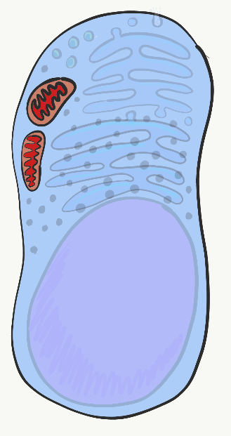
Mitochondria
The mitochondria are where the majority of Adenosine Tri-Phosphate (ATP) is produced. ATP is made of Adenosine, plus three phosphate (PO43-) groups– pay attention to the phosphate part, it is also a major component of bone, enamel, dentin and cementum. ATP powers almost all cellular processes, including the transcription and translation of mucous proteins within a salivary gland, the electrical signals sent by neurons in the tongue when food enters the oral cavity, and the contraction of myo-epithelial cells to cause salivation. Mitochondria burn glucose, using oxygen, and harness some of the energy released in the form of ATP. Mitochondria are different from other organelles in that they contain a little bit of their own DNA, which is inherited just from mother. Mitochondria also contain two phospholipid bilayer membranes, not one like other organelles. They use this extra membrane to generate ATP. Perhaps you covered glycolysis and the citric acid cycle before. The part to remember is that mitochondria use a proton (or H+) gradient, which makes the inside of mitochondria acidic, and therefore potentially toxic to the rest of the cell.

Lysosomes
Lysosomes are small compartments surrounded by the same phospholipids as in the plasma membrane. Inside lysosomes are acids and digestive enzymes that can be used to destroy stuff inside the cell when it wears out, or materials that the cell has gobbled up from outside (e.g. debris, bacteria). When a cell dies and begins to break apart, neighboring cells are in danger of being damaged from the acids and enzymes found within lysosomes. In the oral cavity, epithelial cells only have a life-span of days before they wear out, so it is very important for these cells to neutralize acids and enzymes in their lysosomes first, in a process called apoptosis.

Endoplasmic Reticulum
The Endoplasmic Reticulum (ER) is a series of interconnected tubes surrounded by a phospholipid bilayer– similar to lysosomes, only bigger, more tubular, and not full of acid. The smooth Endoplasmic Reticulum (sER) is where cells produce lipids and store calcium. The rough Endoplasmic Reticulum (rER) is covered in ribosomes. Proteins made by these ribosomes wind up inside the rER, then travel to the Golgi apparatus, and either wind up being secreted (such as mucous proteins) or stay within the plasma membrane (such as cell-junction proteins, or receptors for morphogen molecules).

Golgi apparatus
The Golgi apparatus is another set of tubes, similar to the rER. Small membrane-enclosed spheres called vesicles shuttle proteins made in the rER to the Golgi apparatus, where the proteins are modified. Often, these proteins have sugars attached to them, making them glycoproteins. Vesicles take these proteins to the plasma membrane, where they are either secreted or become a part of the plasma membrane. We cover the role the secreted protein collagen plays in enamel and the periodontal ligament. We also cover the shared role of the secreted glycoprotein fibronectin and the membrane-bound protein integrin have in healing damaged gingival tissue.
Cytoskeleton
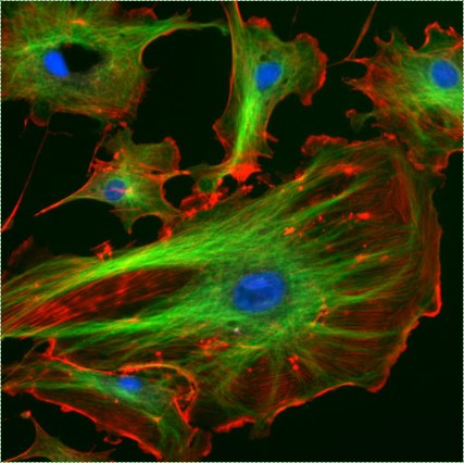
The cytoskeleton is a network of structural proteins within the cytoplasm. This network gives the cell its shape, the ability to change its shape, or to migrate. Shown in Fig 1.10, cells have microtubules and actin filaments stained red and green, respectively. These proteins are not generally visible on more old-fashioned histology images– both would be pink on a H&E stain, as would soluble proteins in the cytoplasm.
Outside the cell
| Extra-cellular matrix: |
|---|
| Ground substance |
| Fibers |
Table 1.1: Two major components of ECM. Extra-Cellular Matrix (ECM) includes all the material found outside of cells. It is usually broken down into the two components listed in Table 1.1.

Ground substance
Ground substance is the name for all of the material outside of the cell that has no particular shape when viewed under traditional microscopy. One part of ground substance is Extra-Cellular Fluid (ECF), which is the water and nutrients surrounding cells that was called plasma when it was inside of a blood vessel. Once fluid exits the blood and surrounds cells, it is called ECF. Unlike plasma, this fluid is held in place by the other major part of ground substance, which includes proteins, glycoproteins, and polysaccharides. These molecules are solutes, and their size makes them immovable, therefore they are good at holding the fluid in place, forming a gel. It doesn’t look like much under a microscope, no more than if you looked really closely at some jello. These proteins, glycoproteins and polysaccharides are made and secreted by cells (these cells probably have a lot of rER and Golgi apparatus). One of the glycoproteins found in ground substance is fibronectin, which is a really long protein. Cell may recognize, bind to and move along fibronectin if they have the correct integrin protein spanning their plasma membrane. Therefore, fibronectin acts not only as a road along which cells travel, it is also a road map. Getting the right cells to the right place at the right time is very important both in healing and in development. In fact, what you learn about development is re-used (recapitulated) in healing. Another important molecule found in ground substance is a large polysaccharide called Hyaluronic Acid (HA). Like fibronectin, cells can bind to and travel over HA (using a different type of plasma membrane protein), which has applications in dentistry, such as helping cells of the gingiva stick to a dental implant and form a bacteria-resistant seal. We can’t see fibronectin or HA without using some modern imaging tricks, which is why they are listed in as ground substance, and not in the next section, fibers. Because cells migrate over ground substance proteins, we say that ECM proteins function as a scaffold. Without scaffolds, tissues grow only from their edges. This is fine if speed is not important, such as when enamel and dentin are forming. However, in wound repair, it is optimal for a wound to heal everywhere at once, rather than from the edges. The body, therefore, often puts down some form of scaffolding first, such as a scab. In dentistry, artificial scaffolds can be created to help the body heal, based on our knowledge of the functions of ground substance. Examples of artificial scaffolds include some bone grafting materials, periodontal membranes and materials. In addition to their structural role as a scaffold, guiding cells to new locations, ground substance molecules provide cells with information. This information tells cells where they are located, and what they should be doing. For instance, when a stem cell binds to fibronectin, fibronectin can instruct the stem cell to express different genes and differentiate into a new type of cell, such as an odontoblast, and begin secreting dentin. Getting cells to the correct location is nice, but they need to know what to do when they get there. This happens during tooth formation and in response to tooth injury. As we learn more about how ground substance instructs stem cells, we get better at helping teeth repair themselves. It is unfortunate that many textbooks gloss over ground substance as just the gelatinous material outside of a cell, which is why we have gone into it in more detail, and instead glossed over mitochondria.
Fibers
Three extracellular proteins were visible under a light microscope a century ago, and they were grouped together as fibers of the extra-cellular matrix. Like fibronectin and other ground-substance proteins, fibers are secreted by cells called fibroblasts.
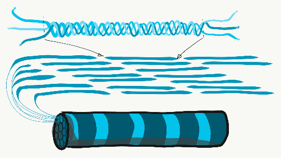
Collagen is the strongest of the three fibers, and is therefore often referred to as a structural protein—it gives many organs their shape. This fiber is made of 3 coiled α-helices, that are in turn coiled and cross-linked together, making a very strong macromolecule with the same basic shape as a rope. It is very strong if you pull on it from the ends, but bends if you apply force from the side. For instance, an area of advanced caries on a tooth can be as soft as wet driftwood, as it contains its collagen fibers without the plentiful minerals that used to surround them. Collagen fibers are found in regions of the oral cavity where the ability to resist force is important, such as in enamel or the periodontal ligament. In fact, it’s found throughout the human body, accounting for 25% of our protein content. In addition to having a structural role, collagen also acts as a scaffold, similar to fibronectin and HA, which allows cells to migrate across the long fibers. Getting collagen from the rER (where it is translated) to the Golgi body for modification, and finally secreted from the cell requires extra-large vesicles for transport. Reticular fibers, which are not shown in Figure 1.11, look like a fine, spider web-like mesh under the microscope. Later it was discovered that reticular fibers are different form of collagen, but they are still called by their own name, and often receive equal footing with collagen on the list of ECM fibers. This web-like network of proteins isn’t as strong, but provides enough of a frame for blood cells to rest throughout organs like the spleen and lymph nodes, as opposed to sinking to the bottom. Elastic fibers are thinner than collagen fibers, and often look like fine hairs in the ECM under a microscope. They are made from a different type of protein (elastin). As their name suggests, these fibers can be stretched and then spring back to their original length. This is not something collagen can do (if you can’t put your palms on the floor while keeping your legs straight, the collagen fibers of your hamstring and gastrocnemious muscles and their tendons limit your range of motion. Regular stretching can activate fibroblast activity and lengthen them). Elastic fibers are found in higher quantity in regions of the oral cavity that change shape during speech or swallowing, such as the soft palate.
Neither in nor out:

Mitosis
Cell division, or mitosis, the process by which one cell makes a copy of itself, producing two identical daughter cells. Early in development, as we are growing from a single cell to a trillion cells, lots of mitosis occurs. When a cell is not undergoing mitosis, it is said to be in interphase. This is the time where a cell might be doing its job, such as producing fibers for new ECM, or a cell might be preparing for mitosis. Before cell division can occur, a cell must have roughly double of everything. During mitosis, everything is divided in half between two new daughter cells. Not all cells are capable of mitosis– in fact, most cells in an adult have differentiated and are performing tasks, they are too busy to reproduce. We say these terminally differentiated cells have exited the cell cycle. To repair damage, tissues have stem cells. Stem cells are undifferentiated cells that are capable of dividing and differentiating into one or more different types of cells. A stem cell divides into two daughter cells, one daughter typically remains a stem cell, and the other differentiates into whatever cell type it is instructed to. A tissue can therefore have a constant supply of stem cells, as long as the stem cells don’t die before they undergo mitosis. As we get older, our tissues don’t heal as well because we have lost stem cells. Generally speaking, when a stem cell dies it is gone, another stem cell does not undergo mitosis to produce two stem cells and replace it. Stem cells are named based on how many different types of cells they can potentially become. The uni-potent stem cells of the oral epithelium become keratinocytes, and only keratinocytes. The multi-potent neuro-mesenchymal stem cells turn into dentin, pulp, cementum and periodontal ligament. The omni-potent fertilized egg becomes every cell in a human, plus more.

To go through interphase and prepare for another round of mitosis, cells go through a series of cell cycle checkpoints. These are enzyme-catalyzed reactions which prevent progression to the next phase until a certain amount of a product build This sets up the base rate for cell division, which helps ensure that the correct amount of tissue growth occurs. The passage through cell cycle checkpoints is regulated by the phosphorylation of proteins called cyclins. Cyclins are transcription factors that activate genes that allow progression through the checkpoint. The speed of this process can be sped up or slowed down by external signals, such as growth factors. Growth factors are hormones that are secreted into the ground substance of a tissue. The density and stickiness of the ground substance influences how far the growth factor diffuses. If diffusion is limited, the growth factor only speeds up growth in a localized area. This is important in the formation of new organs, such as teeth. Other growth factors might spread over a wide area, especially if they are secreted into the bloodstream. Secretion of Growth Hormone into the blood, for instance, allows different organs to grow at roughly the same speed. Mutation in a gene for a growth factor, or the receptor for a growth factor, can lead to a gain-of-function. If cells receive a constant on-signal for passage through the cell cycle as result, we call these genes oncogenes. It takes just one mutated copy of a receptor gene to gain a function. The cyclins and other genes that regulate the cell cycle, however, are called tumor suppressor genes. For a cell to lose its ability to regulate cell cycle checkpoints, both copies of a tumor suppressor gene must be mutated. In your car, it would take one foot to step on the gas too hard, but you’d have to be missing both feet to not be able to hit the brakes. Usually, cancers form when a single cell acquires mutations to both oncogenes and pairs of tumor suppressor gene alleles. An allele is one of the two copies of a gene found in the same position on two homologous chromosomes. Even if this occurs, there is another layer of protection covered is the next section.

Apoptosis
All cells contain a group of cell-surface receptor proteins and intra-cellular enzymes that allow them to undergo programmed cell death, or apoptosis, when instructed. Programmed cell death is critical to multi-cellular life, which is an odd thing to say. Without it, our bodies would be full of very old, non-functioning cells. Or worse, as cells reached the end of their lifespan– which for epithelial and blood cells is really short– they release the contents of their lysosomes and mitochondria. As you may recall, the contents of these organelles are highly acidic, which damage or kill neighboring cells. If those neighboring cells died as a result, they too would release their lysosomal and mitochondrial contents, causing even more damage. When this happens in the human body it is called tissue necrosis. That’s not all, a dead cell spews out DNA, and DNA is really long and stringy and tends to be very sticky. This can trap other cells and prevent them from migrating properly. This is great if great if a cell is trapping and killing bacteria and doesn’t mind dying in the process, but not something good to do to neighboring human cells. Therefore, cells are instructed to undergo apoptosis as they are reaching the end of their lifespan, if the immune system has determined them to be infected or cancerous, or just aren’t needed anymore. Apoptosis ensures that before a cell dies, it neutralizes the pH of its lysosomes and mitochondria, and chops up its DNA into safe, small bits. During development, more cells are produced than needed, and the extra cells are removed later in an organized fashion– similar to the way construction of a large building involves building scaffolding first, and the scaffolding is removed towards the end of the project. The process of wound repair also involves an over-production of cells followed by their organized removal. Hopefully, this will seem logical by the time you finish this course. During wound repair, DNA instructions are turned on that were used by cells when they were developing embryonically. If you want to sound fancy, and don’t we all, you can say wound healing recapitulates embryonic development. Recapitulate means to state again (repeat). The process of apoptosis begins either with an internal or an external signal. For instance, when a cell’s DNA becomes too mutated, or if there are an odd number of chromosomes during mitosis, this triggers apoptosis. Alternatively, a cell can be instructed to undergo apoptosis from an extracellular signal called a Tumor Necrosis Factor (TNF). A series of enzymes called caspases are activated, which ultimately leads to the neutralization of acids, destruction of DNA, and cause the cell to explode into numerous small bits, which can be cleaned up by macrophages.
Cell-to-cell junctions
| Types of cell junctions | Examples |
|---|---|
| Cell-to-cell | Desmosomes, tight junctions, gap junctions |
| Cell-to-ECM | Hemi-desmosomes |
Table 1.2: Major types of cell junctions. Junctions are specialized groups of proteins on or near the cell surface that make connections to some other structure. These connections can be to other cells, or to the ECM, as listed in Table 1.2.

Desmosomes
Anchoring junctions (desmosomes) are strong connections between two cells. The anchoring junctions pair up and anchor the cytoskeleton of one cell to its neighbor. A large group of cells anchored together by these junctions are much stronger as a group. “A single twig breaks, but the bundle of twigs is strong” —Tecumseh. Hemi-desmosomes are half of a desmosome anchored to the ECM, such as the seal between the gingival epithelium and the non-cellular surface of a tooth. One of the many proteins in a desmosome is an integrin. This protein recognizes and binds to proteins in the ECM such as fibronectin. When the integrin protein of a cell connects to fibronectin, this not only anchors the cell’s cytoskeleton to the ECM and anchor the cell in place, the integrin also signals to the inside of the cell, allowing the nucleus to know what type of tissue the cell is located in. Before a cell can migrate to a new location, it must first remove its anchoring junctions. During development, cells migrate to new locations and form new structures. During wound healing, stem cells detach from their neighbors, migrate into the injured area, and begin cell division to create enough cells to fix the injury.

Tight junctions
Tight junctions are smaller junctions between cells. Tight junctions completely encircle a cell, and create a water-tight seal between that cell and another cell. This serves to create barriers between one part of the body and another, allowing the cells to regulate what goes across and what does not. This also gives cells apical-to-basolateral polarity (or a difference between top and bottom), which is especially important to an epithelium. The apical side of an epithelial cell faces the lumen (hollow center) of an organ, while the basolateral side is closest to underlying connective tissue. Proteins synthesized on the rER can be sent to either the apical or basolateral portion of the plasma membrane. Once there, trans-membrane proteins of the apical side of the cell cannot diffuse to the plasma membrane on the basolateral side, because the ring of tight junctions blocks their movement.

Gap junctions
Gap junctions (or connexons) are a group of proteins that form a small passage between cells, and can be opened or closed. This gap allows cells to communicate directly with one another. Because of the way epithelial cells are connected to each other– in a sheet– this communication occurs across a plane. This is one way that cells know their position relative to one of the body’s axes, and is a process called Planar Cell Polarity (PCP) (more reading can be found here). PCP is side-to-side polarity, while apical-to-basolateral polarity is top-to-bottom. PCP allows cells to know what direction they are facing in the body, ensuring that the structures they are forming are not only in the correct location, but in the correct orientation. For instance, this type of signaling allows teeth to form so that a tooth’s lingual side is facing the lingual side, not facing the buccal, mesial or distal sides. Planar cell polarity also helps teeth to erupt straight into the oral cavity, rather than at an angle.
Chapter 0 * Chapter 2