
9.9: Solitary Lung Nodule
- Page ID
- 14841

ACR – Chest – Radiographically Detected Solitary Pulmonary Nodule
Case
Incidental, Solitary Pulmonary Nodule
Clinical:
History – As part of a General Physical Exam, a routine chest x-ray was ordered on this 53 year old male. His last physical examination was 3 years ago. Chronic smoker. Mild COPD. Insulin dependent diabetic.
Symptoms – Dry cough, mild chronic dyspnea. Nil acute.
Physical – Non-contributory.
DDx:
Routine screening, smoker.
Solitary pulmonary nodule detected.
Imaging Recommendation
ACR – Chest – Radiographically Detected Solitary Pulmonary Nodule, Variant 1
Chest X-ray
CT Chest without and with IV contrast
ODIN Link for Chest x-ray images, Figure 9.24A and B: mistr.usask.ca/odin/?caseID=20170202123906863 ODIN Link for Chest CT images, 9.24: mistr.usask.ca/odin/?caseID=20170202131226013

Imaging Assessment
Findings:
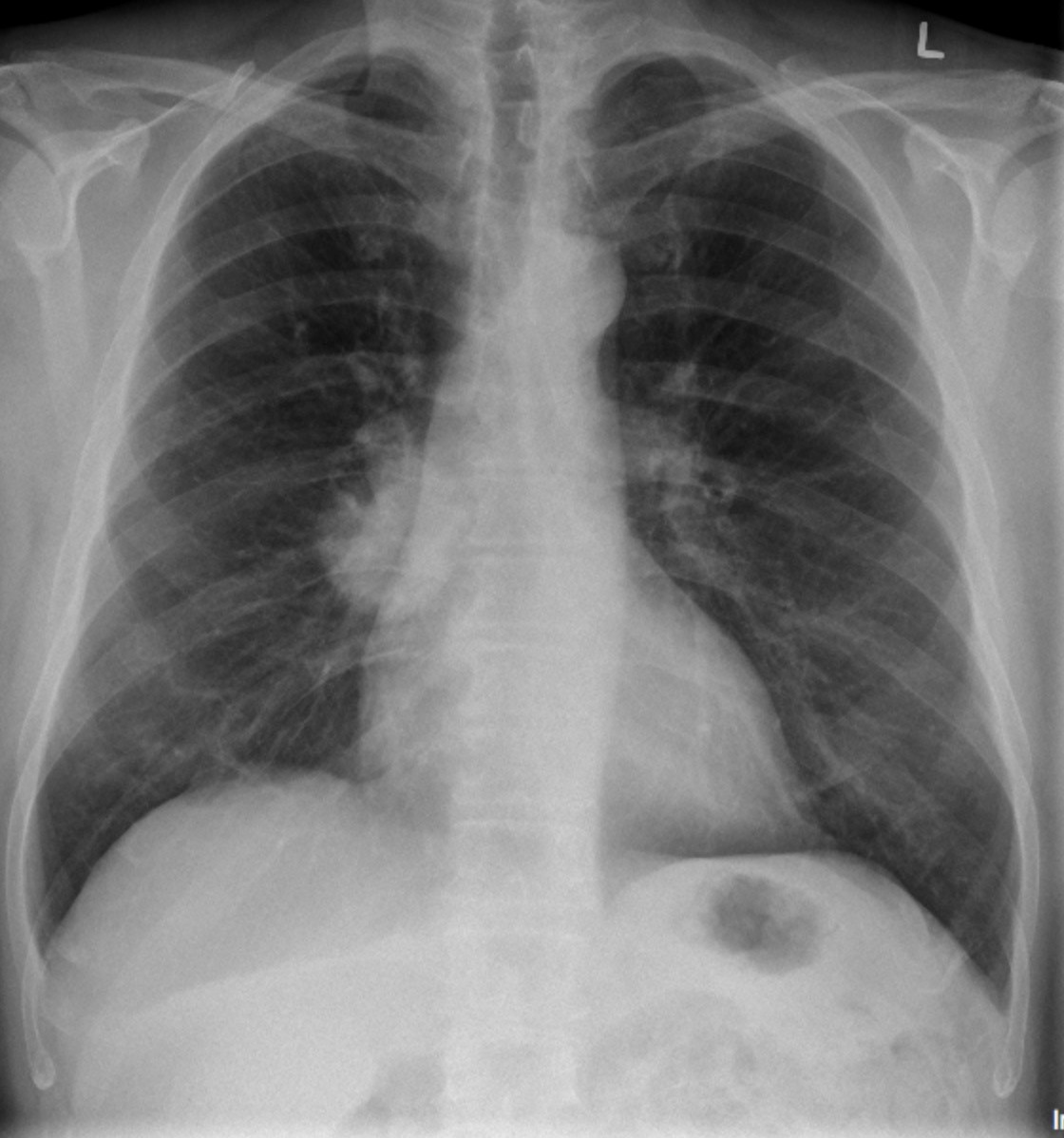
There were changes of mild COPD. A 2.5 – 3 cm diameter, circumscribed, opacity was seen in the right lower lobe. The opacity overlapped the right hilum and the thoracic spine. No other findings.
Interpretation:
Solitary pulmonary nodule. This abnormality was suspicious for a malignancy. CT of the chest was recommended for further assessment. If concern persists for malignancy after this examination an imaging guided needle biopsy of the lung nodule should be considered.
Diagnosis:
Solitary Pulmonary Nodule – Suspicious for Malignancy
Discussion:
A nodule is defined as a circumscribed opacity < 3 cm in diameter. The differential is broad. There are imaging features that suggest benignity, while for others there are imaging features that require imaging follow-up or biopsy.
The Fleischner Society has established nodule follow-up guidelines:
fleischnersociety.org
X-ray findings may include:
- A nodule or mass is an opacity with smooth or lobulated margins.
- The margins of the nodule or mass may be smooth, lobulated, or spiculated.
- There may be calcification in the nodule or mass.
- Masses may grow to invade the pleura, chest wall, hilum, mediastinum, and may cross fissures.
- The mass may have adenopathy or pleural effusion associated with it.
Attributions
Figure 9.24A PA Chest x-ray displaying a solitary lung nodule by Dr. Brent Burbridge MD, FRCPC, University Medical Imaging Consultants, College of Medicine, University of Saskatchewan is used under a CC-BY-NC-SA 4.0 license.
Figure 9.24B Lateral Chest x-ray displaying a solitary lung nodule by Dr. Brent Burbridge MD, FRCPC, University Medical Imaging Consultants, College of Medicine, University of Saskatchewan is used under a CC-BY-NC-SA 4.0 license.