
10.4: Intestinal Obstruction
- Page ID
- 14847

ACR – Gastrointestinal – Suspected Small Bowel Obstruction
Case 1
Adhesions
Clinical:
History – This patient has had numerous ventriculo-peritoneal shunts. He has had many abdominal surgeries for revisions of the shunt tubing. No other significant history.
Symptoms – Abdominal pain and bloating. Diminished appetite. No flatus or bowel movements for 24 hours. Vomiting watery green fluid for 12 hours.
Physical – The abdomen was distended and mildly, diffusely tender. The bowel sounds were infrequent and high-pitched. The abdomen was tympanitic. No rebound or guarding. Scars were seen from his previous surgeries.
DDx:
Suspected Small Bowel Obstruction
Imaging Recommendation
ACR–Gastrointestinal – Suspected Small Bowel Obstruction, Variant 1
Three views of the Abdomen
CT of the Abdomen
ODIN Link for Small Bowel Obstruction images (3 Views of the Abdomen), Figure 10.6A and B: mistr.usask.ca/odin/?caseID=20170410113920578
Imaging Assessment
Findings:
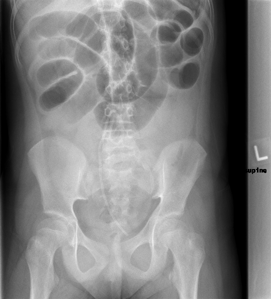
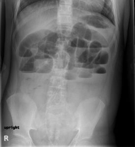
There was a paucity of bowel gas in the colon. The small bowel was dilated and there were multiple air-fluid levels (> 3 in number) in the small intestine. No calculi seen. The ventriculo-peritoneal shunt tubing was noted.
Interpretation:
Suspicious for high-grade, or complete, small bowel obstruction.
Diagnosis:
Adhesions, small bowel obstruction from restrictive band.
Discussion:
An abnormality in the small bowel lumen, the small bowel wall, or an abnormality extrinsic to the small bowel can cause a blockage of the lumen. This prevents antegrade passage of gas and fluid. Initially, the small bowel increases peristaltic effort to move the contents forward (hyperperistalsis, high-pitched bowel sounds), then later the peristalsis ceases.
The hyperperistalsis clears the bowel downstream of the obstruction resulting in a relatively clear distal bowel (sigmoid colon, rectum).
Causes of small bowel obstruction include:
- Adhesions after previous abdominal or pelvic surgery
- Inflammatory bowel disease
- Hernias
- Gallstones
- Malignancy
- Ingested foreign body
- Intussusception – more common in children
X-ray findings may include:
- Dilated bowel – small bowel > 2.5 – 3 cm, cecum > 10 cm, transverse colon > 6 cm, sigmoid colon > 4cm
- Gas and fluid in the bowel.
- Air-fluid levels in small bowel > 3 in number
- Less gas and fluid in the bowel downstream of obstruction.
Case 2
Ventral Abdominal Hernia
Clinical:
History – This patient had a previous laparotomy for a perforated diverticulum 10 years ago. A noticeable hernia was present in the ventral abdomen and it was noted to have enlarged over the last two years.
Symptoms – Abdominal pain and bloating. Diminished appetite. No flatus or bowel movements for 48 hours. Vomiting watery green fluid for 24 hours.
Physical – The abdomen was distended and mildly, diffusely tender. The bowel sounds were infrequent and high-pitched. The abdomen was tympanitic. A large, firm, mass was noted in the ventral abdominal wall. It was not particularly tender to palpation. No rebound or guarding.
DDx:
Suspected Small Bowel Obstruction
Ileus
Bowel Perforation
Imaging Recommendation
ACR–Gastrointestinal – Suspected Small Bowel Obstruction, Variant 1
Three views of the Abdomen
CT of the Abdomen
ODIN Link to Abdominal Wall Mass with Small Bowel Obstruction images (Ultrasound, 2 Views of the Abdomen, and CT), Figure 10.7A and B: mistr.usask.ca/odin/?caseID=20170410112613962
Imaging Assessment
Findings:
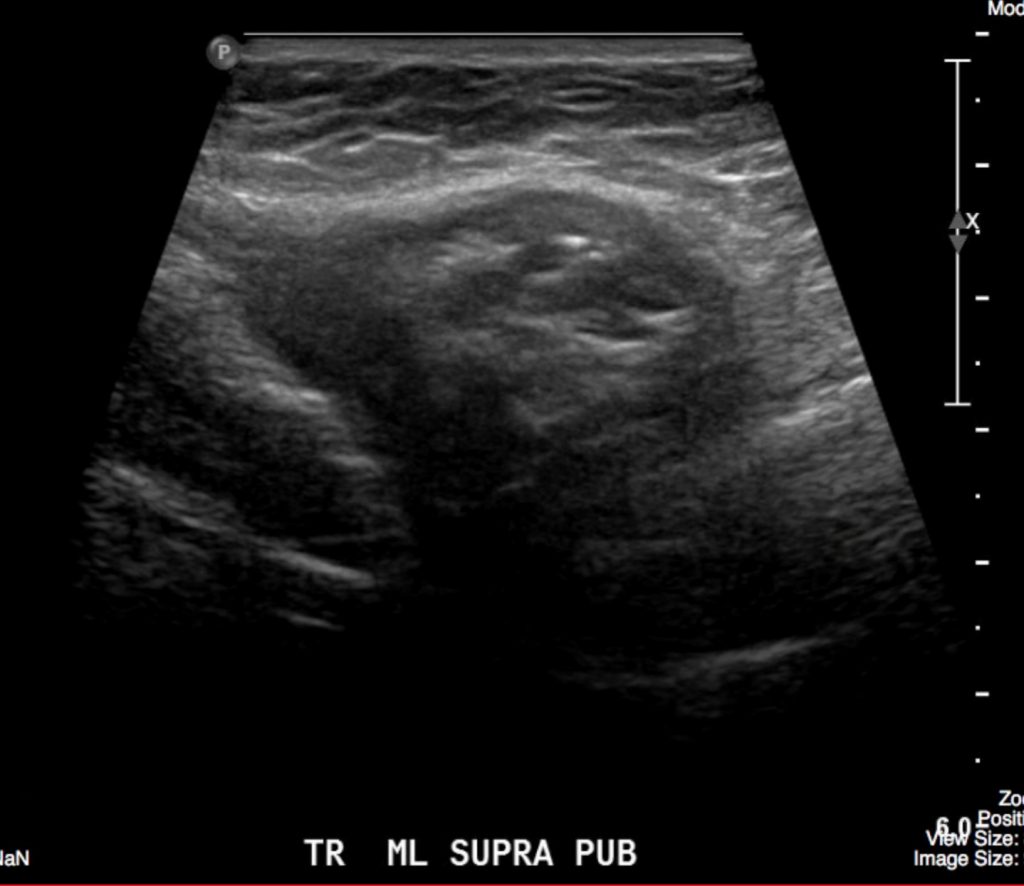
Ultrasound – A mixed echogenicity mass is seen in the lower mid abdomen. There is suspicion for a fat containing intestine within the hernia sac. No calculi seen.
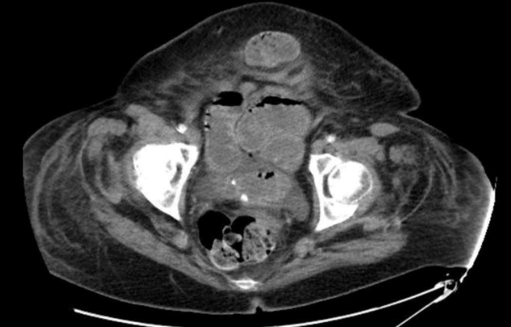
CT – There was a soft tissue mass in the lower, ventral, abdominal wall, subcutaneous fat. This has the appearance of a hernia. There was intestine in the hernia sac. The visualized small intestine was dilated, consistent with a small bowel obstruction.
Interpretation:
Moderate grade small bowel obstruction.
Diagnosis:
The patient had a ventral, bowel containing hernia, with small bowel obstruction secondary to entrapment of the bowel in the hernia sac (incarceration).
Attributions
Figure 10.6A Abdominal x-ray, supine, suspicious for SBO by Dr. Brent Burbridge MD, FRCPC, University Medical Imaging Consultants, College of Medicine, University of Saskatchewan is used under a CC-BY-NC-SA 4.0 license.
Figure 10.6B Abdominal x-ray, decubitus, suspicious for SBO by Dr. Brent Burbridge MD, FRCPC, University Medical Imaging Consultants, College of Medicine, University of Saskatchewan is used under a CC-BY-NC-SA 4.0 license.
Figure 10.7A Ultrasound of Abdomen displaying a mass in the abdominal wall fat by Dr. Brent Burbridge MD, FRCPC, University Medical Imaging Consultants, College of Medicine, University of Saskatchewan is used under a CC-BY-NC-SA 4.0 license.
Figure 10.7B CT Scan of the Abdomen displaying a mass in the abdominal wall by Dr. Brent Burbridge MD, FRCPC, University Medical Imaging Consultants, College of Medicine, University of Saskatchewan is used under a CC-BY-NC-SA 4.0 license.