
12.1: Thyroid – Palpable Nodule
- Page ID
- 14860

Case
Thyroid, Palpable Nodule, Benign Follicular Nodule
Clinical:
History – This 55 year old female presented for an annual physical.
Symptoms – None.
Physical – Neck examination detected a hard nodule in the right thyroid lobe. The thyroid gland was not enlarged.
DDx:
Thyroid Cyst
Thyroid Nodule
Adenopathy
Parathyroid Nodule
Imaging Recommendation
Palpable Thyroid Nodule
Thyroid Ultrasound
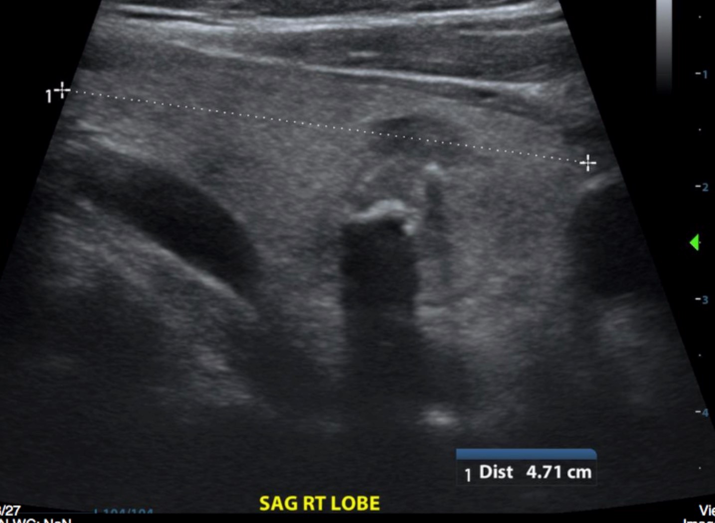
ODIN Link for Thyroid Nodule images (Ultrasound), Figure 12.1A and B: mistr.usask.ca/odin/?caseID=20161101145508526

Imaging Assessment
Findings:
There was a mixed echogenicity nodule in the right thyroid lobe with at least two foci of very dense calcification. There was minimally increased Doppler signal in this nodule. The nodule measures 1.9 x 1.7 x 1.9 cm. No other obvious nodes or nodules.
Interpretation:
Benign appearing right thyroid nodule.
Diagnosis:
Benign Follicular Nodule
Discussion:
The risk factors associated with an increased likelihood of a malignancy in thyroid nodules include:
- A previous history of irradiation
- A family history of medullary thyroid carcinoma or multiple endocrine neoplasia (MEN) type II,
- Patients younger than 20 years or older than 60 years
- Male patients
- Rapid growth of a nodule
- A nodule with a rim and hard consistency
- An inconspicuous margin of the nodule on palpation
- The presence of enlarged cervical lymph nodes.
Among the modern imaging modalities, high-resolution US is the most sensitive diagnostic modality for the detection of thyroid abnormalities and it is necessary to perform US for the nodules found after palpation. In addition, US can guide FNA for thyroid nodules and it can diagnose lymph node metastasis.
In order to image and describe thyroid abnormalities on ultrasound in a more scientific and logical manner the ACR has established a, “Thyroid Imaging Reporting and Data System – TI-RADS.” One of the important elements of this strategy is to create a lexicon that communicates the relative risk of malignancy for a thyroid nodule based upon objective descriptors and agreed upon ultrasound criteria.
A detailed discussion of the scoring system and nodule risk stratification can be found at the ACR TI-RADS Atlas. This atlas provides detailed examples of nodule imaging features and a methodology for risk of malignancy scoring.
ACR TI-RADS Atlas
There is no consensus in the literature regarding optimal spacing of follow-up sonograms for nodules that do not meet the criteria for FNA, as growth rates do not reliably distinguish benign from malignant nodules. Scanning intervals of less than 1 year are not warranted, except for proven cancers under active surveillance, that are imaged at the discretion of the referring physician. Follow-up imaging timing should be on the basis of a nodule’s ACR, TI-RADS score.
For More Information:
ACR – Thyroid Imaging Reporting and Data System (TI-RADS) – www.acr.org/Quality-Safety/Resources/TIRADS
Attributions
Figure 12.1A Sagittal Right Lobe Ultrasound of Thyroid by Dr. Brent Burbridge MD, FRCPC, University Medical Imaging Consultants, College of Medicine, University of Saskatchewan is used under a CC-BY-NC-SA 4.0 license.
Figure 12.1B Transverse Right Lobe Ultrasound of Thyroid by Dr. Brent Burbridge MD, FRCPC, University Medical Imaging Consultants, College of Medicine, University of Saskatchewan is used under a CC-BY-NC-SA 4.0 license.