
14.9: Knee Trauma – Acute Fractures
- Page ID
- 14888

ACR – MSK – Acute Knee Trauma
Case
Comminuted, Intra-articular, Tibial Fractures
Clinical:
History– This 18 year old male injured his knee in a trampoline accident.
Symptoms – Painful, swollen knee. Unable to weight bear.
Physical – The knee was swollen and there was tenderness of the tibia below the joint line. An effusion was present. There was a sense of crepitation with knee range of motion.
DDx:
Femur fracture
Tibia fracture
Knee dislocation
Imaging Recommendation
ACR – MSK – Acute Knee Trauma, Variant 2
X-rays
CT will be required if there is intra-articular bone injury.
MR is the best modality for suspected ligamentous injury.
ODIN Link for Knee Trauma images (X-rays and CT), Figure 14.13A and B: mistr.usask.ca/odin/?caseID=20150320080231242
Imaging Assessment
Findings:
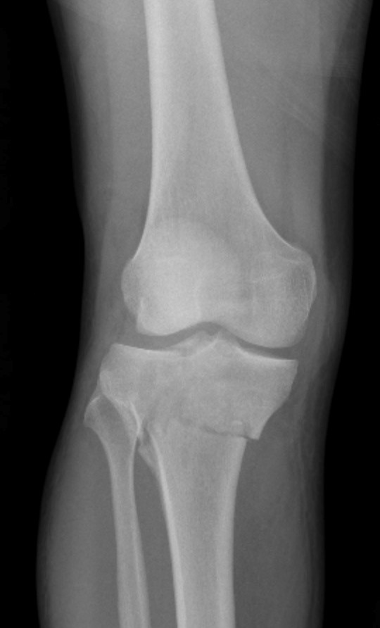
X-ray – There was a comminuted, intra-articular fracture of the tibia. A fat-fluid level was seen in the knee joint due to marrow from the tibia communicating with the joint fluid. Alignment was satisfactory.
Interpretation:
Comminuted Tibial fracture. CT was recommended.
Diagnosis:
Comminuted, Intra-articular, Tibial Injury
CT Findings –
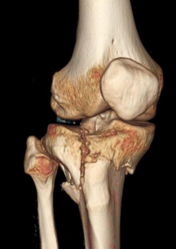
There was an intra-articular fracture of the tibia with joint fluid, fat, and blood. The fracture was complex and therefore it required surgical fixation in the operating room.
Discussion:
Clinical decision rules for the acutely injured knee suggest that radiographic examination of the knee following acute injury can be eliminated in many instances by applying specific clinical guidelines. A commonly used clinical decision rule is the Ottawa Knee Rule.
The Ottawa Knee Rule states that patients ≥18 years old with acute knee pain should have knee radiographs if they meet any of the following criteria:
- Are 55 years of age or older,
- Have palpable tenderness over the head of the fibula,
- Have isolated patellar tenderness,
- Cannot flex the knee to 90°,
- Cannot weight bear immediately following the injury, or
- Cannot walk in the emergency room i.e. can’t take 4 steps.
X-ray findings may include:
- Visible fractures of the patella, femur, or tibia (multiple bones fractured possible).
- Generalized soft tissue swelling.
- Joint effusion may be present.
- If fat has entered the joint space from the marrow of a bone a fat-fluid level will be present.
- Angulation of the bones should be assessed.
- The knee joint, and patella, may be subluxated or dislocated.
Clinical decision rules, such as the Ottawa Knee Rule, are well validated and have been shown to reduce the number of radiographs obtained for acute knee trauma while still identifying almost all patients who have a fracture. It is generally agreed that radiographs should be obtained and the clinical decision rule should not be applied for patients with gross deformity, a palpable mass, a penetrating injury, prosthetic hardware, an unreliable clinical history or physical examination secondary to multiple injuries, altered mental status (e.g., head injury, drug or alcohol use, dementia), neuropathy (e.g., paraplegia, diabetes), or a history suggesting increased risk of fracture. The physician’s judgment and common sense, however, should supersede clinical guidelines.
Attributions
Figure 14.13A X-ray of the knee displaying a complex tibial fracture by Dr. Brent Burbridge MD, FRCPC, University Medical Imaging Consultants, College of Medicine, University of Saskatchewan is used under a CC-BY-NC-SA 4.0 license.
Figure 14.13B 3D image of the knee, displaying a complex tibial fracture by Dr. Brent Burbridge MD, FRCPC, University Medical Imaging Consultants, College of Medicine, University of Saskatchewan is used under a CC-BY-NC-SA 4.0 license.