
8.3: Skeletal Muscle Organ Structure
- Page ID
- 63421

\( \newcommand{\vecs}[1]{\overset { \scriptstyle \rightharpoonup} {\mathbf{#1}} } \)
\( \newcommand{\vecd}[1]{\overset{-\!-\!\rightharpoonup}{\vphantom{a}\smash {#1}}} \)
\( \newcommand{\dsum}{\displaystyle\sum\limits} \)
\( \newcommand{\dint}{\displaystyle\int\limits} \)
\( \newcommand{\dlim}{\displaystyle\lim\limits} \)
\( \newcommand{\id}{\mathrm{id}}\) \( \newcommand{\Span}{\mathrm{span}}\)
( \newcommand{\kernel}{\mathrm{null}\,}\) \( \newcommand{\range}{\mathrm{range}\,}\)
\( \newcommand{\RealPart}{\mathrm{Re}}\) \( \newcommand{\ImaginaryPart}{\mathrm{Im}}\)
\( \newcommand{\Argument}{\mathrm{Arg}}\) \( \newcommand{\norm}[1]{\| #1 \|}\)
\( \newcommand{\inner}[2]{\langle #1, #2 \rangle}\)
\( \newcommand{\Span}{\mathrm{span}}\)
\( \newcommand{\id}{\mathrm{id}}\)
\( \newcommand{\Span}{\mathrm{span}}\)
\( \newcommand{\kernel}{\mathrm{null}\,}\)
\( \newcommand{\range}{\mathrm{range}\,}\)
\( \newcommand{\RealPart}{\mathrm{Re}}\)
\( \newcommand{\ImaginaryPart}{\mathrm{Im}}\)
\( \newcommand{\Argument}{\mathrm{Arg}}\)
\( \newcommand{\norm}[1]{\| #1 \|}\)
\( \newcommand{\inner}[2]{\langle #1, #2 \rangle}\)
\( \newcommand{\Span}{\mathrm{span}}\) \( \newcommand{\AA}{\unicode[.8,0]{x212B}}\)
\( \newcommand{\vectorA}[1]{\vec{#1}} % arrow\)
\( \newcommand{\vectorAt}[1]{\vec{\text{#1}}} % arrow\)
\( \newcommand{\vectorB}[1]{\overset { \scriptstyle \rightharpoonup} {\mathbf{#1}} } \)
\( \newcommand{\vectorC}[1]{\textbf{#1}} \)
\( \newcommand{\vectorD}[1]{\overrightarrow{#1}} \)
\( \newcommand{\vectorDt}[1]{\overrightarrow{\text{#1}}} \)
\( \newcommand{\vectE}[1]{\overset{-\!-\!\rightharpoonup}{\vphantom{a}\smash{\mathbf {#1}}}} \)
\( \newcommand{\vecs}[1]{\overset { \scriptstyle \rightharpoonup} {\mathbf{#1}} } \)
\(\newcommand{\longvect}{\overrightarrow}\)
\( \newcommand{\vecd}[1]{\overset{-\!-\!\rightharpoonup}{\vphantom{a}\smash {#1}}} \)
\(\newcommand{\avec}{\mathbf a}\) \(\newcommand{\bvec}{\mathbf b}\) \(\newcommand{\cvec}{\mathbf c}\) \(\newcommand{\dvec}{\mathbf d}\) \(\newcommand{\dtil}{\widetilde{\mathbf d}}\) \(\newcommand{\evec}{\mathbf e}\) \(\newcommand{\fvec}{\mathbf f}\) \(\newcommand{\nvec}{\mathbf n}\) \(\newcommand{\pvec}{\mathbf p}\) \(\newcommand{\qvec}{\mathbf q}\) \(\newcommand{\svec}{\mathbf s}\) \(\newcommand{\tvec}{\mathbf t}\) \(\newcommand{\uvec}{\mathbf u}\) \(\newcommand{\vvec}{\mathbf v}\) \(\newcommand{\wvec}{\mathbf w}\) \(\newcommand{\xvec}{\mathbf x}\) \(\newcommand{\yvec}{\mathbf y}\) \(\newcommand{\zvec}{\mathbf z}\) \(\newcommand{\rvec}{\mathbf r}\) \(\newcommand{\mvec}{\mathbf m}\) \(\newcommand{\zerovec}{\mathbf 0}\) \(\newcommand{\onevec}{\mathbf 1}\) \(\newcommand{\real}{\mathbb R}\) \(\newcommand{\twovec}[2]{\left[\begin{array}{r}#1 \\ #2 \end{array}\right]}\) \(\newcommand{\ctwovec}[2]{\left[\begin{array}{c}#1 \\ #2 \end{array}\right]}\) \(\newcommand{\threevec}[3]{\left[\begin{array}{r}#1 \\ #2 \\ #3 \end{array}\right]}\) \(\newcommand{\cthreevec}[3]{\left[\begin{array}{c}#1 \\ #2 \\ #3 \end{array}\right]}\) \(\newcommand{\fourvec}[4]{\left[\begin{array}{r}#1 \\ #2 \\ #3 \\ #4 \end{array}\right]}\) \(\newcommand{\cfourvec}[4]{\left[\begin{array}{c}#1 \\ #2 \\ #3 \\ #4 \end{array}\right]}\) \(\newcommand{\fivevec}[5]{\left[\begin{array}{r}#1 \\ #2 \\ #3 \\ #4 \\ #5 \\ \end{array}\right]}\) \(\newcommand{\cfivevec}[5]{\left[\begin{array}{c}#1 \\ #2 \\ #3 \\ #4 \\ #5 \\ \end{array}\right]}\) \(\newcommand{\mattwo}[4]{\left[\begin{array}{rr}#1 \amp #2 \\ #3 \amp #4 \\ \end{array}\right]}\) \(\newcommand{\laspan}[1]{\text{Span}\{#1\}}\) \(\newcommand{\bcal}{\cal B}\) \(\newcommand{\ccal}{\cal C}\) \(\newcommand{\scal}{\cal S}\) \(\newcommand{\wcal}{\cal W}\) \(\newcommand{\ecal}{\cal E}\) \(\newcommand{\coords}[2]{\left\{#1\right\}_{#2}}\) \(\newcommand{\gray}[1]{\color{gray}{#1}}\) \(\newcommand{\lgray}[1]{\color{lightgray}{#1}}\) \(\newcommand{\rank}{\operatorname{rank}}\) \(\newcommand{\row}{\text{Row}}\) \(\newcommand{\col}{\text{Col}}\) \(\renewcommand{\row}{\text{Row}}\) \(\newcommand{\nul}{\text{Nul}}\) \(\newcommand{\var}{\text{Var}}\) \(\newcommand{\corr}{\text{corr}}\) \(\newcommand{\len}[1]{\left|#1\right|}\) \(\newcommand{\bbar}{\overline{\bvec}}\) \(\newcommand{\bhat}{\widehat{\bvec}}\) \(\newcommand{\bperp}{\bvec^\perp}\) \(\newcommand{\xhat}{\widehat{\xvec}}\) \(\newcommand{\vhat}{\widehat{\vvec}}\) \(\newcommand{\uhat}{\widehat{\uvec}}\) \(\newcommand{\what}{\widehat{\wvec}}\) \(\newcommand{\Sighat}{\widehat{\Sigma}}\) \(\newcommand{\lt}{<}\) \(\newcommand{\gt}{>}\) \(\newcommand{\amp}{&}\) \(\definecolor{fillinmathshade}{gray}{0.9}\)- Describe the layers of connective tissues and how they package skeletal muscle
- Explain how muscles work with tendons to move the body
- Identify and describe the microscopic anatomy of a muscle fiber and a sarcomere
The best-known feature of skeletal muscle is its ability to contract and cause movement. Skeletal muscles act not only to produce movement but also to stop movement, such as resisting gravity to maintain posture. Small, constant adjustments of skeletal muscles are needed to hold a body upright or balanced in any position. Muscles also prevent excess movement of the bones and joints, maintaining skeletal stability and preventing structural damage or deformation. Joints can become misaligned or dislocated by excessive or inappropriate forces applied to associated bones; muscles work to keep joints stable. Skeletal muscles are located throughout the body at the openings of internal tracts to control the movement of various substances. These muscles allow functions, such as swallowing, urination, and defecation, to be under voluntary control. Skeletal muscles also protect internal organs (particularly abdominal and pelvic organs) by acting as an external barrier or shield to external trauma and by supporting the weight of the organs.
Skeletal muscles contribute to the maintenance of homeostasis in the body by generating heat. Muscle contraction requires energy, and when ATP is broken down, heat is produced. This heat is very noticeable during exercise, when sustained muscle movement causes body temperature to rise, and in cases of extreme cold, when shivering produces random skeletal muscle contractions to generate heat.
Organ Structure
Each skeletal muscle is an organ consisting of multiple integrated tissues acting together to cause movement: skeletal muscle and three layers of proper connective tissue. The connective tissue layers (called “mysia”) provide structure to the muscle as a whole and also compartmentalize the muscle fibers within the muscle (Figure \(\PageIndex{1}\)) and (Figure \(\PageIndex{2}\)). Wrapping around the organ is a sheath of dense irregular connective tissue called the epimysium that allows a muscle to contract and move powerfully while maintaining its structural integrity. The epimysium also separates muscle from other tissues and organs in the area, allowing the muscle to move independently. Within the epimysium skeletal muscle fibers are organized into bundles, called fascicles, by a middle layer of dense irregular connective tissue making up the perimysium. This fascicular organization is common in muscles of the limbs; it allows the nervous system to trigger a specific movement of a muscle by activating a subset of muscle fibers within a bundle, or fascicle, of the muscle. Inside each fascicle, each muscle fiber is encased in a thin areolar connective tissue layer called the endomysium. The endomysium contains the extracellular fluid and nutrients to support the muscle fiber. These nutrients are supplied via blood capillaries located in the areolar tissue.
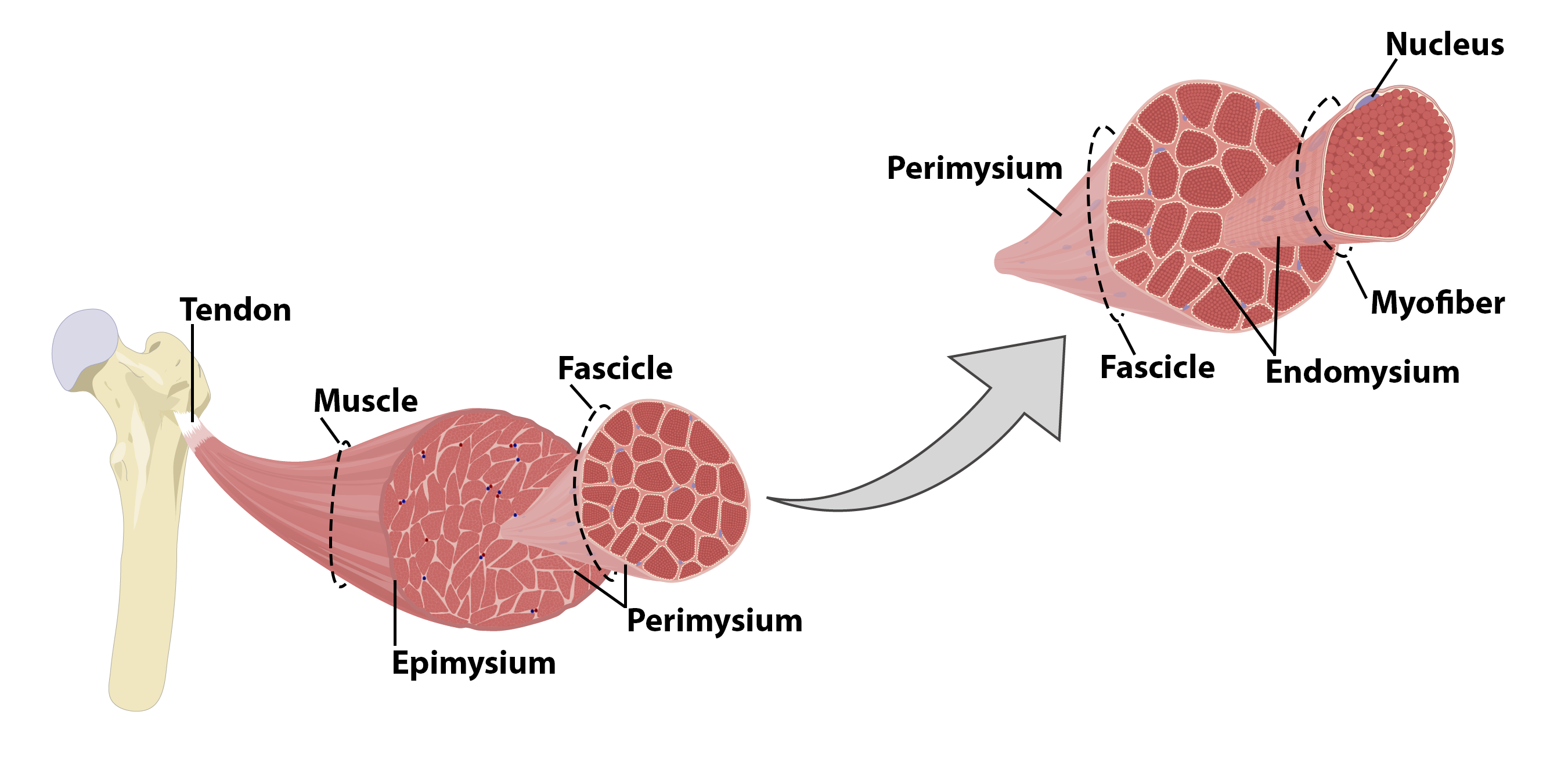
Figure \(\PageIndex{1}\): Skeletal Muscle Organ Structure. Each level of the organization of a skeletal muscle is wrapped in a connective tissue layer: the cell (myofiber) is surrounded by the endomysium, the bundle of cells (muscle fascicle) is surrounded by the perimysium, and the organ (muscle) is surrounded by the epimysium. (Image credit: "Skeletal Muscle Organ Structure" by Justin Greene is licensed under CC BY-NC-SA 4.0).


Skeletal muscles have tendons that attach to bones and transfer the force of contraction to the skeleton. The collagen in the three tissue layers intertwines and integrates with the collagen of a tendon, straightening to form a cord of dense regular connective tissue. At the other end of the tendon, the collagen incorporates into periosteum becoming dense irregular connective tissue once more. Tension created by contraction of the muscle fibers is transferred though the mysia, to the tendon, and then to the periosteum to pull on the bone to move the skeleton. In other places, the mysia may fuse with a broad, tendon-like sheet called an aponeurosis, or to fascia, the connective tissue between skin and bones. The broad sheet of connective tissue in the lower back that the latissimus dorsi muscles (the “lats”) fuse into is an example of an aponeurosis.
Every skeletal muscle is richly supplied by blood vessels for nourishment, oxygen delivery, and waste removal. In addition, every muscle fiber in a skeletal muscle is supplied by the axon branch of a somatic motor neuron, which signals the fiber to contract. Unlike cardiac and smooth muscle, the only way to functionally contract a skeletal muscle is through signaling from the nervous system.
Skeletal Muscle Fibers
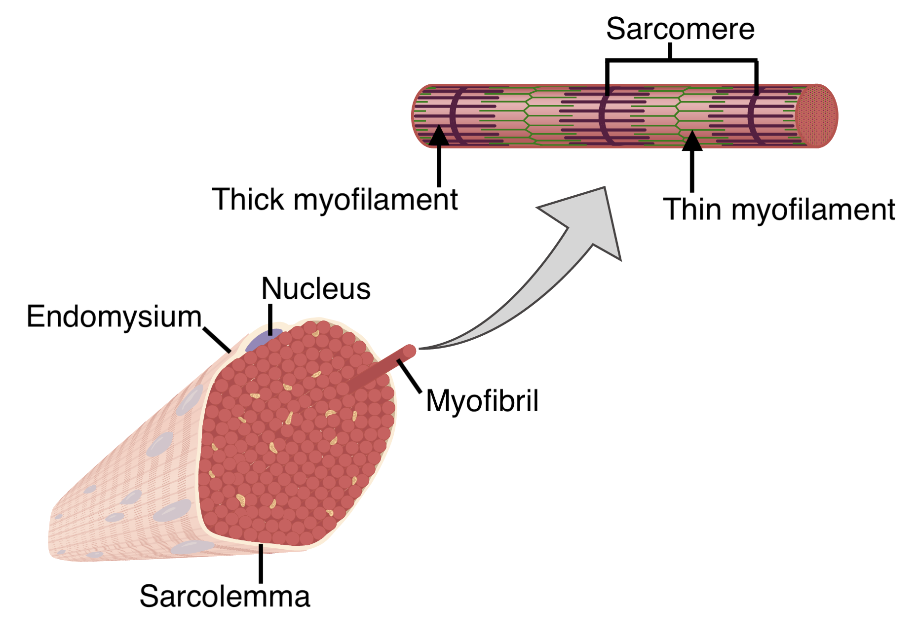
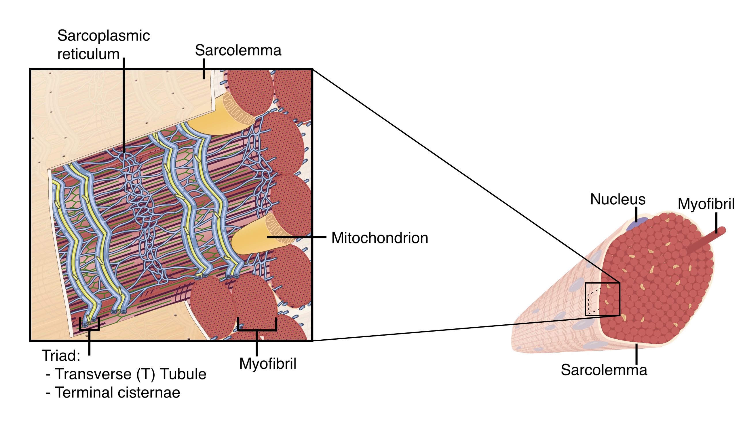
Because skeletal muscle cells are long and cylindrical, they are commonly referred to as skeletal muscle fibers or skeletal myofibers. Specific terminology associated with myofibers is rooted in the Greek sarco, which means “flesh.” The plasma membrane of muscle fibers is called the sarcolemma and the cytoplasm is referred to as sarcoplasm. Skeletal myofibers can be quite large for human cells, with diameters up to 100 μm and lengths up to 30 cm (11.8 in). During early development, embryonic myoblasts, each with its own nucleus, fuse with up to hundreds of other myoblasts to form the multinucleated myofibers. Multiple nuclei means multiple copies of genes, permitting the production of the large amounts of proteins and enzymes needed for muscle contraction along the length of the entire muscle fiber. Multiple nuclei also means mitosis would be extremely complicated, so skeletal myofibers do not divide.
Within the sarcoplasm of each muscle fiber are myofibrils, rods of protein that run the entire length of the muscle fiber and attach to the sarcolemma at its end (Figure \(\PageIndex{3}\)). Each myofibril is composed of a bundle of contractile thin and thick myofilaments, along with other structural and elastic proteins. As myofibrils contract, the entire muscle cell contracts. Because myofibrils are only approximately 1.2 μm in diameter, hundreds to thousands can be found inside one muscle fiber.
The Sarcomere, Myofilaments and Protein Organization
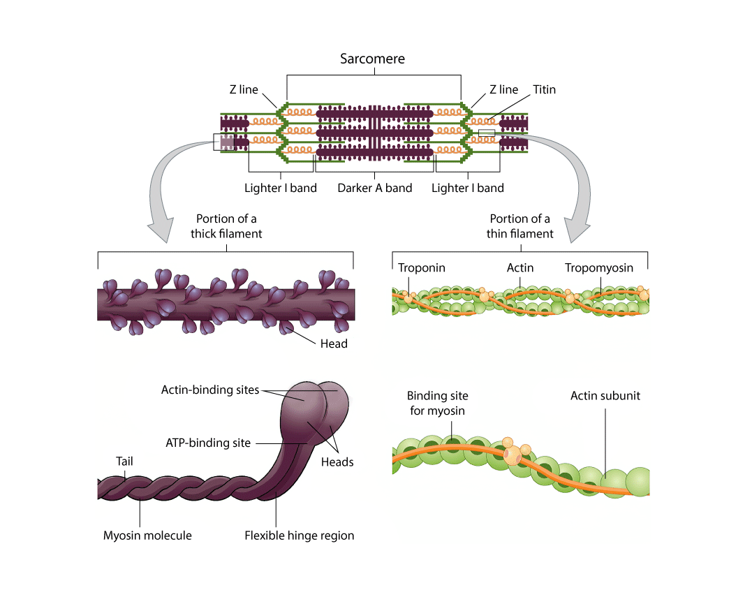
The striated appearance of skeletal muscle fibers is due to the arrangement of the thin and thick myofilaments in sequential order from one end of the muscle fiber to the other. Each segment of these myofilaments and their regulatory proteins, connected on each end to a protein network called the Z-line (or Z-disc), is called a sarcomere. The sarcomere is the functional unit of the muscle fiber and runs from one Z-line to the next Z-line. A chain of thousands of repeating sarcomeres forms the long tube-like myofibrils.
Each sarcomere is approximately 2 μm in length with a three-dimensional cylinder-like arrangement. The repeating pattern of the myofilaments, along with other proteins, within the sarcomere gives rise to very distinctive regions and structures (Figure \(\PageIndex{5}\)). Each sarcomere is bordered by Z-lines to which the thin myofilaments are anchored. The central M-line anchors the thick filaments, which are composed of bundles of a single contractile protein called myosin. Each thick filaments contains approximately 300 myosin strands with their mobile heads protruding on the outer surface. Thin filaments contain two strands of the contractile protein actin that are twisted around each other and two regulatory proteins, troponin and tropomyosin, whose position determines if myosin heads can bind to actin. The difference in protein mass between the two filaments is what causes the striated appearance of skeletal muscle fibers as their positions alternate along the myofibril (Figure \(\PageIndex{4}\)).
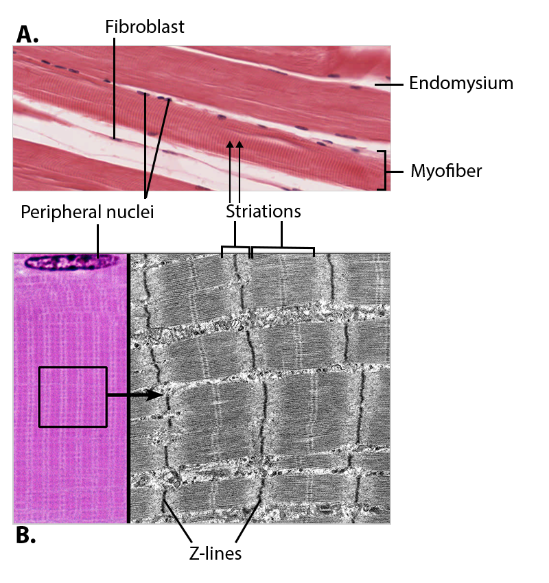
The zone immediately surrounding the Z-lines is known as the I band, or light band, because only thin filaments are present (Figure \(\PageIndex{5}\)). The thin composition of actin makes it difficult to see, hence the name the light band. The myosin is anchored to the central M line, from whence it projects toward, but not all to way to, the Z-lines. The area of the sarcomere where myosin is present is known as the A band, or dark band. There are some areas where thin and thick myofilaments are both present, known as the zone of overlap. The area in which no overlap occurs is the H zone, where only myosin is present. The alternation of I bands and A bands along the length of the myofibril, and thus the myofiber, is what gives the cell its striated appearance.
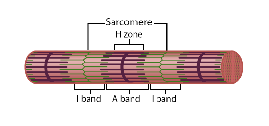
Figure \(\PageIndex{5}\): Myofibril and the Sarcomere. Myofibrils are composed of chains of thousands of repeating sarcomeres. The alternating I bands and A bands give the myofibril (and the myofiber) its striated appearance. (Image credit: "Myofibril and the Sarcomere" by Justin Greene is licensed under CC BY-NC-SA 4.0, based on original by Openstax.)
As mentioned above, actin and myosin are the proteins that are responsible for muscular contraction. The structural differences between these two myofilaments not only leads to the striated appearance of skeletal muscle tissue, but also to the ability of the two to work together to produce the overall function of muscle contraction.
Actin strands are formed by multiple spherical actin subunits, each of which contain a myosin binding site (Figure \(\PageIndex{6}\)). In a relaxed muscle, the binding sites remain shielded by the troponin-tropomyosin regulatory complex. The tropomyosin filaments run the length of the actin, covering all of the binding sites. These filaments are held in place by troponin, which is also responsible for shifting the position of tropomyosin to uncover the binding sites in the presence of calcium ions.
Each myosin filament has two specific regions, the tail and the head (Figure \(\PageIndex{6}\)). Connecting these two regions is a flexible hinge region. The tails are linked together into a bundle, while the heads take an active role in the muscle contraction process by binding to and pulling on actin. Each head contains an actin binding site as well as a binding site for the ATP molecules that will provide the energy needed for the contraction cycle to occur.
Surrounding each myofibril is the sarcoplasmic reticulum (SR), the specialized smooth endoplasmic reticulum of myofibers, which stores, releases, and retrieves the calcium ions (Ca++) needed for contraction to occur (Figure \(\PageIndex{7}\)). The signal from a motor neuron stimulates the release of calcium ions (Ca++) from its storage in the cell’s sarcoplasmic reticulum. For the neuron's signal to reach the membrane of the SR there are periodic invaginations in the sarcolemma, called T-tubules (“T” stands for “transverse”). You will recall that the diameter of a muscle fiber can be up to 100 μm, so these T-tubules ensure that the cell membrane can get close to the SR in the sarcoplasm. On each side of the T-tubule is a terminal cisterna (plural - cisternae), an enlarged region of the SR that contains high concentrations of calcium ions. The arrangement of a T-tubule with the terminal cisternae of the SR on either side is called a triad (Figure \(\PageIndex{7}\)). The triad encircles the cylindrical structure of the myofibril.
The Neuromuscular Junction
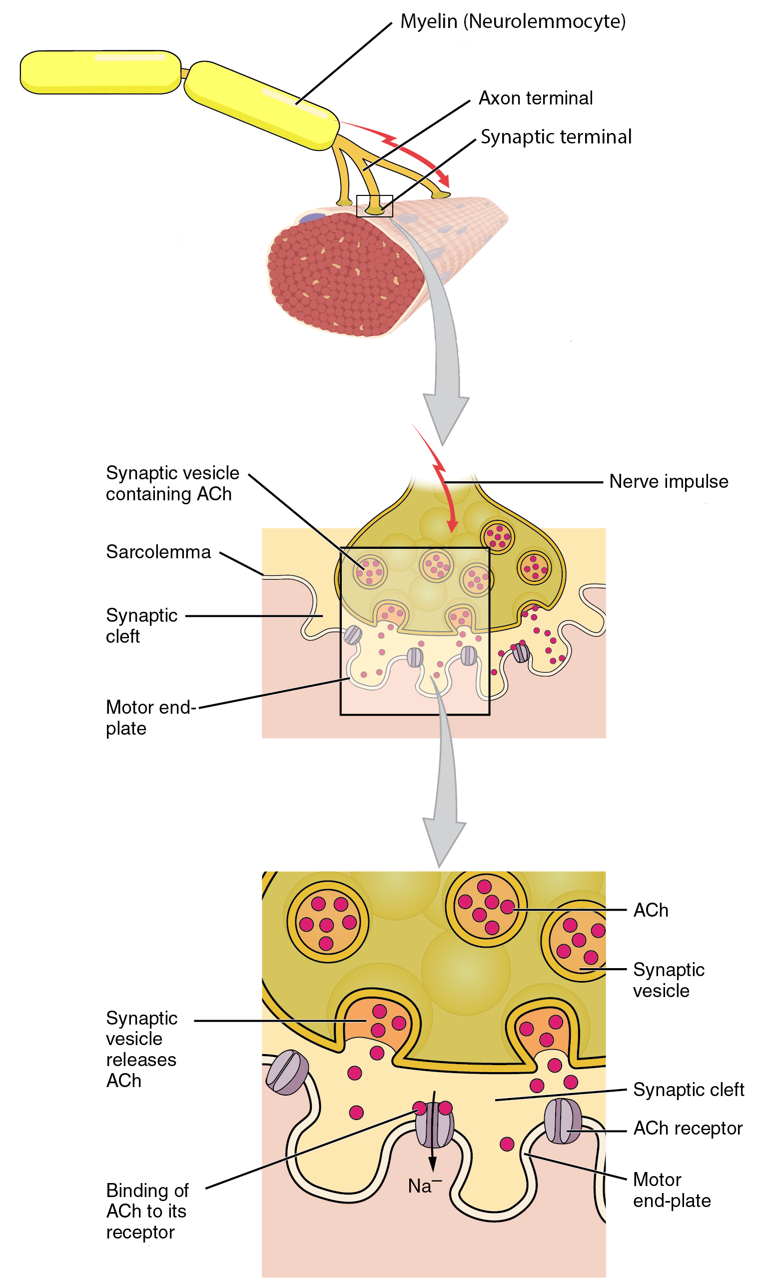
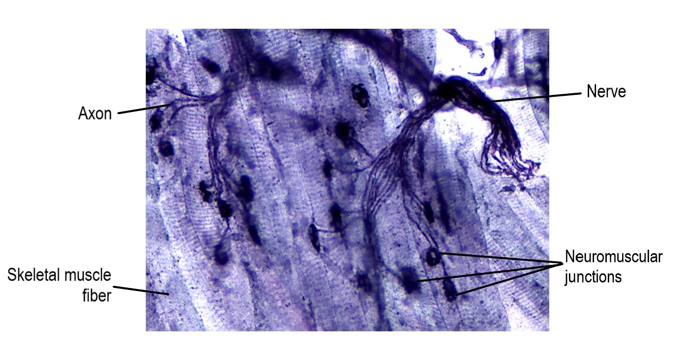
The specialized site of the skeletal muscle where a motor neuron’s axon terminal meets the muscle fiber is the neuromuscular junction (NMJ) (Figure \(\PageIndex{8}\)). The axon terminal releases a chemical messenger, acetylcholine (ACh), that diffuses across the minute space between the two cells and bind to receptors embedded in the sarcolemma of the myofiber. These ACh receptors are only located within the NMJ region called the motor end-plate. Every myofiber in every skeletal muscle is innervated by a motor neuron at a NMJ. Excitation signals from the neuron are the only way to functionally activate the fiber to contract by allowing the myosin heads to bind to actin. (Although you could contract the muscles using electrical current or molecules, such as nicotine, that mimic the molecular structure of Ach.)
Muscle Shortening and Lengthening
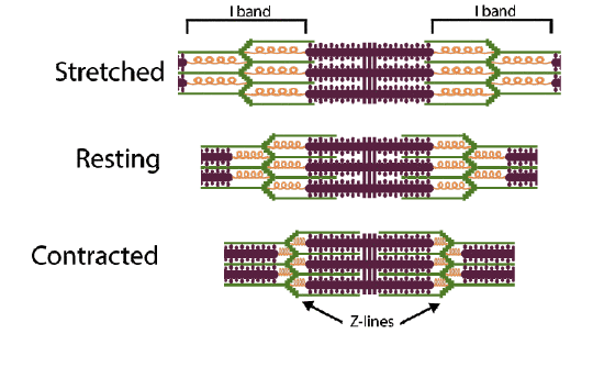
When you contract your muscles you will feel them tightening up and they will bulge a bit in the middle, this is because the muscle is getting shorter and, therefore, fatter. (In physiology you will learn about lengthening contractions, which is an oxymoron since the definition of "to contract" means to shorten.) When you stretch out your muscles before you exercise or to try to ease muscle tension, you are physically lengthening the muscle. These alterations in the size and dimensions of the entire muscle are reflected in each of its structural levels: when the muscle shortens the fascicles, myofibers, and myofibrils also become shorter. The structure that stays the same size is the myofilaments. Recall from above that the functional unit of a muscle is the sarcomere and that the function of a muscle is to contract/relax/stretch, this means the sarcomere is the contractile unit that is physically becoming shorter.
During shortening contractions the Z-lines of the sarcomere are drawn closer together by the interaction of myosin and actin while the protein filaments stay the same length but shift relative positions, with the the area of overlap of the two filaments increasing. When stretching the muscle, the Z-lines are drawn farther apart and have a smaller area of overlapping filaments. The I band, the distance from the end of one thick filament to the next, shortens during a contraction and lengthens when stretched.
Concept Review
Skeletal muscles contain connective tissue, blood vessels, and nerves. There are three layers of connective tissue: epimysium, perimysium, and endomysium. Skeletal muscle fibers are organized into groups called fascicles. Blood vessels and nerves enter the connective tissue and branch. Muscles attach to bones directly or through tendons or aponeuroses. Skeletal muscles maintain posture, stabilize joints, support organs, control internal movement, and generate heat.
Skeletal muscle fibers are long, multinucleated cells. The membrane of the cell is the sarcolemma; the cytoplasm of the cell is the sarcoplasm. The sarcoplasmic reticulum (SR) is a form of endoplasmic reticulum that specializes in storing and releasing calcium. Muscle fibers are composed of myofibrils. The banding pattern along the myofibrils, called striations, are created by the organization of actin and myosin proteins in functional units called sarcomeres.
Review Questions
Query \(\PageIndex{1}\)
Query \(\PageIndex{2}\)
Critical Thinking Questions
Query \(\PageIndex{3}\)
Query \(\PageIndex{4}\)
Query \(\PageIndex{5}\)
Query \(\PageIndex{6}\)
Glossary
Query \(\PageIndex{7}\)
Contributors and Attributions
OpenStax Anatomy & Physiology (CC BY 4.0). Access for free at https://openstax.org/books/anatomy-and-physiology