
8.2: Overview of Muscle Tissues
- Page ID
- 63420
\( \newcommand{\vecs}[1]{\overset { \scriptstyle \rightharpoonup} {\mathbf{#1}} } \)
\( \newcommand{\vecd}[1]{\overset{-\!-\!\rightharpoonup}{\vphantom{a}\smash {#1}}} \)
\( \newcommand{\id}{\mathrm{id}}\) \( \newcommand{\Span}{\mathrm{span}}\)
( \newcommand{\kernel}{\mathrm{null}\,}\) \( \newcommand{\range}{\mathrm{range}\,}\)
\( \newcommand{\RealPart}{\mathrm{Re}}\) \( \newcommand{\ImaginaryPart}{\mathrm{Im}}\)
\( \newcommand{\Argument}{\mathrm{Arg}}\) \( \newcommand{\norm}[1]{\| #1 \|}\)
\( \newcommand{\inner}[2]{\langle #1, #2 \rangle}\)
\( \newcommand{\Span}{\mathrm{span}}\)
\( \newcommand{\id}{\mathrm{id}}\)
\( \newcommand{\Span}{\mathrm{span}}\)
\( \newcommand{\kernel}{\mathrm{null}\,}\)
\( \newcommand{\range}{\mathrm{range}\,}\)
\( \newcommand{\RealPart}{\mathrm{Re}}\)
\( \newcommand{\ImaginaryPart}{\mathrm{Im}}\)
\( \newcommand{\Argument}{\mathrm{Arg}}\)
\( \newcommand{\norm}[1]{\| #1 \|}\)
\( \newcommand{\inner}[2]{\langle #1, #2 \rangle}\)
\( \newcommand{\Span}{\mathrm{span}}\) \( \newcommand{\AA}{\unicode[.8,0]{x212B}}\)
\( \newcommand{\vectorA}[1]{\vec{#1}} % arrow\)
\( \newcommand{\vectorAt}[1]{\vec{\text{#1}}} % arrow\)
\( \newcommand{\vectorB}[1]{\overset { \scriptstyle \rightharpoonup} {\mathbf{#1}} } \)
\( \newcommand{\vectorC}[1]{\textbf{#1}} \)
\( \newcommand{\vectorD}[1]{\overrightarrow{#1}} \)
\( \newcommand{\vectorDt}[1]{\overrightarrow{\text{#1}}} \)
\( \newcommand{\vectE}[1]{\overset{-\!-\!\rightharpoonup}{\vphantom{a}\smash{\mathbf {#1}}}} \)
\( \newcommand{\vecs}[1]{\overset { \scriptstyle \rightharpoonup} {\mathbf{#1}} } \)
\( \newcommand{\vecd}[1]{\overset{-\!-\!\rightharpoonup}{\vphantom{a}\smash {#1}}} \)
\(\newcommand{\avec}{\mathbf a}\) \(\newcommand{\bvec}{\mathbf b}\) \(\newcommand{\cvec}{\mathbf c}\) \(\newcommand{\dvec}{\mathbf d}\) \(\newcommand{\dtil}{\widetilde{\mathbf d}}\) \(\newcommand{\evec}{\mathbf e}\) \(\newcommand{\fvec}{\mathbf f}\) \(\newcommand{\nvec}{\mathbf n}\) \(\newcommand{\pvec}{\mathbf p}\) \(\newcommand{\qvec}{\mathbf q}\) \(\newcommand{\svec}{\mathbf s}\) \(\newcommand{\tvec}{\mathbf t}\) \(\newcommand{\uvec}{\mathbf u}\) \(\newcommand{\vvec}{\mathbf v}\) \(\newcommand{\wvec}{\mathbf w}\) \(\newcommand{\xvec}{\mathbf x}\) \(\newcommand{\yvec}{\mathbf y}\) \(\newcommand{\zvec}{\mathbf z}\) \(\newcommand{\rvec}{\mathbf r}\) \(\newcommand{\mvec}{\mathbf m}\) \(\newcommand{\zerovec}{\mathbf 0}\) \(\newcommand{\onevec}{\mathbf 1}\) \(\newcommand{\real}{\mathbb R}\) \(\newcommand{\twovec}[2]{\left[\begin{array}{r}#1 \\ #2 \end{array}\right]}\) \(\newcommand{\ctwovec}[2]{\left[\begin{array}{c}#1 \\ #2 \end{array}\right]}\) \(\newcommand{\threevec}[3]{\left[\begin{array}{r}#1 \\ #2 \\ #3 \end{array}\right]}\) \(\newcommand{\cthreevec}[3]{\left[\begin{array}{c}#1 \\ #2 \\ #3 \end{array}\right]}\) \(\newcommand{\fourvec}[4]{\left[\begin{array}{r}#1 \\ #2 \\ #3 \\ #4 \end{array}\right]}\) \(\newcommand{\cfourvec}[4]{\left[\begin{array}{c}#1 \\ #2 \\ #3 \\ #4 \end{array}\right]}\) \(\newcommand{\fivevec}[5]{\left[\begin{array}{r}#1 \\ #2 \\ #3 \\ #4 \\ #5 \\ \end{array}\right]}\) \(\newcommand{\cfivevec}[5]{\left[\begin{array}{c}#1 \\ #2 \\ #3 \\ #4 \\ #5 \\ \end{array}\right]}\) \(\newcommand{\mattwo}[4]{\left[\begin{array}{rr}#1 \amp #2 \\ #3 \amp #4 \\ \end{array}\right]}\) \(\newcommand{\laspan}[1]{\text{Span}\{#1\}}\) \(\newcommand{\bcal}{\cal B}\) \(\newcommand{\ccal}{\cal C}\) \(\newcommand{\scal}{\cal S}\) \(\newcommand{\wcal}{\cal W}\) \(\newcommand{\ecal}{\cal E}\) \(\newcommand{\coords}[2]{\left\{#1\right\}_{#2}}\) \(\newcommand{\gray}[1]{\color{gray}{#1}}\) \(\newcommand{\lgray}[1]{\color{lightgray}{#1}}\) \(\newcommand{\rank}{\operatorname{rank}}\) \(\newcommand{\row}{\text{Row}}\) \(\newcommand{\col}{\text{Col}}\) \(\renewcommand{\row}{\text{Row}}\) \(\newcommand{\nul}{\text{Nul}}\) \(\newcommand{\var}{\text{Var}}\) \(\newcommand{\corr}{\text{corr}}\) \(\newcommand{\len}[1]{\left|#1\right|}\) \(\newcommand{\bbar}{\overline{\bvec}}\) \(\newcommand{\bhat}{\widehat{\bvec}}\) \(\newcommand{\bperp}{\bvec^\perp}\) \(\newcommand{\xhat}{\widehat{\xvec}}\) \(\newcommand{\vhat}{\widehat{\vvec}}\) \(\newcommand{\uhat}{\widehat{\uvec}}\) \(\newcommand{\what}{\widehat{\wvec}}\) \(\newcommand{\Sighat}{\widehat{\Sigma}}\) \(\newcommand{\lt}{<}\) \(\newcommand{\gt}{>}\) \(\newcommand{\amp}{&}\) \(\definecolor{fillinmathshade}{gray}{0.9}\)- Compare and contrast the different types of muscle tissue
- Explain the four functional properties of muscle tissue
Muscle is one of the four primary tissue types of the body, and the body contains three types of muscle tissue: skeletal muscle, cardiac muscle, and smooth muscle (Figure \(\PageIndex{1}\)). All muscle tissues have four functional properties in common which include excitability, contractility, extensibility, and elasticity.
- Excitability - the plasma membrane can change electrical states and send an electrical wave called an action potential along the entire surface of each cell.
- Contractility - allows muscle tissue to pull on its attachment points as the cells shorten.
- Extensibility - the cells of muscle tissue can stretch or elongate.
- Elasticity - following contraction or extension, a muscle can recoil to its original length when relaxed due to elastic proteins.
Muscle Tissue Types
Differences among the three muscle types include the microscopic organization of the intracellular contractile proteins—actin and myosin. The actin and myosin proteins are arranged very regularly in the cytoplasm of individual muscle cells (referred to as myofibers) in both skeletal muscle and cardiac muscle, which creates a striped pattern across the width of the cell called striations. The striations are visible with a light microscope under high magnification (see Figure \(\PageIndex{1.A,B}\)) as an alternating pattern of light - dark - light - dark - etc. along the entire length of the myofiber. These striations are more prominent in skeletal muscle fibers than in cardiac muscle fibers. Because the actin and myosin are not arranged in such regular fashion in smooth muscle, the cytoplasm of a smooth muscle fiber has a uniform, nonstriated appearance (resulting in the name smooth muscle).
Skeletal Muscle Tissue
Skeletal myofibers are long, striated rods that range from a few millimeters to multiple centimeters, extending beyond the sides of the images in Figure \(\PageIndex{1.A}\) below (individual cell is outlined). The longest muscle in the body is the sartorius that has myofibers that are, on average, 42 centimeters/16.5 inches long! Skeletal muscle comprises approximately 40% of our body weight and contains 50 to 75% of all body proteins. Due to the need to conduct the functioning of such a large cell, each myofiber has many nuclei. The longer the fiber, the more nuclei it has. These nuclei are pushed to the edges of the cell to accommodate all of the intracellular proteins (as indicated by the arrows in Figure \(\PageIndex{1.A2}\)). Skeletal muscle is under voluntary control, so any muscular contraction that you can consciously decide to contract or relax has skeletal muscle in it. This includes all of the body's muscles that move the skeleton and the skin, including your muscles of respiration, as well as some of the body's tubular organs: anal sphincters, urinary sphincters, and your throat. Your tongue also contains skeletal muscle!
|
A. Skeletal Muscle @ 100x and @ 400x |
 |
 |
| B. Cardiac Muscle @ 400x (longitudinal section) |
 |
 |
| C. Smooth Muscle @ 400x (longitudinal and cross sections) |
 |
 |
Figure \(\PageIndex{1}\). The Three Types of Muscle Tissue. The body contains three types of muscle tissue: A. skeletal muscle, B. cardiac muscle, C. smooth muscle. (Image credits: Types of Muscle Tissue by Jennifer Lange are licensed under CC-BY-SA-NC 4.0. Micrographs A1, A2, and C1 provided by the Regents of University of Michigan Medical School, micrographs B1, B2, and C2 provided by Virginia Commonwealth University.)
Cardiac Muscle Tissue
Cardiac muscle tissue is only found in the heart. Highly coordinated contractions of cardiac muscle pump blood into the vessels of the circulatory system. Cardiac muscle fibers are shorter than skeletal muscle fibers and usually contain only one nucleus, which is located in the central region of the cell. Cardiac muscle fibers also possess many mitochondria and myoglobin, as ATP is produced primarily through aerobic metabolism. Cardiac muscle fibers cells also are extensively branched and are physically and functionally connected to one another at their ends by specialized cell junctions call intercalated disks (indicated by arrows in Figure \(\PageIndex{1.B}\)). Intercalated disks are part of the sarcolemma and contain two structures important in cardiac muscle contraction: gap junctions and desmosomes (Figure \(\PageIndex{2}\)). A gap junction forms channels between adjacent cardiac muscle fibers that allows the electrical signal to flow quickly from one cardiac myofiber to the next. This joining of cells allows coordinated contraction of the entire heart. The remainder of the intercalated disk is composed of desmosomes. A desmosome is a cell structure that anchors the ends of cardiac muscle fibers together so the cells do not pull apart during the stress of individual fibers contracting.

Smooth Muscle Tissue
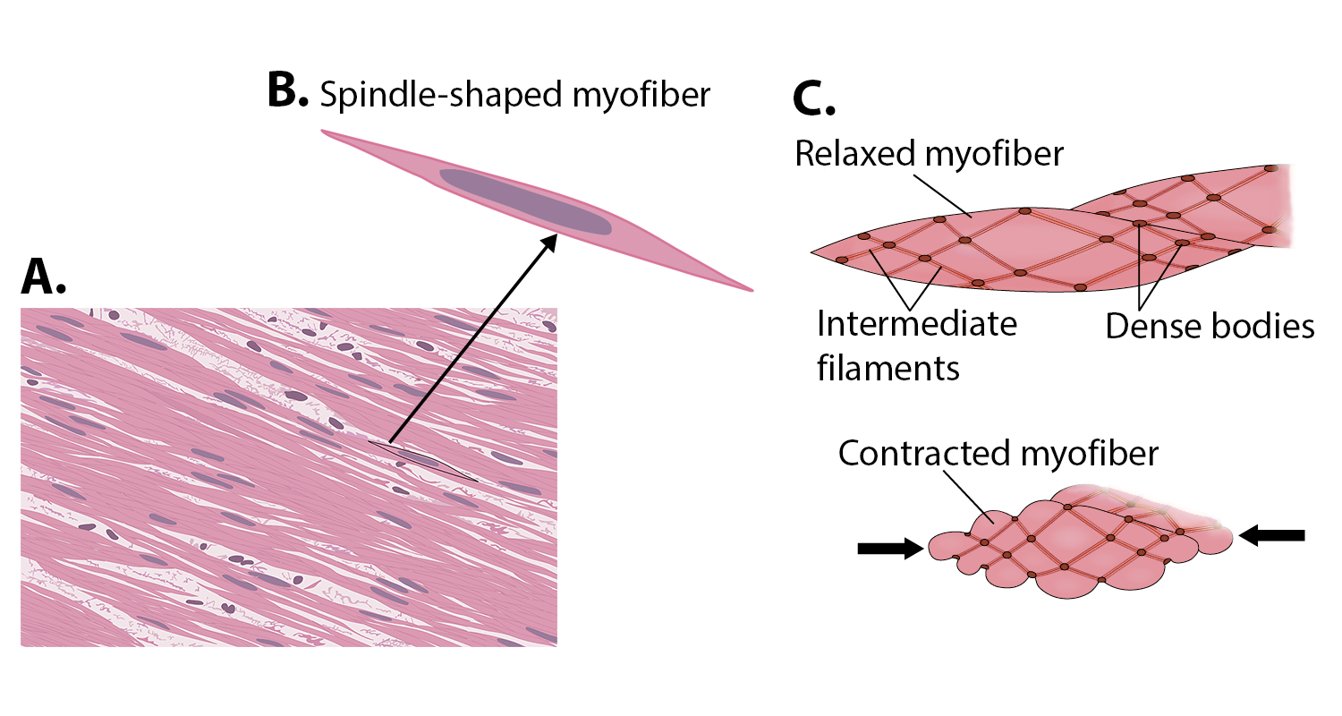
Smooth muscle (so-named because the cells do not have striations) is present in the walls of hollow organs like the urinary bladder, uterus, stomach, intestines, and in the walls of passageways, such as the arteries and veins of the circulatory system, and the tracts of the respiratory, urinary, and reproductive systems. Smooth muscle is also present in the eyes, where it functions to change the size of the iris and alter the shape of the lens; and in the skin where it causes hair to stand erect in response to cold temperature or fear. Smooth muscle fibers are spindle-shaped (wide in the middle and tapered at both ends, somewhat like a football) and have a single nucleus (see outlined cells in (Figure \(\PageIndex{1.C}\) and in (Figure \(\PageIndex{3}\)). They range from about 30 to 200 μm (thousands of times shorter than skeletal muscle fibers), and they produce their own connective tissue, endomysium.


Although they do not have striations, smooth muscle fibers do have actin and myosin contractile proteins. These contractile proteins are connected to dense bodies that are fastened to the sarcolemma. Dense bodies also have cord-like intermediate filaments attached to them (Figure \(\PageIndex{4}\)). When contraction occurs the contractile proteins pull on the dense bodies, which then pull on the intermediate filaments networks throughout the sarcoplasm. This arrangement causes the entire muscle fiber to contract in a manner whereby the ends are pulled toward the center, causing the midsection to bulge in a corkscrew motion.
Concept Review
Muscle is the tissue in animals that allows for active movement of the body or materials within the body. There are three types of muscle tissue: skeletal muscle, cardiac muscle, and smooth muscle.
- Most of the body’s skeletal muscle produces voluntary movement by acting on the skeleton. It has:
- striations
- long, rod-shaped cells
- multiple peripheral nuclei
- Cardiac muscle is found in the wall of the heart and involuntarily pumps blood through the circulatory system. It has:
- striations
- short, rectangular, branched cells
- single, central nucleus
- intercalated discs
- Smooth muscle is found in the skin and in the walls of internal tubular organs where it involuntarily assists in moving materials. It has:
- no striations
- spindle-shaped cells
- single, central nucleus
Review Questions
Q. Muscle that has a striped appearance is described as being ________.
A. elastic
B. nonstriated
C. excitable
D. striated
- Answer
-
Answer: D
Q. Which of the following properties is not common to all three muscle tissues?
A. excitability
B. the need for ATP
C. polarity
D. elasticity
- Answer
-
Answer: C
Critical Thinking Questions
Q. Why is elasticity an important quality of muscle tissue?
- Answer
-
A. It allows muscle to return to its original length during relaxation after contraction.
Glossary
- cardiac muscle
- striated muscle found in the heart; joined to one another at intercalated discs and under the regulation of pacemaker cells, which contract as one unit to pump blood through the circulatory system. Cardiac muscle is under involuntary control.
- contractility
- ability to shorten (contract) forcibly
- dense body
- anchoring site for actin filaments in smooth muscle tissue
- desmosome
- a type of intercellular junction that holds cells tightly to each other
- elasticity
- ability to stretch and rebound
- excitability
- ability to undergo neural stimulation
- extensibility
- ability to lengthen (extend)
- gap junction
- a type of intercellular junction that allows ions to from from one cell to the next
- intercalated disk
- specialized cell junction found only in cardiac muscle tissue
- intermediate filaments
- structural proteins connecting dense bodies that assist in smooth muscle contraction
- myofiber
- an elongated cell found in muscle tissues
- skeletal muscle
- striated, multinucleated muscle that requires signaling from the nervous system to trigger contraction; most skeletal muscles are referred to as voluntary muscles that move bones and produce movement
- smooth muscle
- nonstriated, mononucleated muscle in the skin that is associated with hair follicles; assists in moving materials in the walls of internal organs, blood vessels, and internal passageways
- striations
- visible light/dark alternating bands in cardiac and skeletal muscle
Contributors and Attributions
OpenStax Anatomy & Physiology (CC BY 4.0). Access for free at https://openstax.org/books/anatomy-and-physiology