
11.5: Spinal Cord
- Page ID
- 64064

\( \newcommand{\vecs}[1]{\overset { \scriptstyle \rightharpoonup} {\mathbf{#1}} } \)
\( \newcommand{\vecd}[1]{\overset{-\!-\!\rightharpoonup}{\vphantom{a}\smash {#1}}} \)
\( \newcommand{\dsum}{\displaystyle\sum\limits} \)
\( \newcommand{\dint}{\displaystyle\int\limits} \)
\( \newcommand{\dlim}{\displaystyle\lim\limits} \)
\( \newcommand{\id}{\mathrm{id}}\) \( \newcommand{\Span}{\mathrm{span}}\)
( \newcommand{\kernel}{\mathrm{null}\,}\) \( \newcommand{\range}{\mathrm{range}\,}\)
\( \newcommand{\RealPart}{\mathrm{Re}}\) \( \newcommand{\ImaginaryPart}{\mathrm{Im}}\)
\( \newcommand{\Argument}{\mathrm{Arg}}\) \( \newcommand{\norm}[1]{\| #1 \|}\)
\( \newcommand{\inner}[2]{\langle #1, #2 \rangle}\)
\( \newcommand{\Span}{\mathrm{span}}\)
\( \newcommand{\id}{\mathrm{id}}\)
\( \newcommand{\Span}{\mathrm{span}}\)
\( \newcommand{\kernel}{\mathrm{null}\,}\)
\( \newcommand{\range}{\mathrm{range}\,}\)
\( \newcommand{\RealPart}{\mathrm{Re}}\)
\( \newcommand{\ImaginaryPart}{\mathrm{Im}}\)
\( \newcommand{\Argument}{\mathrm{Arg}}\)
\( \newcommand{\norm}[1]{\| #1 \|}\)
\( \newcommand{\inner}[2]{\langle #1, #2 \rangle}\)
\( \newcommand{\Span}{\mathrm{span}}\) \( \newcommand{\AA}{\unicode[.8,0]{x212B}}\)
\( \newcommand{\vectorA}[1]{\vec{#1}} % arrow\)
\( \newcommand{\vectorAt}[1]{\vec{\text{#1}}} % arrow\)
\( \newcommand{\vectorB}[1]{\overset { \scriptstyle \rightharpoonup} {\mathbf{#1}} } \)
\( \newcommand{\vectorC}[1]{\textbf{#1}} \)
\( \newcommand{\vectorD}[1]{\overrightarrow{#1}} \)
\( \newcommand{\vectorDt}[1]{\overrightarrow{\text{#1}}} \)
\( \newcommand{\vectE}[1]{\overset{-\!-\!\rightharpoonup}{\vphantom{a}\smash{\mathbf {#1}}}} \)
\( \newcommand{\vecs}[1]{\overset { \scriptstyle \rightharpoonup} {\mathbf{#1}} } \)
\(\newcommand{\longvect}{\overrightarrow}\)
\( \newcommand{\vecd}[1]{\overset{-\!-\!\rightharpoonup}{\vphantom{a}\smash {#1}}} \)
\(\newcommand{\avec}{\mathbf a}\) \(\newcommand{\bvec}{\mathbf b}\) \(\newcommand{\cvec}{\mathbf c}\) \(\newcommand{\dvec}{\mathbf d}\) \(\newcommand{\dtil}{\widetilde{\mathbf d}}\) \(\newcommand{\evec}{\mathbf e}\) \(\newcommand{\fvec}{\mathbf f}\) \(\newcommand{\nvec}{\mathbf n}\) \(\newcommand{\pvec}{\mathbf p}\) \(\newcommand{\qvec}{\mathbf q}\) \(\newcommand{\svec}{\mathbf s}\) \(\newcommand{\tvec}{\mathbf t}\) \(\newcommand{\uvec}{\mathbf u}\) \(\newcommand{\vvec}{\mathbf v}\) \(\newcommand{\wvec}{\mathbf w}\) \(\newcommand{\xvec}{\mathbf x}\) \(\newcommand{\yvec}{\mathbf y}\) \(\newcommand{\zvec}{\mathbf z}\) \(\newcommand{\rvec}{\mathbf r}\) \(\newcommand{\mvec}{\mathbf m}\) \(\newcommand{\zerovec}{\mathbf 0}\) \(\newcommand{\onevec}{\mathbf 1}\) \(\newcommand{\real}{\mathbb R}\) \(\newcommand{\twovec}[2]{\left[\begin{array}{r}#1 \\ #2 \end{array}\right]}\) \(\newcommand{\ctwovec}[2]{\left[\begin{array}{c}#1 \\ #2 \end{array}\right]}\) \(\newcommand{\threevec}[3]{\left[\begin{array}{r}#1 \\ #2 \\ #3 \end{array}\right]}\) \(\newcommand{\cthreevec}[3]{\left[\begin{array}{c}#1 \\ #2 \\ #3 \end{array}\right]}\) \(\newcommand{\fourvec}[4]{\left[\begin{array}{r}#1 \\ #2 \\ #3 \\ #4 \end{array}\right]}\) \(\newcommand{\cfourvec}[4]{\left[\begin{array}{c}#1 \\ #2 \\ #3 \\ #4 \end{array}\right]}\) \(\newcommand{\fivevec}[5]{\left[\begin{array}{r}#1 \\ #2 \\ #3 \\ #4 \\ #5 \\ \end{array}\right]}\) \(\newcommand{\cfivevec}[5]{\left[\begin{array}{c}#1 \\ #2 \\ #3 \\ #4 \\ #5 \\ \end{array}\right]}\) \(\newcommand{\mattwo}[4]{\left[\begin{array}{rr}#1 \amp #2 \\ #3 \amp #4 \\ \end{array}\right]}\) \(\newcommand{\laspan}[1]{\text{Span}\{#1\}}\) \(\newcommand{\bcal}{\cal B}\) \(\newcommand{\ccal}{\cal C}\) \(\newcommand{\scal}{\cal S}\) \(\newcommand{\wcal}{\cal W}\) \(\newcommand{\ecal}{\cal E}\) \(\newcommand{\coords}[2]{\left\{#1\right\}_{#2}}\) \(\newcommand{\gray}[1]{\color{gray}{#1}}\) \(\newcommand{\lgray}[1]{\color{lightgray}{#1}}\) \(\newcommand{\rank}{\operatorname{rank}}\) \(\newcommand{\row}{\text{Row}}\) \(\newcommand{\col}{\text{Col}}\) \(\renewcommand{\row}{\text{Row}}\) \(\newcommand{\nul}{\text{Nul}}\) \(\newcommand{\var}{\text{Var}}\) \(\newcommand{\corr}{\text{corr}}\) \(\newcommand{\len}[1]{\left|#1\right|}\) \(\newcommand{\bbar}{\overline{\bvec}}\) \(\newcommand{\bhat}{\widehat{\bvec}}\) \(\newcommand{\bperp}{\bvec^\perp}\) \(\newcommand{\xhat}{\widehat{\xvec}}\) \(\newcommand{\vhat}{\widehat{\vvec}}\) \(\newcommand{\uhat}{\widehat{\uvec}}\) \(\newcommand{\what}{\widehat{\wvec}}\) \(\newcommand{\Sighat}{\widehat{\Sigma}}\) \(\newcommand{\lt}{<}\) \(\newcommand{\gt}{>}\) \(\newcommand{\amp}{&}\) \(\definecolor{fillinmathshade}{gray}{0.9}\)- Describe the structures that protect the spinal cord
- Describe the regions and structures of the spinal cord and their functions
- Explain the arrangement of gray and white matter in the spinal cord and the regional variations
The spinal cord is continuous with the brain and transmits sensory information from the periphery of the body to the brain, and motor information from the brain to the periphery. It is also a center for coordination of reflexes, as well as control of reflexes independently from the brain. In a typical adult the length of the spinal cord ranges from 42-45 cm (16-18 inches), depending on the person's height. It extends inferiorly from the medulla oblongata, at the level of the foramen magnum, to approximately the level of the L1 vertebra. The spinal cord is divided into cervical, thoracic, lumbar, sacral and coccygeal regions. The location of these regions within the vertebral column does not match up with the location of the similarly named vertebrae (except for C1). For example, the lumbar region of the spinal cord aligns with the inferior thoracic vertebrae. This occurs because the bone growth period of the vertebra lasts longer than the growth period of the spinal cord, thus the spinal cord stops lengthening before the vertebrae stop gaining height.
Protective Coverings of the Spinal Cord
The spinal cord, as the brain, is protected by meninges, which are continuous with the cranial meninges. From outermost to innermost, the spinal meninges are: dura mater, arachnoid mater and pia mater (Figure \(\PageIndex{1}\) and Figure \(\PageIndex{2}\)). The spinal meninges are similar to the cranial ones, except for the dura mater. The spinal dura mater consists of a single layer of connective tissue, differing from the cranial dura mater. Moreover, at each intervertebral foramen, the dura mater extends and fuses with the connective tissue that covers spinal nerves.
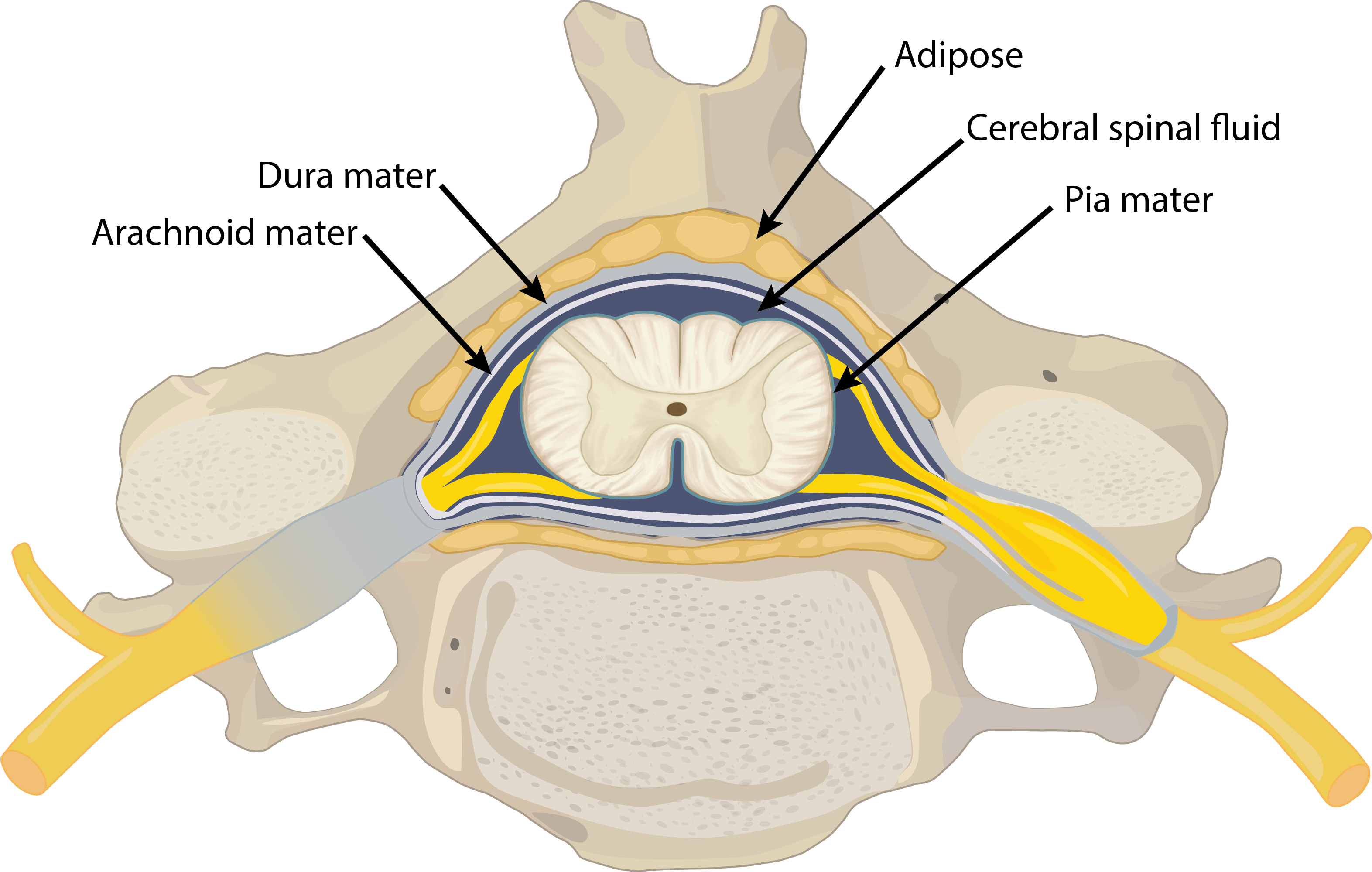

One space between the spinal meninges also differs slightly from the ones present around the brain. The epidural space between the periosteum and dura mater is a real space around the spinal cord and houses areolar and adipose connective tissues (shown in Figure \(\PageIndex{3}\)). Epidural anesthesia is injected in this space.


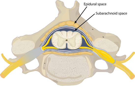
The subarachnoid space is filled with circulating CSF, which also provides a liquid cushion to the brain and spinal cord (Figure \(\PageIndex{3}\)). Similar to clinical blood work, a sample of CSF can be withdrawn to find chemical evidence of neuropathology or metabolic traces of the biochemical functions of nervous tissue. Because the spinal cord does not extend through the lower lumbar region of the vertebral column, a needle can be inserted through the dura and arachnoid layers to withdraw CSF. This procedure is called a lumbar puncture and avoids the risk of damaging the central tissue of the spinal cord.
Spinal Cord Gross Anatomy
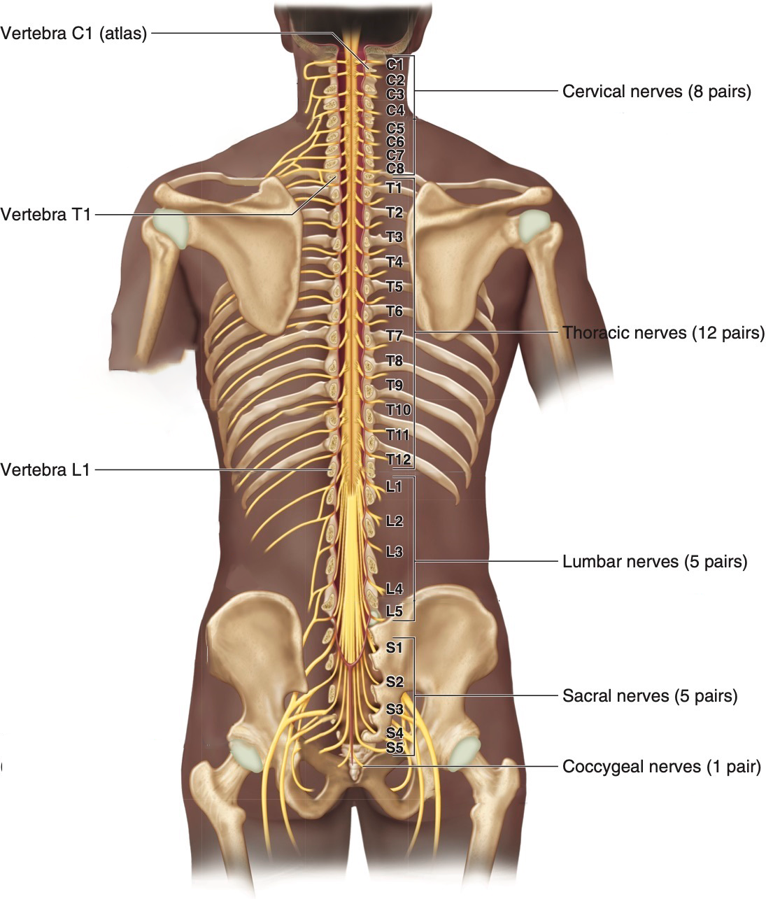
The description of the CNS is concentrated on the structures of the brain, but the spinal cord is another major organ of the system. The length of the spinal cord is divided into regions that correspond to the regions of the vertebral column. As shown in Figure \(\PageIndex{4}\), immediately inferior to the medulla oblongata is the cervical region of the spinal cord that contains motor neurons whose axons, together with sensory axons, extend into cervical spinal nerves. Inferior to the cervical region is the thoracic region, that contains neurons for the thoracic spinal nerves. The lumbar region is a short segment that contains neurons for the lumbar spinal nerves. Finally the sacral region contain neurons for the sacral nerves. The spinal cord is not the full length of the vertebral column because the spinal cord does not grow significantly longer after the first or second year, but the skeleton continues to grow. For this reason, the names of the spinal cord regions and vertebral regions to not match up. The lumbar and sacral spinal cord segments are found between vertebral levels T9 and L2. The spinal cord ends around the L1/L2 vertebral level, forming a tapered structure known as the conus medullaris (or medullary cone), which corresponds to the sacral spinal cord. The nerves that emerge from the spinal cord pass through the intervertebral foramina at the corresponding vertebral segments. For example, lumbar nerves exit the vertebral canal at the respective lumbar vertebrae. As the vertebral column grows, these nerves grow with it and result in a long bundle of nerves inferior to the conus medullaris that resembles a horse’s tail and therefore is named the cauda equina. Within the cauda equina, there is a thin strand of pia mater called filum terminale that helps anchoring the conus medullaris to the coccyx. At each intervertebral foramen, the spinal dura mater extends between vertebrae and surrounds the spinal nerves.
The diameter of the spinal cord changes between the different regions because the amounts of gray and white matter change to reflect the functions of the different regions. The cervical and lumbar regions of the spinal cord show a larger diameter compared to the rest of the regions. The cervical enlargement (C5-T1) and lumbar enlargement (L2-S3) represent an increased amount of neurons in the gray matter that serve the upper limbs and lower limbs, respectively (see Figure \(\PageIndex{5}\)).
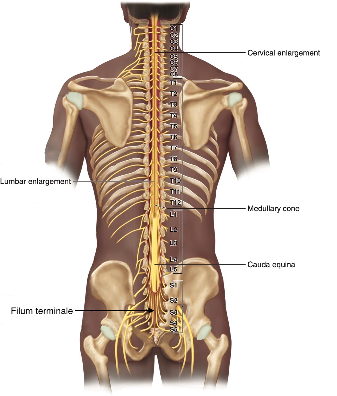
Figure \(\PageIndex{5}\): Gross Anatomy of the Spinal Cord. Compared to the superior cervical and thoracic regions, the cervical and lumbar enlargements have a larger diameter. The inferior end of the cord is anchored to the coccyx by the filum terminale, a thin strand of pia mater. (Image credit: "Spinal Cord Gross Anatomy" by Gabrielle Spurlock is licensed under CC BY-NC-SA 4.0, modification of original by KnowledgeWorks.)
Similar to the homunculi of the pre-central and post-central gyri, motor control and sensory input of the spinal cord can be mapped to specific segments of the spinal cord. Each skeletal muscle is usually innervated by multiple spinal segments, whereas each sensory region tends to correspond to a single spinal cord segment (with some overlap along the edges). (Table \(\PageIndex{1}\)) provides an overview of the muscle groups controlled by different spinal cord segments.
Table \(\PageIndex{1}\): Spinal Segment Control of Muscle Groups
| Spinal Cord Segments | Major Muscle Groups |
|---|---|
| C1-C3 | Cervical muscles - head and neck movements |
| C2-T1 | Muscles moving the scapula and shoulder |
| C3-C5 | Diaphragm |
| C5-C8 | Arm muscles |
| C5-T1 | Forearm muscles |
| T5-L1 | Abdominal muscles |
| T12-L3 | Iliopsoas (hip flexion) |
| L4-S2 | Hip extension and rotation |
| L2-S2 | Thigh muscles |
| L4-S3 | Leg and foot muscles |
| S3-Co1 | Pelvic floor muscles |
Spinal Cord Cross Sectional Anatomy
Whereas the brain develops out of expansions of the neural tube into primary and then secondary vesicles, the spinal cord maintains the tube structure and is only specialized into certain regions. As the spinal cord continues to develop in the newborn, anatomical features mark its surface. Viewed in cross section, the anterior midline is marked by the anterior median fissure, and the posterior midline is marked by the posterior median sulcus (Figure \(\PageIndex{6}\)). In the central region of the spinal cord, a central canal is the continuation of the fourth ventricle of the brain and contains cerebrospinal fluid (CSF). Surrounding the central canal, a horizontal line of gray matter called the gray commissure connects the left and right sides of the spinal cord.
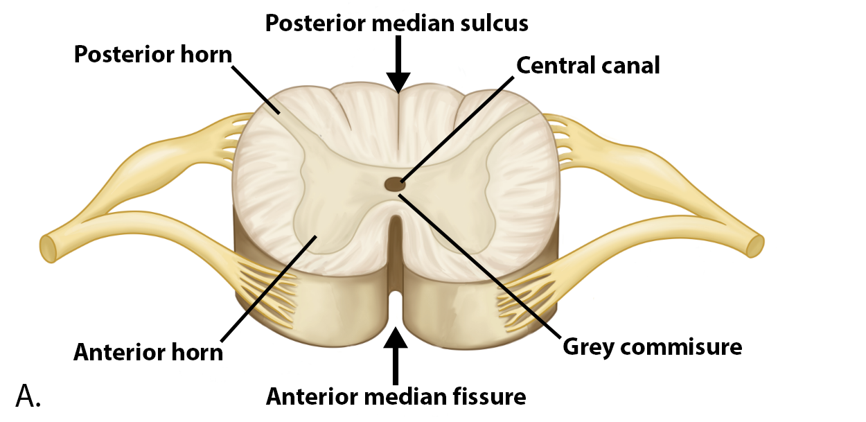
In cross-section, the gray matter of the spinal cord has the appearance of an ink-blot test, with the spread of the gray matter on one side replicated on the other—a shape reminiscent of a bulbous capital “H.” As shown in Figure \(\PageIndex{6}\) and Figure \(\PageIndex{8}\), the gray matter is subdivided into regions that are referred to as horns:
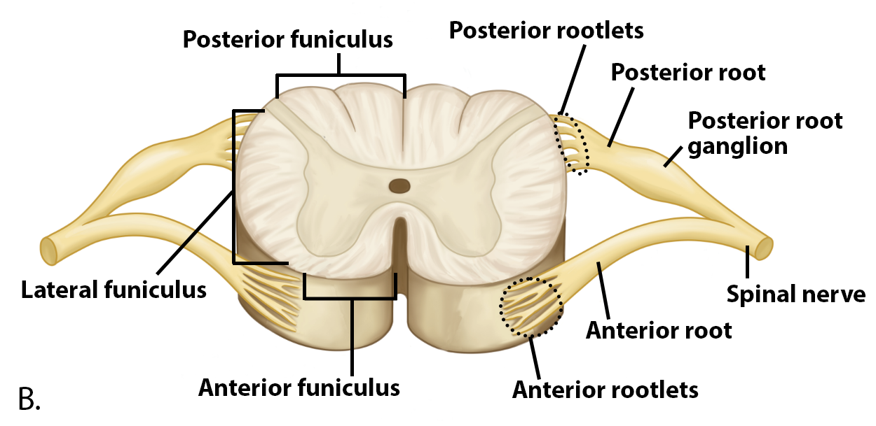
- The posterior horn is responsible for sensory processing and contain the axons of sensory neurons and the cell bodies of interneurons. The central processes of the bipolar sensory neurons are bundled into the posterior root and are coming directly from the posterior root ganglion which contains cell bodies of sensory neurons.
- The anterior horn contains the cell bodies of somatic motor neurons that control skeletal muscles. The somatic motor neurons are the largest neurons of the body and are classified as multipolar neurons. Their axons exit the spinal cord through the anterior nerve root.
- The lateral horn, which is only found in the thoracic, upper lumbar, and sacral regions, is a central component of the autonomic nervous system and contains cell bodies of autonomic motor neurons which innervate cardiac muscle, smooth muscle and glands (Figure \(\PageIndex{7}\)).

Just as the gray matter is separated into horns, the white matter of the spinal cord is separated into funiculi (Figure \(\PageIndex{6}\) and Figure \(\PageIndex{8}\)). Ascending tracts of nervous system fibers in these funiculi carry sensory information up to the brain, whereas descending tracts carry motor commands from the brain. Looking at the spinal cord longitudinally, the funiculi are further divided into columns that extend along its length as continuous bands of white matter. Between the two posterior horns of gray matter are the posterior funiculi composed of axons of ascending tracts. Between the two anterior horns, and bounded by the axons of motor neurons emerging from that gray matter area, are the anterior funiculi. The white matter on either side of the spinal cord, between the posterior horn and the axons of the anterior horn neurons, are the lateral funiculi. The anterior and lateral funiculi are composed of many different column groups of axons of both ascending and descending tracts.
 A. Spinal cord cross section at 15x
B. Spinal cord cross section at 20x
 C. Spinal cord cross section at 40x
 Regional Variations
Cross sectional views of the spinal cord vary, depending on the region from which the section was taken (Figure \(\PageIndex{8}\). The amount of white matter decreases between superior and inferior regions. The axons for all regions are present in the cervical spinal cord but only those for the sacral region are still present in the lumbar spinal cord, as many axons entered/exited in the intervening segments. You can imagine that the white matter for each spinal nerve is a "lane". At the level of C1 all 31 lanes of the road are present, but as you travel inferiorly a lane exits/enters at each intervertebral foramen. At the inferior end of the cervical region 8 lanes will have headed out, leaving 23 lanes. At the inferior end of the thoracic region another 12 lanes will have headed out, leaving only 11 lanes in the lumbar region.
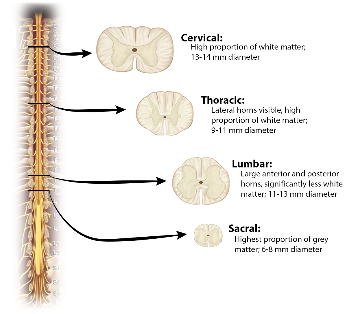
As discussed above, the volume of grey matter in each region varies by how many neurons are needed to serve each region. The grey matter in both the anterior and posterior horns is larger in both the cervical and lumbar regions to incorporate the large numbers of neurons needed for motor control and sensation in the upper and lower limbs.
Review Questions
Query \(\PageIndex{1}\)
Glossary
Query \(\PageIndex{1}\)
Contributors and Attributions
OpenStax Anatomy & Physiology (CC BY 4.0). Access for free at https://openstax.org/books/anatomy-and-physiology