
1.2: Anatomical Position and Planes
- Page ID
- 52717

\( \newcommand{\vecs}[1]{\overset { \scriptstyle \rightharpoonup} {\mathbf{#1}} } \)
\( \newcommand{\vecd}[1]{\overset{-\!-\!\rightharpoonup}{\vphantom{a}\smash {#1}}} \)
\( \newcommand{\dsum}{\displaystyle\sum\limits} \)
\( \newcommand{\dint}{\displaystyle\int\limits} \)
\( \newcommand{\dlim}{\displaystyle\lim\limits} \)
\( \newcommand{\id}{\mathrm{id}}\) \( \newcommand{\Span}{\mathrm{span}}\)
( \newcommand{\kernel}{\mathrm{null}\,}\) \( \newcommand{\range}{\mathrm{range}\,}\)
\( \newcommand{\RealPart}{\mathrm{Re}}\) \( \newcommand{\ImaginaryPart}{\mathrm{Im}}\)
\( \newcommand{\Argument}{\mathrm{Arg}}\) \( \newcommand{\norm}[1]{\| #1 \|}\)
\( \newcommand{\inner}[2]{\langle #1, #2 \rangle}\)
\( \newcommand{\Span}{\mathrm{span}}\)
\( \newcommand{\id}{\mathrm{id}}\)
\( \newcommand{\Span}{\mathrm{span}}\)
\( \newcommand{\kernel}{\mathrm{null}\,}\)
\( \newcommand{\range}{\mathrm{range}\,}\)
\( \newcommand{\RealPart}{\mathrm{Re}}\)
\( \newcommand{\ImaginaryPart}{\mathrm{Im}}\)
\( \newcommand{\Argument}{\mathrm{Arg}}\)
\( \newcommand{\norm}[1]{\| #1 \|}\)
\( \newcommand{\inner}[2]{\langle #1, #2 \rangle}\)
\( \newcommand{\Span}{\mathrm{span}}\) \( \newcommand{\AA}{\unicode[.8,0]{x212B}}\)
\( \newcommand{\vectorA}[1]{\vec{#1}} % arrow\)
\( \newcommand{\vectorAt}[1]{\vec{\text{#1}}} % arrow\)
\( \newcommand{\vectorB}[1]{\overset { \scriptstyle \rightharpoonup} {\mathbf{#1}} } \)
\( \newcommand{\vectorC}[1]{\textbf{#1}} \)
\( \newcommand{\vectorD}[1]{\overrightarrow{#1}} \)
\( \newcommand{\vectorDt}[1]{\overrightarrow{\text{#1}}} \)
\( \newcommand{\vectE}[1]{\overset{-\!-\!\rightharpoonup}{\vphantom{a}\smash{\mathbf {#1}}}} \)
\( \newcommand{\vecs}[1]{\overset { \scriptstyle \rightharpoonup} {\mathbf{#1}} } \)
\(\newcommand{\longvect}{\overrightarrow}\)
\( \newcommand{\vecd}[1]{\overset{-\!-\!\rightharpoonup}{\vphantom{a}\smash {#1}}} \)
\(\newcommand{\avec}{\mathbf a}\) \(\newcommand{\bvec}{\mathbf b}\) \(\newcommand{\cvec}{\mathbf c}\) \(\newcommand{\dvec}{\mathbf d}\) \(\newcommand{\dtil}{\widetilde{\mathbf d}}\) \(\newcommand{\evec}{\mathbf e}\) \(\newcommand{\fvec}{\mathbf f}\) \(\newcommand{\nvec}{\mathbf n}\) \(\newcommand{\pvec}{\mathbf p}\) \(\newcommand{\qvec}{\mathbf q}\) \(\newcommand{\svec}{\mathbf s}\) \(\newcommand{\tvec}{\mathbf t}\) \(\newcommand{\uvec}{\mathbf u}\) \(\newcommand{\vvec}{\mathbf v}\) \(\newcommand{\wvec}{\mathbf w}\) \(\newcommand{\xvec}{\mathbf x}\) \(\newcommand{\yvec}{\mathbf y}\) \(\newcommand{\zvec}{\mathbf z}\) \(\newcommand{\rvec}{\mathbf r}\) \(\newcommand{\mvec}{\mathbf m}\) \(\newcommand{\zerovec}{\mathbf 0}\) \(\newcommand{\onevec}{\mathbf 1}\) \(\newcommand{\real}{\mathbb R}\) \(\newcommand{\twovec}[2]{\left[\begin{array}{r}#1 \\ #2 \end{array}\right]}\) \(\newcommand{\ctwovec}[2]{\left[\begin{array}{c}#1 \\ #2 \end{array}\right]}\) \(\newcommand{\threevec}[3]{\left[\begin{array}{r}#1 \\ #2 \\ #3 \end{array}\right]}\) \(\newcommand{\cthreevec}[3]{\left[\begin{array}{c}#1 \\ #2 \\ #3 \end{array}\right]}\) \(\newcommand{\fourvec}[4]{\left[\begin{array}{r}#1 \\ #2 \\ #3 \\ #4 \end{array}\right]}\) \(\newcommand{\cfourvec}[4]{\left[\begin{array}{c}#1 \\ #2 \\ #3 \\ #4 \end{array}\right]}\) \(\newcommand{\fivevec}[5]{\left[\begin{array}{r}#1 \\ #2 \\ #3 \\ #4 \\ #5 \\ \end{array}\right]}\) \(\newcommand{\cfivevec}[5]{\left[\begin{array}{c}#1 \\ #2 \\ #3 \\ #4 \\ #5 \\ \end{array}\right]}\) \(\newcommand{\mattwo}[4]{\left[\begin{array}{rr}#1 \amp #2 \\ #3 \amp #4 \\ \end{array}\right]}\) \(\newcommand{\laspan}[1]{\text{Span}\{#1\}}\) \(\newcommand{\bcal}{\cal B}\) \(\newcommand{\ccal}{\cal C}\) \(\newcommand{\scal}{\cal S}\) \(\newcommand{\wcal}{\cal W}\) \(\newcommand{\ecal}{\cal E}\) \(\newcommand{\coords}[2]{\left\{#1\right\}_{#2}}\) \(\newcommand{\gray}[1]{\color{gray}{#1}}\) \(\newcommand{\lgray}[1]{\color{lightgray}{#1}}\) \(\newcommand{\rank}{\operatorname{rank}}\) \(\newcommand{\row}{\text{Row}}\) \(\newcommand{\col}{\text{Col}}\) \(\renewcommand{\row}{\text{Row}}\) \(\newcommand{\nul}{\text{Nul}}\) \(\newcommand{\var}{\text{Var}}\) \(\newcommand{\corr}{\text{corr}}\) \(\newcommand{\len}[1]{\left|#1\right|}\) \(\newcommand{\bbar}{\overline{\bvec}}\) \(\newcommand{\bhat}{\widehat{\bvec}}\) \(\newcommand{\bperp}{\bvec^\perp}\) \(\newcommand{\xhat}{\widehat{\xvec}}\) \(\newcommand{\vhat}{\widehat{\vvec}}\) \(\newcommand{\uhat}{\widehat{\uvec}}\) \(\newcommand{\what}{\widehat{\wvec}}\) \(\newcommand{\Sighat}{\widehat{\Sigma}}\) \(\newcommand{\lt}{<}\) \(\newcommand{\gt}{>}\) \(\newcommand{\amp}{&}\) \(\definecolor{fillinmathshade}{gray}{0.9}\)Information
When anatomists or health professionals identify the location of a structure in the human body, they do so in reference to a body in anatomical position. That is, they figure out the location based on the assumption that the body is starting out in anatomical position.
Anatomical position for a human is when the human stands up, faces forward, has arms extended, and has palms facing out.
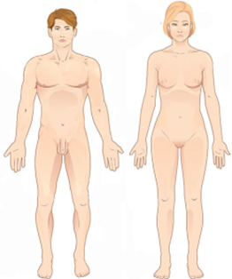
When referencing a structure that is on one side of the body or the other, we use the terms “anatomical right” and “anatomical left.” Anatomical right means that the structure is on the side that a person in anatomical position would consider their right-hand side (not necessarily on the right of the viewer) and anatomical left means that the structure is the side that a person in anatomical position would consider their left-hand side (which likewise is not necessarily the left side of the viewer.)
Anatomical planes
Information
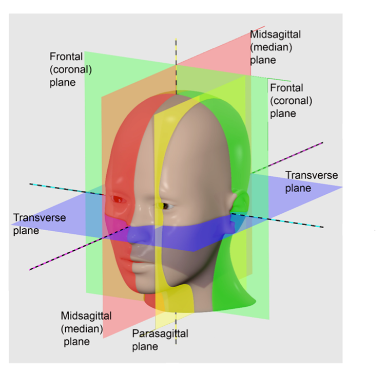
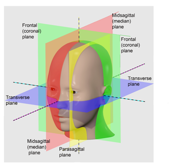
To view the interior of a body, we expose the organs and structures that are visible when that body is cut open along one of four commonly used sectional planes. These planes are the different directions a body is cut to reveal different views of its internal structures.
- Frontal plane—A vertical cut that separates the front from the back of the specimen. Also known as a coronal plane.
- Transverse plane—A horizontal cut that separates the top from the bottom of the specimen. Also known as a cross-sectional plane.
- Midsagittal plane—A vertical cut down the exact center line of the specimen that separates the left half from the right half.
- Parasagittal plane—A vertical cut that is off-center that separates the left of the specimen from the right in unequal portions. It does not matter whether it is the left side or the right side that is larger, as long as they are not equal.
LICENSES AND ATTRIBUTIONS
CC LICENSED CONTENT, ORIGINAL
- A&P Labs. Authored by: Ross Whitwam. Provided by: Mississippi University for Women. Located at: http://www.muw.edu/. License: CC BY-SA: Attribution-ShareAlike
- Figure 1-3. A banana person prior to being cut along transverse, frontal, and midsagittal or parasagittal planes.. Authored by: Ross Whitwam. Provided by: Mississippi University for Women. Located at: http://www.muw.edu/. License: CC BY-SA: Attribution- ShareAlike
CC LICENSED CONTENT, SHARED PREVIOUSLY
Figure 1-1. These two people are both in anatomical position.. Provided by: OpenStax College. Located at: http://cnx.org/contents/Gko70fNo@1/Anatomical-Terms. License: CC BY-SA: Attribution-ShareAlike
Figure 1-2. The different sectional planes used to expose internal structures.. Authored by: JonRichfield. Provided by:
Wikipedia. Located at: https://commons.wikimedia.org/wiki/F...nnotations.jpg. License: CC BY-SA: Attribution-ShareAlike
Anatomical Vocabulary
Anatomical nouns and adjective for external body parts
Information
Like all areas of science, there is a lot of jargon associated with anatomy and physiology. Often terms are used within the field that differ from what we would name things in everyday conversation. Such jargon usually allows the specialist in the field to be more precise in what exactly they are referring to, but the jargon also can be intimidating and exclusionary. If you don’t know it, you are not in the club.
LAB 1 EXERCISE 1-1
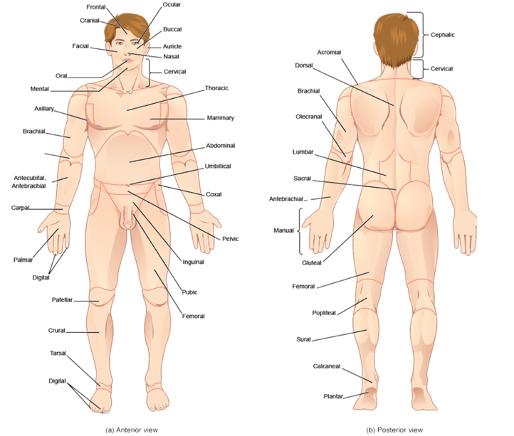
Here are a bunch of anatomical adjectives (followed in parentheses by the noun version of the same term). For each, use your smart phone or laptop or whatever is most convenient to you to find what body part the term refers to. (Shortcut hint: the Google search engine will return definitions for words if you type “define: word” in the search box, leaving out the quotation marks.)
Write down the body part or body region next to each term. Use Figure 1.4 to help you make sure you have the correct definition but look up each definition to make sure you are being accurate.
1. Find the body part or region indicated by each of the following terms. Use everyday language to describe the part or region. (Forearm, belly, etc.)
|
Abdominal (abdomen) |
Acromial (acromion) |
||
|
Antebrachial (antebrachium) |
Antecubital (antecubitis) |
||
|
Auricle (auris) |
Axillary (axilla) |
||
|
Brachial (brachium) |
Buccal (bucca) |
||
|
Carpal (carpus) |
Cephalic (cephalus) |
||
|
Cervical (cervicis) |
Coxal (coxa) |
||
|
Cranial (cranium) |
Crural (crus) |
||
|
Digital (digit) |
Dorsal (dorsa) |
||
|
Facial (facies) |
Femoral (femur) |
||
|
Frontal (frons) |
Gluteal (gluteus) |
||
|
Inguinal (inguen) |
Lumbar (lumbus) |
||
|
Mammary (mamma) |
Manual (manus) |
||
|
Mental (mentis) |
Nasal (nasus) |
||
|
Olecranal (olecranon) |
Oral (oris) |
||
|
Ocular (oculus) |
Palmar (palma) |
||
|
Patellar (patella) |
Pelvic (pelvis) |
||
|
Plantar (planta) |
Popliteal (popliteus) |
||
|
Pubic (pubis) |
Sacrum (sacral) |
||
|
Sural (sura) |
Tarsal (tarsus) |
||
|
Thoracic (thorax) |
Umbilical (umbilicus) |
LICENSES AND ATTRIBUTIONS
CC LICENSED CONTENT, ORIGINAL
A&P Labs. Authored by: Ross Whitwam. Provided by: Mississippi University for Women. Located at: . License: CC BY-SA: Attribution-ShareAlike
CC LICENSED CONTENT, SPECIFIC ATTRIBUTION
Figure 1-4. Anatomical adjectives for common surface features.. Provided by: OpenStax College. Located at: http://cnx.org/contents/FPtK1zmh@6.2...al-Terminology. License: CC BY-SA: Attribution- ShareAlike
Anatomical Orientation and Directions
Information
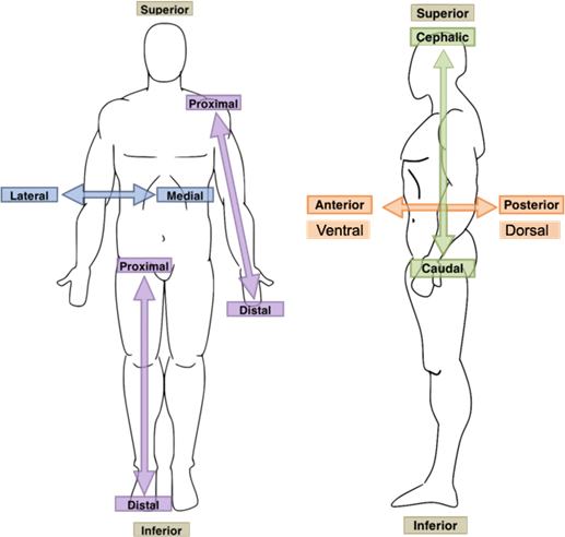
To be able to direct others to specific anatomical structures, or to find structures based on someone else’s directions, it is useful to have specific pairs of terms that allow you to orient your search with respect to the location of another, known structures. The following pairs of terms are used to make comparisons. Each term is used to orient a first structure or feature with respect to the position of a second structure or feature.
- Superior/Inferior–Equivalent to above and below when moving along the long axis of a body in anatomical position. The structure that is superior to another is above the second structure when the body is in anatomical position. A feature that is inferior to another is below the second feature when the body is in anatomical position.
- Proximal/Distal–Equivalent to near and far. Usually used to orient the positions of structures and features along the limbs with respect to the trunk of the body. A feature that is proximal to something else is closer to the limb’s point of attachment to the trunk. A structure that is distal to something else is farther away from the limb’s point of attachment. Less precisely but still occasionally used in the trunk of the body itself to indicate whether something is closer to (proximal) or farther away from (distal) something else.
- Medial/Lateral–Equivalent to towards the middle or towards the edge. Used with respect to the midline of the trunk of a body in anatomical position. A structure that medial to another is closer to the midline of the body’s trunk. A feature that is lateral to another is farther away from the midline of the trunk.
- Anterior/Posterior–Equivalent to the front and back of a body in anatomical position. A structure that is anterior to another is closer to the front of the body when the body is in anatomical position. A feature that is posterior to another is closer to the back of the body when the body is in anatomical position.
- Ventral/Dorsal–Equivalent to belly-side and back-side of a body in anatomical position. For a human in anatomical position, this pair of terms is equivalent to anterior and posterior. However, for four-legged animals in what is considered their anatomical position, the belly-side is not equivalent to the front of the animal. A structure that is ventral to another is closer to the belly-side of the body. A feature that is dorsal to another is closer to the back of the body.
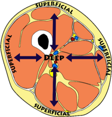
- Superficial/Deep–Equivalent to closer to the surface and farther from the surface.
- Cephalic/Caudal–Equivalent to closer to the head and closer to the tail. This is more useful for four-legged animals with tails than for upright humans with only a vestigial tail.
LAB 1 EXERCISE 1-2
1. Fill in the blank with the appropriate directional term to complete the following sentences. More than one answer may be correct.
- The heart is to the lungs.
- The knee is to the hip.
- The wrist is to the hand.
- The mouth is to the nose.
- The thorax is to the abdomen.
- The thumb is to the ring finger.
- The sternum is to the heart.
- The skull is to the scalp.
- The ears are to the nose.
- Dorsal refers to the of the human body, while ventral refers to the of the human body.
2. Find the indicated structures in the diagrams provided, based on the directional terms given. The structure to find will be one of those at the end of an unlabeled line.
A. Label the extensor digitorum (ED) muscle in the figure below. It is:
- Distal to the anconeus muscle
- Lateral to the extensor digiti minimi muscle

- Superficial to the Extensor pollicis brevis muscle
B. Label the Incus in the figure below. Label only one of the empty boxes. It is:
- Superior to the lateral end of the cochlear nerve
- Medial to the malleus

- Lateral to the stapes
3. Using your knowledge of the different body planes shown in Figure 1-2 (shown again below), fill in the blanks with the appropriate body plane for each of the following descriptions.
- The plane that divides the body into anterior and posterior parts is the plane.
- A transverse plane divides the body into and regions.
- A or plane divides the body into right and left parts.
LICENSES AND ATTRIBUTIONS
CC LICENSED CONTENT, ORIGINAL
A&P Labs. Authored by: Ross Whitwam. Provided by: Mississippi University for Women. Located at: http://www.muw.edu/. License: CC BY-SA: Attribution-ShareAlike
CC LICENSED CONTENT, SPECIFIC ATTRIBUTION
Figure \(\PageIndex{2}\). The different sectional planes used to expose internal structures.. Authored by: JonRichfield. Provided by: Wikipedia. Located at: https://commons.wikimedia.org/wiki/F...nnotations.jpg. License: CC BY-SA: Attribution-ShareAlike
Figure \(\PageIndex{4}\). Pairs of terms providing anatomical direction or orientation.. Authored by: Osteomyoamare. Provided by:Wikipedia. Located at: https://commons.wikimedia.org/wiki/F...Directions.png. License: CC BY-SA: Attribution- ShareAlike
Figure \(\PageIndex{4}\). Pairs of terms providing anatomical direction or orientation.. Authored by: Osteomyoamare. Provided by: Wikpedia. Located at: https://commons.wikimedia.org/wiki/F...rections_2.png. License: CC BY-SA: Attribution-ShareAlike
Figure \(\PageIndex{5}\). Cross-section of the thigh..Authored by: Marshall Strother. Provided by: Wikipedia. Located at: https://commons.wikimedia.org/wiki/F...ss_section.svg. License: CC BY-SA: Attribution-ShareAlike
Figure \(\PageIndex{6}\). Muscles of the forearm. Provided by: OpenStax College. Locate at: http://cnx.org/contents/FPtK1zmh@6.2...ectoral-Girdle. Project: Anatomy & Physiology. License: CC BY-SA: Attribution-ShareAlike
Figure \(\PageIndex{7}\).. Anatomy of the human ear.. Authored by: Chittka L, Brockmann. Provided by: Wikipedia. Located at: https://commons.wikimedia.org/wiki/F...man_Ear_en.svg. License: CC BY-SA: Attribution- ShareAlike