
13.5: Exercises
- Page ID
- 52791

Lab 13 Exercise \(\PageIndex{1}\)
 |
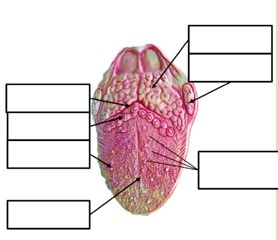
Draw the following:
Label the following
|
 |
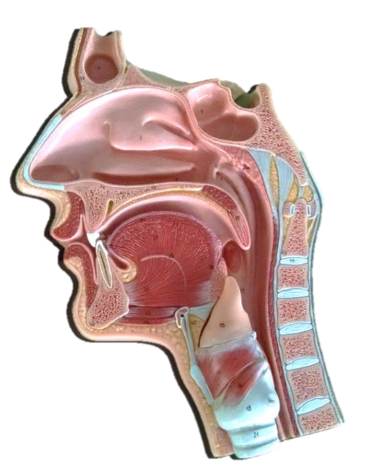
Label the following:
|
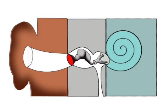
Lab 13 Exercise \(\PageIndex{2}\)
Basic Ear Anatomy
|
|
Match the following items to their proper location
|
Label the following on this stylized diagram: Auricle, Auditory canal, Pharyngotympanic tube, Stapes, Incus, Malleus, Tympanic membrane, Cochlea, Outer ear, Middle ear, Inner ear
|
|
|
Lab 13 Exercise \(\PageIndex{3}\)
Inner Ear Anatomy
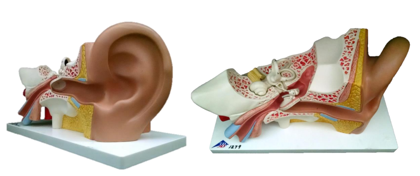
Label the following: Auricle, External Acoustic meatus, Pharyngotympanic tube, Cochlea, Vestibule, Semicircular canals, Tympanic membrane, Middle ear
|
Label the following:
|
Lab 13 Exercise \(\PageIndex{4}\)
Inner Ear Anatomy
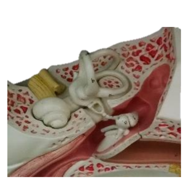 |
Label the following:
|
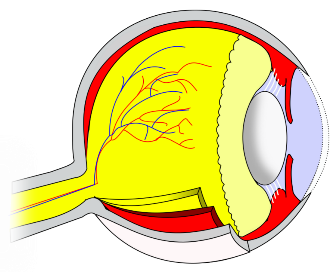
Lab 13 Exercise \(\PageIndex{5}\)
Label the following: Lens, Suspensory ligaments, Ciliary body, Cornea, Iris, Choroid, Vitreous humor
 |
Label the following:
|
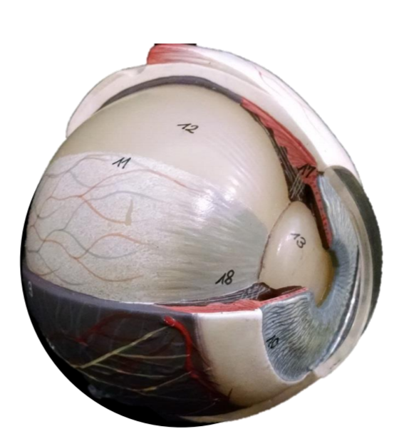
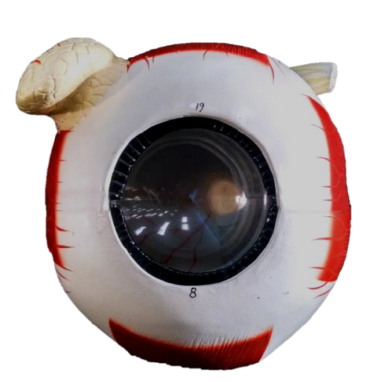
Lab 13 Exercise \(\PageIndex{6}\)
|
|
Identify Internal regions and layers of the eye:
|
Lab 13 Exercise \(\PageIndex{7}\)
Eye Anatomy
|
|
Label the following:
|
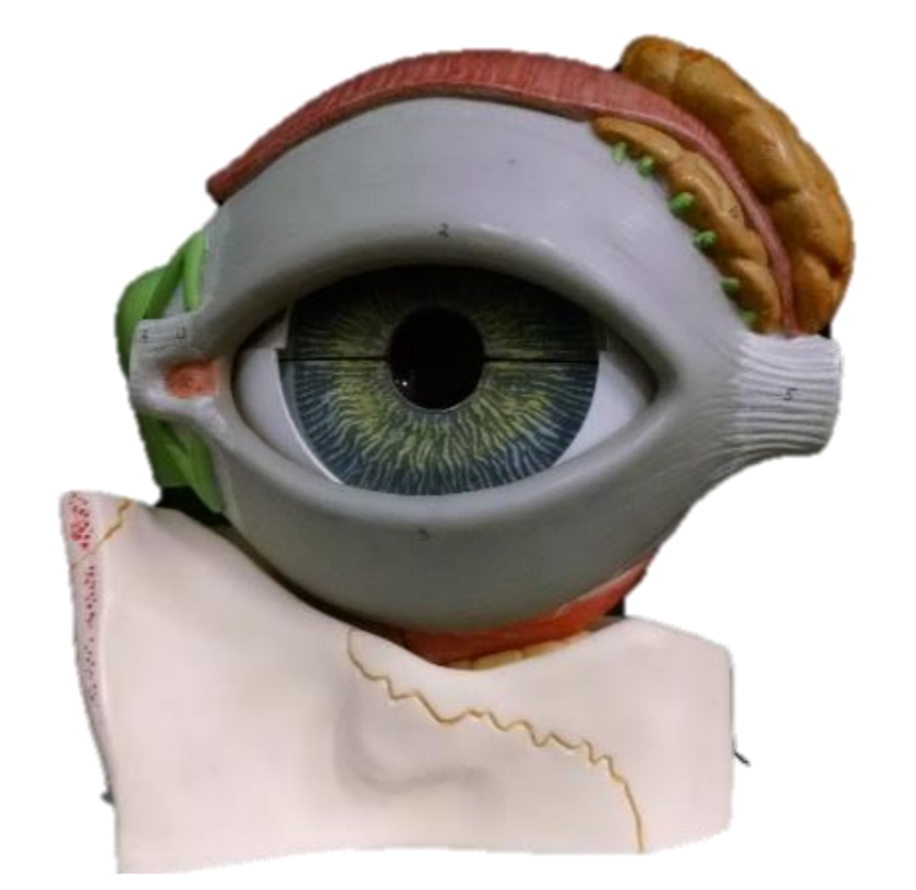
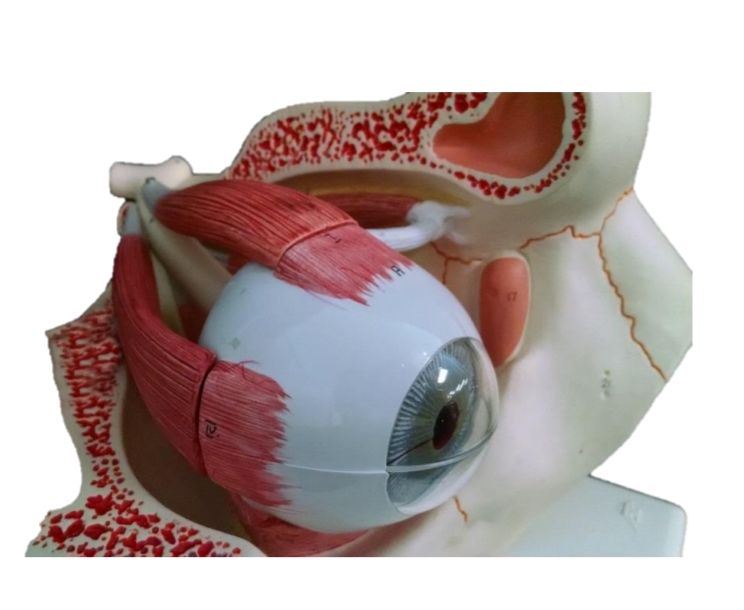
Lab 13 Exercise \(\PageIndex{8}\)
Extrinsic Eye Muscles
|
|
Identify the extrinsic eye muscles (4 are clearly visible)
|
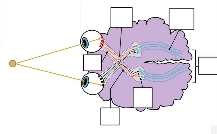
Lab 13 Exercise \(\PageIndex{9}\)
Optical Chiasm
Label the following:
- Conscious visual sensation;
- Beginning of axons of retinal ganglion cells;
- 3rd order neuron;
- synapse between 2nd & 3rd order neuron;
- Half of nerve fibers decussate here;
- First region with axons from only the left side of both eyes
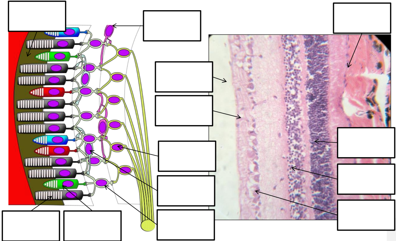
Lab 13 Exercise \(\PageIndex{10}\)
Retina Histology
A B
Label the following
| 1 | Rod | 6 | Rods and cones |
| 2 | Cone | 7 | Bipolar cells |
| 3 | Horizontal Cell | 8 | Ganglion cells |
| 4 | Bipolar Cell | 9 | Optic nerve axons |
| 5 | Ganglion cell | 10 | Pigmented epithelium |
| 11 | Vitreous humor |