
11.1: Folate and Folic Acid
- Page ID
- 21031

Folate is a B vitamin that exists in either its reduced form (folate) or oxidized form (folic acid). When folate is used in this section, we are referring to the reduced form, not the vitamin itself. Another key distinction between the 2 terms is that folic acid refers to the synthetic form, while folate refers to the natural form. Folic acid is only found in certain foods because they have been fortified with it, not because they produce it. The structure of folic acid is shown below.
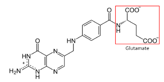
Another key difference between folate and folic acid is the number of glutamates in their tails. Notice that glutamate is boxed in the structure of folic acid above. Folic acid always exists as a monoglutamate, meaning it only contains 1 glutamate. On the other hand, about 90% of the folate found in foods are polyglutamates, meaning there is more than 1 glutamate in their tail. Folic acid is more stable than folate, which can be destroyed by heat, oxidation, and light2. Table \(\PageIndex{1}\) summarizes the key differences between folate and folic acid.
| Folate | Folic Acid |
|---|---|
| Reduced Form | Oxidized Form |
| Natural | Synthetic |
| Polyglutamate | Monoglutamate |
| More Stable |
The bioavailability of folate was believed to be much lower than folic acid.3 To account for these differences, the DRI committee created dietary folate equivalents (DFEs) to set the DRIs4. DFEs are defined as follows:
1 DFE = 1 ug food folate = 0.6 ug food folic acid = 0.5 ug folic acid on an empty stomach
DFE = ug food folate + (ug folic acid X 1.7)
The 1.7 came from research suggesting that folic acid from food was 85% bioavailable, compared to 50% for folate (85%/50% = 1.7)4. This was established in 1998 by the DRI committee, but there is newer evidence suggesting folate's bioavailability from food is higher (80% of folic acid) than previously believed3. With this data, a revised conversion factor for folic acid would be 1.25 (100%/80%). This conversion factor means that food folate levels are probably contributing more towards our dietary needs than currently being estimated by the DFE, but the DRI for folate/folic acid has not been updated.
Recent research investigating various food with different folate levels found variable responses in human subjects to both the food and the folate bioavailability. Thus, this research suggests that the bioavailability of folate is dependent on the individual as well as the food source5.
Before folate (polyglutamates) can be taken up into the enterocyte, the extra glutamates must be cleaved prior to uptake into the enterocyte by the Proton-Coupled Folate Transporter (PCFT). Folic acid because it is a monoglutamate, requires no cleavage before uptake. PCFT is believed to be the primary transporter for uptake. Once inside the enterocyte, the monoglutamate form is methylated and transported into circulation through Multidrug Associated Protein 3 (MAP3) or (MAP5)6. This series of events is depicted in the figure below6.
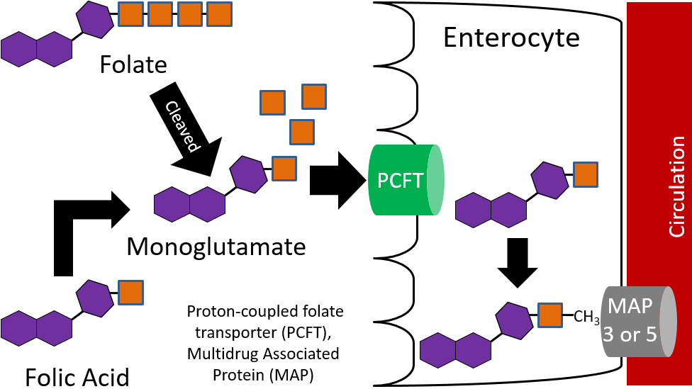
Thus, the methylated monoglutamate form is the circulating form. This is transported to the liver where it is converted back to the polyglutamate forms THF and 5-methyl THF for storage. Folate is excreted in both the urine and feces7.
Folate Functions
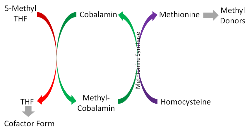
The major function of folate is that it participates in 1-carbon metabolism. As described earlier, this is the transfer of 1-carbon units from 1 compound to another. The cofactor form of folate is tetrahydrofolate (\(\ce{THF}\)). As is shown in the figure below, in order for THF to be formed, a methyl group is transferred to cobalamin (vitamin \(B_{12}\)) from 5-methyl \(\ce{THF}\) (\(\ce{THF}\) plus a methyl group), forming methyl-cobalamin. You can see this on the left side of the figure below.
There are 2 major cofactor functions of \(\ce{THF}\)8:
DNA Base Synthesis
THF is required for the synthesis of DNA bases (purines and pyrimidines)8. \(N^{10}\)-formyl-\(\ce{THF}\) (a form of \(\ce{THF}\)) is needed in 2 reactions in purine synthesis.
Amino Acid Metabolism
\(\ce{THF}\) is a cofactor for enzymes that metabolize histidine, serine, glycine, and methionine8. \(\ce{THF}\) is a cofactor for serine hydroxymethyltransferase, the enzyme that converts serine to glycine.
Folate Deficiency & Toxicity
Folate deficiency affects some Americans. The hallmark symptom of folate deficiency is macrocytic, megaloblastic anemia. Macrocytic anemia is characterized by large red blood cells and can be megaloblastic or non-megaloblastic. Megaloblastic anemia is characterized by large, nucleated (most red blood cells do not have a nucleus), immature red blood cells. This occurs because folate is needed for DNA synthesis; without it red blood cells are not able to divide properly2. As a result, fewer and poorer functioning red blood cells are produced that cannot carry oxygen as efficiently as normal red blood cells10.
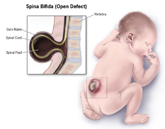
A maternal folate deficiency can lead to neural tube defects in infants. The exact cause of neural tube defects is unknown, but folate/folic acid supplementation has been shown to decrease the incidence of neural tube defects8. The most common of these neural tube defects is spina bifida (1 out of 2500 babies born in the United States), which is a failure of the neural tube to close and the spinal cord and its fluid protrude out the infant's back, as shown below11,12. Recent research suggests the defect may not be a result of a deficiency, rather a need for extra folate for some women at the time of increased cell division during embryonic development13.
The following video illustrates and describes spina bifida.
The neural tube closes 24-28 days after conception13, and with 50% of pregnancies estimated to be unplanned, many women are not aware they are pregnant during this period2,9. It is recommended that women of childbearing age consume 400 ug of folic acid daily8. However, to expect all women to do this through supplements would likely be most difficult for those at most risk (women of low socioeconomic status, young mothers) because they might not be able to afford or not know to take the supplement. Thus, in 1998 the FDA mandated that all refined cereals and grains be fortified with 140 ug folic acid /100 grams of product8. As you can see below, spina bifida prevalence rates declined during the optional fortification years and declined further once fortification became mandatory in the United States.
However, more recent research has found that folic acid supplementation begun before conception reduced the occurrence and severity of neural tube defects16.
It is not clear whether folic acid fortification was fully responsible for the decrease in spina bifida rates shown above, but the rates are lower than they were pre-fortification. However, you would think that the hope was that the impact would be greater than it has been thus far. The link below is to the announcement that in 2016 the FDA approved the fortification of corn masa flour. This may be beneficial for Hispanic/Latinx populations that might not consume much fortified refined cereals or grains.
Folate/Folic acid is not toxic, but it can mask a vitamin \(B_{12}\) deficiency and prevent its diagnosis. This effect will be discussed further in the vitamin \(B_{12}\) deficiency section. To put this in perspective, oral intakes of several hundred times the DRI are safe to consume due to the specificity and limited absorption of folic acid17.
References
- http://en.Wikipedia.org/wiki/File:Folat.svg
- Byrd-Bredbenner C, Moe G, Beshgetoor D, Berning J. (2009) Wardlaw's perspectives in nutrition. New York, NY: McGraw-Hill.
- Winkels R, Brouwer I, Siebelink E, Katan M, Verhoef P. (2007) Bioavailability of food folates is 80% of that of folic acid. Am J Clin Nutr 85(2): 465-473.
- Anonymous. (1998) Dietary reference intakes for thiamin, riboflavin, niacin, vitamin B6, folate, vitamin B12, pantothenic acid, biotin, and choline. Washington D.C.: National Academies Press.
- Mönch S, Netzel M, Netzel G, Ott U, Frank T, Rychlik M. (2015). Folate bioavailability from foods rich in folates assessed in a short term human study using stable isotope dilution assays. Food Func 6, 242-248.
- Visentin M, Diop-Bove N, Zhao R, Goldman ID. (2014) The intestinal absorption of folates. Annu Rev Physiol. 2014;76:251-274. doi:10.1146/annurev-physiol-020911-153251
- Shils ME, Shike M, Ross AC, Caballero B, Cousins RJ, editors. (2006) Modern nutrition in health and disease. Baltimore, MD: Lippincott Williams & Wilkins.
- Gropper SS, Smith JL, Groff JL. (2008) Advanced nutrition and human metabolism. Belmont, CA: Wadsworth Publishing.
- Stipanuk MH. (2006) Biochemical, physiological, & molecular aspects of human nutrition. St. Louis, MO: Saunders Elsevier.
- Whitney E, Rolfes SR. (2008) Understanding nutrition. Belmont, CA: Thomson Wadsworth.
- http://www.cdc.gov/ncbddd/birthdefec...pinaBifida.htm
- http://www.nlm.nih.gov/medlineplus/e...ages/19087.htm
- Mann J, Truswell AS. (2017) Essentials of Human Nutrition, Oxford University Press
- https://www.cdc.gov/ncbddd/spinabifida/facts.html
- http://www.cdc.gov/mmwr/preview/mmwrhtml/mm6401a2.htm
- Bergman JEH, Otten E, Verheij JBGM, de Walle HEK. (2016) Folic acid supplementation influences the distribution of neural tube defect subtypes: A registry-based study. Reprod Toxicol. 59:96-100.
- Essentials of Human Nutrition, Fifth Edition. 2017. Oxford University Press: New York, NY.