
12.5A: Vitamin K Functions
- Page ID
- 1561

Vitamin K is a cofactor for carboxylation reactions that add a CO2 to the amino acid, glutamic acid (glutamate), in certain proteins. The structure of glutamic acid is shown below.
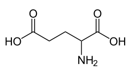
Figure 12.511 Structure of glutamic acid1
The enzyme, gamma-glutamyl carboxylase, uses a vitamin K cofactor to convert glutamic acid to gamma-carboxyglutamic acid (Gla). Gla proteins are those that contain glutamic acid(s) that have been converted to gamma-carboxyglutamic acid(s). The formation of Gla proteins allows the 2 positive charges of calcium to bind between the 2 negative charges on the carboxylic acid groups (COO-) in the Gla. The binding of calcium activates these proteins2-4.
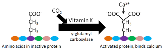
Figure 12.512 Gamma-glutamyl carboxylase converts glutamic acid to gamma-carboxyglutamic acid (Gla).
Gla proteins are important in blood clotting. Blood clotting occurs through a cascade of events, as shown in the following 2 videos. The animation below gives an overview of blood clotting, the video is a fun depiction of the blood clotting cascade.
Video: The Clotting Cascade (1:20)
|
Web Links Hemostasis Animation |
Within the blood clotting cascade, there a number of potential Gla proteins, as shown in the figure below.
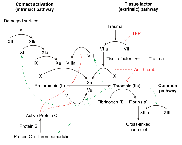
Figure 12.513 Blood clotting cascade with potential Gla proteins circled5
If these proteins within the blood clotting cascade are not activated to Gla, the cascade does not proceed as normal, leading to impaired blood clotting. After being used as a cofactor by gamma-glutamyl carboxylase to produce a Gla protein, vitamin K becomes vitamin K epoxide. Vitamin K epoxide needs to be converted back to vitamin K to serve as a cofactor again. Warfarin (Coumadin) and dicumarol are a couple of blood thinning drugs that inhibit this regeneration of vitamin K. This reduces the amount of Gla in the blood clotting proteins ,thus reducing the clotting response. The structure of warfarin and dicumarol are shown below5.
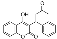
Figure 12.514 Structure of warfarin7
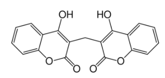
Figure 12.515 Structure of dicumarol8
The following coumadin rap song video gives further information on warfarin.
|
Web Link Video: Coumadin Rap Song (3:44) |
Vitamin K may also be important for bone health. There are 3 Gla proteins found in bone: osteocalcin, matrix Gla protein (MGP), and protein S6. Osteocalcin is a major bone protein, constituting 15-20% of all non-collagen proteins in bone. However, overall, the function of these 3 proteins in bone is not known2,3. Some research suggests that higher vitamin K status or intake decreases bone loss, but it is still not clear whether vitamin K truly is important for bone health9.
References & Links
1. en.Wikipedia.org/wiki/Glutam..._Non-ionic.png
2. Gropper SS, Smith JL, Groff JL. (2008) Advanced nutrition and human metabolism. Belmont, CA: Wadsworth Publishing.
3. Byrd-Bredbenner C, Moe G, Beshgetoor D, Berning J. (2009) Wardlaw's perspectives in nutrition. New York, NY: McGraw-Hill.
4. McGuire M, Beerman KA. (2011) Nutritional sciences: From fundamentals to food. Belmont, CA: Wadsworth Cengage Learning.
5. en.Wikipedia.org/wiki/File:Co...ation_full.svg
6. Shils ME, Shike M, Ross AC, Caballero B, Cousins RJ, editors. (2006) Modern nutrition in health and disease. Baltimore, MD: Lippincott Williams & Wilkins.
7. en.Wikipedia.org/wiki/File:Warfarin.svg
8. en.Wikipedia.org/wiki/File:Dicumarol.svg
9. Shea MK, Booth S. (2008) Update on the role of vitamin K in skeletal health. Nutr Rev 66(10): 549-557.
Videos
- Hemostasis Animation- http://www.mhhe.com/biosci/esp/2002_...7/trm1s7_3.htm
- The Clotting Cascade - https://www.youtube.com/watch?v=NJm4...eature=related
- Coumadin Rap Song - http://www.youtube.com/watch?v=Mfk05...watch_response