
12.2: Support and Protection of the Brain


- Last updated
- May 18, 2021
- Save as PDF
( \newcommand{\kernel}{\mathrm{null}\,}\)
By the end of this section, you will be able to:
- Describe the meninges that protect the brain
- Describe the blood vessels that supply the brain
- Name the components of the ventricular system and the regions of the brain in which each is located
- Explain the production of cerebrospinal fluid and its flow through the ventricles
Protective Coverings of the Brain
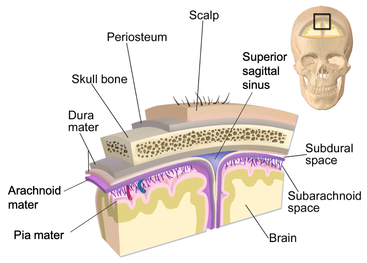
The central nervous system (CNS) is crucial to the operation of the body and any compromise of function in the brain and spinal cord can lead to severe difficulties. The brain is protected by multiple structures. First, the bones of the skull enclose and house the brain. Underneath the skeletal structures, the brain is protected by membranes made by connective tissue, called meninges, that surround, support, stabilize and partition the nervous tissue (Figure 12.2.1). In addition, the brain has a privileged blood supply, as suggested by the blood-brain barrier. The function of the tissue is crucial to the survival of the organism, so the contents of the blood cannot simply pass into the central nervous tissue. To protect this region from the toxins and pathogens that may be traveling through the blood stream, there is strict control over what can move out of the general systems and into the brain. Because of this privilege, the brain needs specialized structures for the maintenance of circulation. This begins with a unique arrangement of blood vessels carrying fresh blood into the brain and venous sinuses carrying deoxygenated blood out of the brain. Beyond the supply of blood, the brain filters the blood into cerebrospinal fluid (CSF), which is then circulated through the cavities of the brain, such as the subarachnoid space and the ventricles.
Cranial Meninges
The outer surface of the CNS is covered by a series of membranes composed of connective tissue called the meninges, which protect, stabilize and partition the brain. From superficial to deep, the meningeal layers are the dura mater, arachnoid mater and pia mater. The dura mater is a thick fibrous layer and a strong protective sheath over the entire brain. It is anchored to the inner surface of the cranium and vertebral cavity. The arachnoid mater is a membrane of thin fibrous tissue that forms a loose sac around the brain. Beneath the arachnoid mater, there is a space called the subarachnoid space where a thin, filamentous mesh form the arachnoid trabeculae, that looking like a spider web give this layer its name. Directly adjacent to the surface of the brain is the pia mater, a thin fibrous membrane that follows the superficial convolutions of the brain and fits into other grooves and indentations (Figure 12.2.2).
Dura Mater
Like a thick cap covering the brain, the dura mater is a tough outer covering. The name comes from the Latin for “tough mother” to represent its physically protective role. It encloses the entire CNS and the major blood vessels that enter the cranium and vertebral cavity. It is directly attached to the inner surface of the bones of the cranium and to the very end of the vertebral cavity. The dura mater of the brain is made by two layers, the periosteal layer which is more superficial and attached to the skull, and the meningeal layer, which lies deep to the first layer. These two layers are usually fused together. However, in some regions of the brain they separate to form large space filled with venous blood called dural sinuses. The dura mater of the spinal cord is composed of only one layer. The region between the bones and the dura mater is called epidural space. In the cranium, the epidural space contains arteries and veins that supply and drain blood from the brain. In normal conditions, the cranial epidural space is a potential space and not a real space. However, trauma can cause the leakage of fluid (hematomas) which accumulates in the epidural space enlarging it and transforming it into a real space. In the vertebral column, the epidural space is a real space that contains areolar and adipose connective tissue for an extra layer of protection, as well as blood vessels. The spinal epidural space at the lumbar level is where epidural injections are administered. Deep to the dura mater, there is another potential space called the subdural space. As the epidural space, this space can be filled with fluid causing a subdural hematoma.
The meningeal layer of the dura mater extends into the cranial cavity at four locations. These flat partitions are called cranial dural septa and separate specific parts of the brain (Figure 12.2.3). The falx cerebri goes through the midline separation of the brain while the falx cerebelli separates the two cerebellar hemispheres. The tentorium cerebelli separates the cerebellum from the superior part of the brain (cerebrum), forming a shelf-like tent. Another partition called diaphragma sellae surrounds the pituitary gland.
Arachnoid Mater
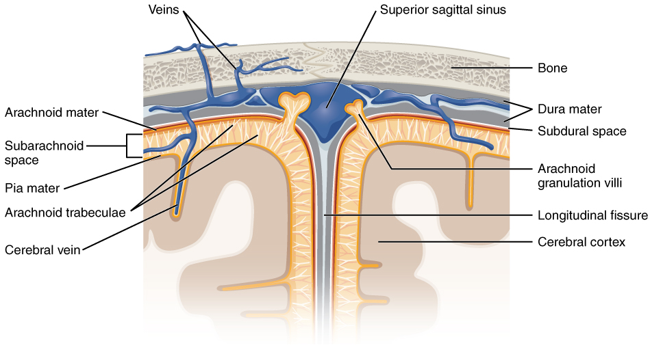
Deep to the subdural space lies the middle layer of the meninges called the arachnoid mater. This layer is named for the spider-web–like projections called arachnoid trabeculae between this layer and the pia mater. The arachnoid defines a sac-like enclosure around the CNS. The trabeculae are found in the subarachnoid space, which is filled with circulating CSF, making it a real space. The arachnoid emerges into the dural sinuses as the arachnoid granulations, where the CSF is filtered back into the blood for drainage from the nervous system.
Pia Mater
The outer surface of the CNS is covered in the thin fibrous membrane of the pia mater. It is thought to have a continuous layer of cells providing a fluid-impermeable membrane. The name pia mater comes from the Latin for “tender mother,” suggesting the thin membrane is a gentle covering for the brain. The pia extends into every convolution of the CNS. Blood vessels that are nourishing the central nervous tissue are between the pia mater and the nervous tissue.
DISORDERS OF THE...
Meninges: Meningitis
Meningitis is an inflammation of the meninges, the three layers of fibrous membrane that surround the CNS. Meningitis can be caused by infection by bacteria or viruses. The particular pathogens are not special to meningitis; it is just an inflammation of that specific set of tissues from what might be a broader infection. Bacterial meningitis can be caused by Streptococcus, Staphylococcus, or the tuberculosis pathogen, among many others. Viral meningitis is usually the result of common enteroviruses (such as those that cause intestinal disorders), but may be the result of the herpes virus or West Nile virus. Bacterial meningitis tends to be more severe.
The symptoms associated with meningitis can be fever, chills, nausea, vomiting, light sensitivity, soreness of the neck, or severe headache. More important are the neurological symptoms, such as changes in mental state (confusion, memory deficits, and other dementia-type symptoms). A serious risk of meningitis can be damage to peripheral structures because of the nerves that pass through the meninges. Hearing loss is a common result of meningitis.
The primary test for meningitis is a lumbar puncture. A needle inserted into the lumbar region of the spinal column through the dura mater and arachnoid membrane into the subarachnoid space can be used to withdraw the fluid for chemical testing. Fatality occurs in 5 to 40 percent of children and 20 to 50 percent of adults with bacterial meningitis. Treatment of bacterial meningitis is through antibiotics, but viral meningitis cannot be treated with antibiotics because viruses do not respond to that type of drug. Fortunately, the viral forms are milder.
Blood Supply to the Brain
A lack of oxygen to the CNS can be devastating, and the cardiovascular system has specific regulatory reflexes to ensure that the blood supply is not interrupted. There are multiple routes for blood to get into the CNS, with specializations to protect that blood supply and to maximize the ability of the nervous tissue to get an uninterrupted perfusion.
Arterial Supply to the Brain
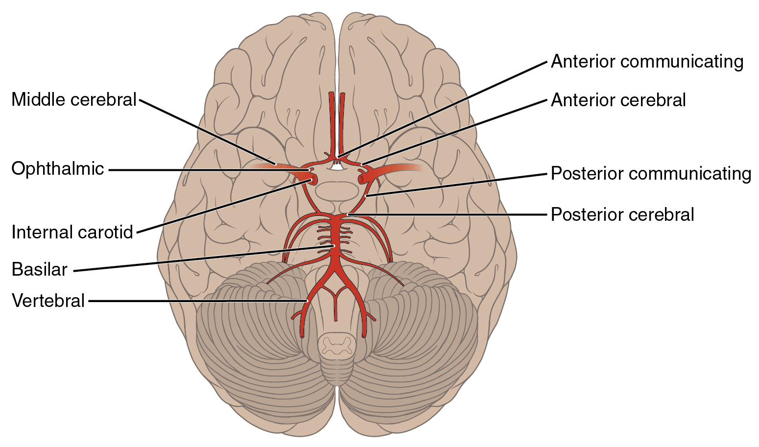
The major artery carrying recently oxygenated blood away from the heart is the aorta. The very first branches off the aorta supply the heart with nutrients and oxygen. The next branches give rise to the common carotid arteries, which further branch into the internal carotid arteries. The external carotid arteries supply blood to the tissues on the surface of the cranium. The internal carotid artery enters the cranium through the carotid canal in the temporal bone.
A second set of vessels that supply the CNS are the vertebral arteries, which are protected as they pass through the neck region by the transverse foramina of the cervical vertebrae. The vertebral arteries enter the cranium through the foramen magnum of the occipital bone. The two vertebral arteries then merge into the basilar artery, which gives rise to branches to the brainstem and cerebellum. The left and right internal carotid arteries and branches of the basilar artery all become the circle of Willis, a confluence of arteries that can maintain perfusion of the brain even if narrowing or a blockage limits flow through one part (Figure 12.2.4).
Interactive Link
Circle of Willis

Watch this animation of the Circle of Willis to see how blood flows to the brain and passes through it before being distributed through the tissues. The circle of Willis is a specialized arrangement of arteries that ensure constant perfusion of the brain even in the event of a blockage of one of the arteries in the circle. The animation shows the normal direction of flow through the circle of Willis to the middle cerebral artery. Where would the blood come from if there were a blockage just posterior to the middle cerebral artery on the left?
- Answer
-
If blood could not get to the middle cerebral artery through the posterior circulation, the blood would flow around the circle of Willis to reach that artery from an anterior vessel. Blood flow would just reverse within the circle.
Venous Return from the Brain
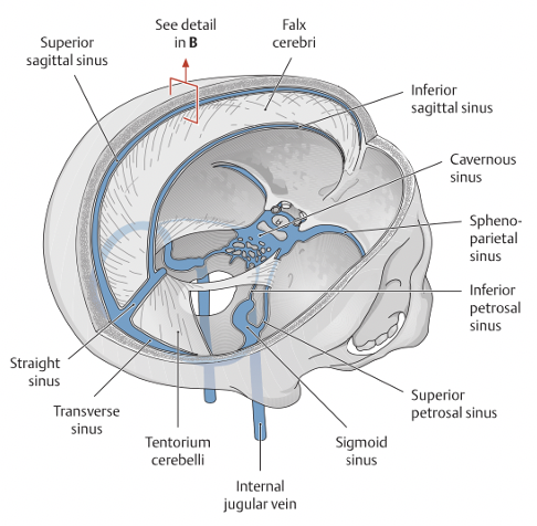
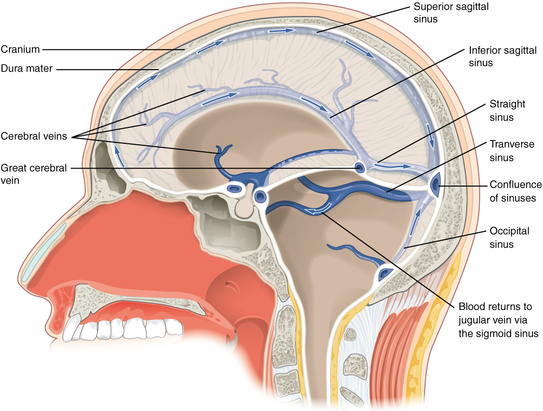
After passing through the CNS, blood returns to the circulation through a series of dural sinuses and cerebral veins (Figure 12.2.5). The dural sinuses are housed within the two layers of the dura mater. The superior sagittal sinus runs in the medial groove of the brain, where it absorbs CSF from the meninges. The superior sagittal sinus drains to the confluence of sinuses, along with the occipital sinuses and straight sinus, to then drain into the transverse sinuses. The transverse sinuses connect to the sigmoid sinuses, which then connect to the jugular veins. From there, the blood continues toward the heart to be pumped to the lungs for reoxygenation.
Ventricular System
Cerebrospinal fluid (CSF) is produced by a type of specialized membrane made of ependymal cells called a choroid plexus. Ependymal cells (one of the types of glial cells described in the introduction to the nervous system) surround blood capillaries and filter the blood to make CSF. The fluid is a clear solution with a limited amount of the constituents of blood. It is essentially water, small molecules, and electrolytes and is continuous with the interstitial fluid. Oxygen and carbon dioxide are dissolved into the CSF, as they are in blood, and can diffuse between the fluid and the nervous tissue. CSF circulates through the nervous tissue to remove metabolic wastes from the interstitial fluids of nervous tissues and return them to the blood stream. The choroid plexus lines open spaces within the brain called ventricles. The CSF circulates through all of the ventricles to eventually emerge into the subarachnoid space where it will be reabsorbed into the blood.
Ventricles
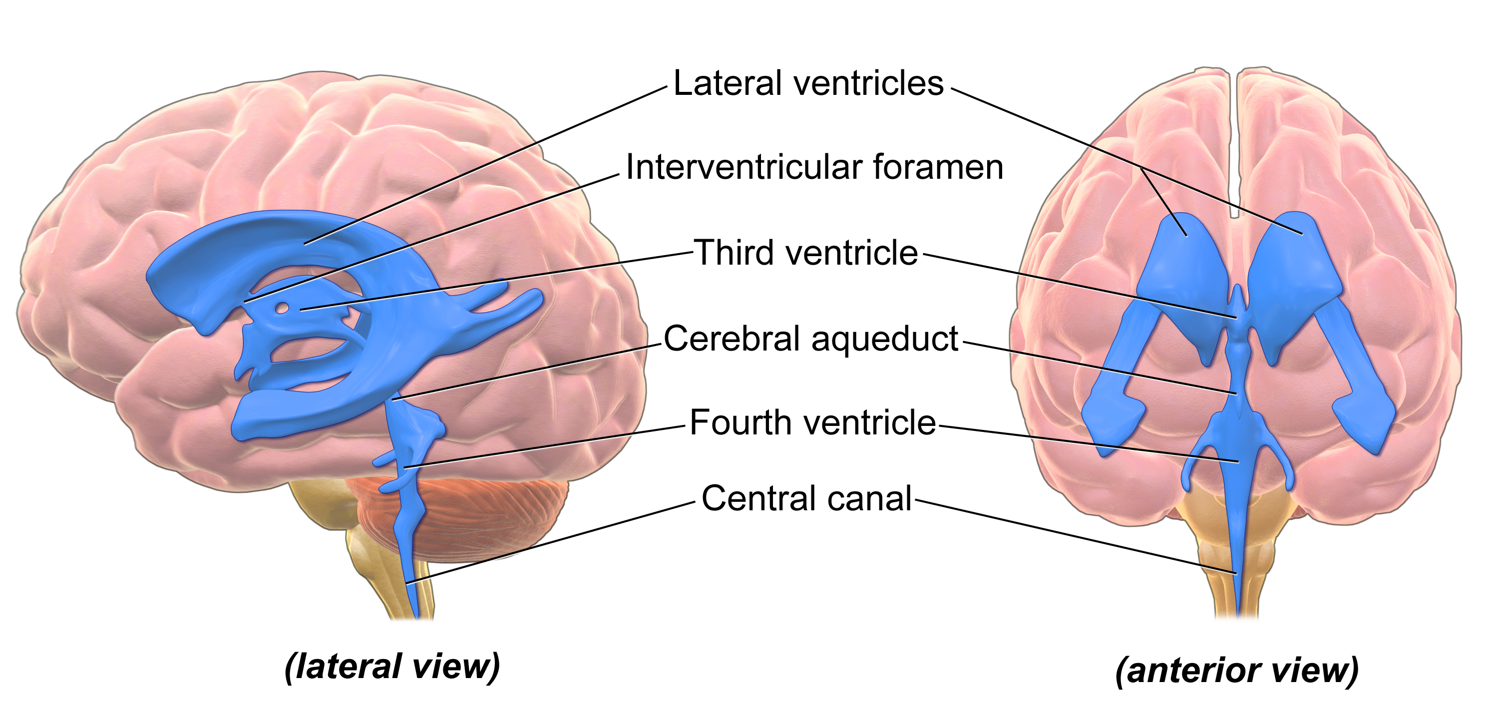
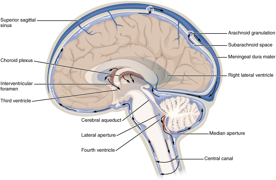
There are four ventricles within the brain, all of which developed from the original hollow space within the neural tube, the central canal. The first two are named the lateral ventricles and are deep within the brain (Figure 12.2.6). The two lateral ventricles are shaped as a C and are located in the left and right hemispheres, and were at one time referred to as the first and second ventricles. These ventricles are connected to the third ventricle by two openings called the interventricular foramina. The third ventricle opens into a canal called the cerebral aqueduct that passes through the midbrain and connects the third ventricle to the fourth ventricle. The fourth ventricle is the space between the cerebellum and the pons and upper medulla. The ventricular system opens up to the subarachnoid space from the fourth ventricle. The single median aperture and the pair of lateral apertures connect to the subarachnoid space so that CSF can flow through the ventricles and around the outside of the CNS (Figure 12.2.7). From the fourth ventricle, CSF can continue down the central canal of the spinal cord.
Cerebrospinal Fluid Circulation
The choroid plexuses are found in all four ventricles. Observed in dissection, they appear as soft, fuzzy structures that may still be pink, depending on how well the circulatory system is cleared in preparation of the tissue. The CSF is produced from components extracted from the blood, so its flow out of the ventricles is tied to the pulse of cardiovascular circulation.
From the lateral ventricles, the CSF flows into the third ventricle, where more CSF is produced, and then through the cerebral aqueduct into the fourth ventricle where even more CSF is produced (Figure 12.2.7). A very small amount of CSF is filtered at any one of the plexuses, for a total of about 500 milliliters daily, but it is continuously made and pulses through the ventricular system, keeping the fluid moving. From the fourth ventricle, CSF can continue down the central canal of the spinal cord, but this is essentially a cul-de-sac, so more of the fluid leaves the ventricular system and moves into the subarachnoid space through the median and lateral apertures.
Within the subarachnoid space, the CSF flows around all of the CNS, providing two important functions. As with elsewhere in its circulation, the CSF picks up metabolic wastes from the nervous tissue and moves it out of the CNS. It also acts as a liquid cushion for the brain and spinal cord. By surrounding the entire system in the subarachnoid space, it provides a thin buffer around the organs within the strong, protective dura mater. The arachnoid granulations are outpocketings of the arachnoid membrane into the dural sinuses so that CSF can be reabsorbed into the blood, along with the metabolic wastes. From the dural sinuses, blood drains out of the head and neck through the jugular veins, along with the rest of the circulation for blood, to be reoxygenated by the lungs and wastes to be filtered out by the kidneys.
DISORDERS OF THE...
Central Nervous System: Strokes
The supply of blood to the brain is crucial to its ability to perform many functions. Without a steady supply of oxygen, and to a lesser extent glucose, the nervous tissue in the brain cannot keep up its extensive electrical activity. These nutrients get into the brain through the blood, and if blood flow is interrupted, neurological function is compromised.
The common name for a disruption of blood supply to the brain is a stroke. It is caused by a blockage to an artery in the brain or by blood leaking out of blood vessels (hemorrhagic stroke), although less common. The blockage is caused by some type of embolus: a blood clot, a fat embolus, or an air bubble. When the blood cannot travel through the artery, the surrounding tissue that is deprived starves and dies. Strokes will often result in the loss of very specific functions. A stroke in the lateral medulla, for example, can cause a loss in the ability to swallow. Sometimes, seemingly unrelated functions will be lost because they are dependent on structures in the same region. Along with the swallowing in the previous example, a stroke in that region could affect sensory functions from the face or extremities because important white matter pathways also pass through the lateral medulla. Loss of blood flow to specific regions of the brain can lead to the loss of specific higher functions, from the ability to recognize faces to the ability to move a particular region of the body. Severe or limited memory loss can be the result of a temporal lobe stroke.
Related to strokes are transient ischemic attacks (TIAs), which can also be called “mini-strokes.” These are events in which a physical blockage may be temporary, cutting off the blood supply and oxygen to a region, but not to the extent that it causes cell death in that region. While the neurons in that area are recovering from the event, neurological function may be lost. TIAs usually resolve spontaneously by the body, that is why it is called "transient".
Recovery from a stroke (or TIA) is strongly dependent on the speed of treatment. Often, the person who is present and notices something is wrong must then make a decision. The mnemonic FAST helps people remember what to look for when someone is dealing with sudden losses of neurological function. If someone complains of feeling “funny,” check these things quickly: Look at the person’s face. Does he or she have problems moving Face muscles and making regular facial expressions? Ask the person to raise his or her Arms above the head. Can the person lift one arm but not the other? Has the person’s Speech changed? Is he or she slurring words or having trouble saying things? If any of these things have happened, then it is Time to call for help.
Sometimes, treatment with blood-thinning drugs can alleviate the problem, and recovery is possible. If the tissue is damaged, the amazing thing about the nervous system is that it is adaptable. With physical, occupational, and speech therapy, victims of strokes can recover, or more accurately relearn, functions.
Concept Review
The CNS has a privileged blood supply established by the blood-brain barrier. Establishing this barrier are anatomical structures that help to protect and isolate the CNS. The skull and vertebral column are the first mean of protection of the brain and spinal cord, respectively. Layers of connective tissue called meninges support and stabilize the brain and spinal cord, as well as partition the brain into specific regions. The outer layer is the dura mater, the middle layer is the arachnoid mater and the inner layer is the pia mater.
The arterial blood to the brain comes from the internal carotid and vertebral arteries, which both contribute to the unique circle of Willis that provides constant perfusion of the brain even if one of the blood vessels is blocked or narrowed. That blood is eventually filtered to make a separate medium, the CSF, that circulates within the spaces of the brain and then into the surrounding space defined by the meninges, the protective covering of the brain and spinal cord.
The blood that nourishes the brain and spinal cord is behind the glial-cell–enforced blood-brain barrier, which limits the exchange of material from blood vessels with the interstitial fluid of the nervous tissue. Thus, metabolic wastes are collected in cerebrospinal fluid that circulates through the CNS. This fluid is produced by filtering blood at the choroid plexuses in the four ventricles of the brain. It then circulates through the ventricles and into the subarachnoid space, between the pia mater and the arachnoid mater. From the arachnoid granulations, CSF is reabsorbed into the blood, removing the waste from the privileged central nervous tissue.
The blood, now with the reabsorbed CSF, drains out of the cranium through the dural sinuses. The dura mater is the tough outer covering of the CNS, which is anchored to the inner surface of the cranial and vertebral cavities. It surrounds the venous space known as the dural sinuses, which connect to the jugular veins, where blood drains from the head and neck.
Review Questions
Q. What blood vessel enters the cranium to supply the brain with fresh, oxygenated blood?
A. common carotid artery
B. jugular vein
C. internal carotid artery
D. aorta
- Answer
-
C
Q. Which layer of the meninges surrounds and supports the sinuses that form the route through which blood drains from the CNS?
A. dura mater
B. arachnoid mater
C. subarachnoid
D. pia mater
- Answer
-
A
Q. What type of glial cell is responsible for filtering blood to produce CSF at the choroid plexus?
A. ependymal cell
B. astrocyte
C. oligodendrocyte
D. Schwann cell
- Answer
-
A
Critical Thinking Questions
Q. Why can the circle of Willis maintain perfusion of the brain even if there is a blockage in one part of the structure?
A. The structure is a circular connection of blood vessels, so that blood coming up from one of the arteries can flow in either direction around the circle and avoid any blockage or narrowing of the blood vessels.
Q. Meningitis is an inflammation of the meninges that can have severe effects on neurological function. Why is infection of this structure potentially so dangerous?
A. The nerves that connect the periphery to the CNS pass through these layers of tissue and can be damaged by that inflammation, causing a loss of important neurological functions.
Glossary
- arachnoid granulation
- outpocket of the arachnoid membrane into the dural sinuses that allows for reabsorption of CSF into the blood
- arachnoid mater
- middle layer of the meninges named for the spider-web–like trabeculae that extend between it and the pia mater
- arachnoid trabeculae
- filaments between the arachnoid and pia mater within the subarachnoid space
- basilar artery
- blood vessel from the merged vertebral arteries that runs along the dorsal surface of the brainstem
- central canal
- hollow space within the spinal cord that is the remnant of the center of the neural tube
- cerebral aqueduct
- connection of the ventricular system between the third and fourth ventricles located in the midbrain
- choroid plexus
- specialized structures containing ependymal cells lining blood capillaries that filter blood to produce CSF in the four ventricles of the brain
- circle of Willis
- unique anatomical arrangement of blood vessels around the base of the brain that maintains perfusion of blood into the brain even if one component of the structure is blocked or narrowed
- common carotid artery
- blood vessel that branches off the aorta (or the brachiocephalic artery on the right) and supplies blood to the head and neck
- confluence of sinuses
- connecting point of the superior sagittal sinus, straight sinus, and occipital sinus
- cranial dura septa
- extensions of meningeal dura mater into the cranial cavity
- diaphragma sellae
- extension of the dura that separates the pituitary from the neural structures located superiorly including the optic chiasm
- dura mater
- tough, fibrous, outer layer of the meninges that is attached to the inner surface of the cranium and vertebral column and surrounds the entire CNS
- dural sinus
- any of the venous structures surrounding the brain, enclosed within the dura mater, which drain blood from the CNS to the common venous return of the jugular veins
- epidural space
- area between the dura mater and the above structures
- falx cerebelli
- extension of dura mater, projecting to the cerebellum
- falx cerebri
- extension of dura mater that lies between the cerebral hemispheres, in the longitudinal fissure
- fourth ventricle
- the portion of the ventricular system that is in the region of the brainstem and opens into the subarachnoid space through the median and lateral apertures
- internal carotid artery
- branch from the common carotid artery that enters the cranium and supplies blood to the brain
- interventricular foramina
- openings between the lateral ventricles and third ventricle allowing for the passage of CSF
- jugular veins
- blood vessels that return “used” blood from the head and neck
- lateral apertures
- pair of openings from the fourth ventricle to the subarachnoid space on either side and between the medulla and cerebellum
- lateral ventricles
- portions of the ventricular system that are deep within the brain
- median aperture
- singular opening from the fourth ventricle into the subarachnoid space at the midline between the medulla and cerebellum
- meningeal layer
- one of the two layers of the cranial dura mater
- meninges
- protective outer coverings of the CNS composed of connective tissue
- occipital sinuses
- dural sinuses along the edge of the most posterior region of the brain
- periosteal layer
- one of the two layers of the cranial dura mater
- pia mater
- thin, innermost membrane of the meninges that directly covers the surface of the CNS
- sigmoid sinuses
- dural sinuses that drain directly into the jugular veins
- straight sinus
- dural sinus that drains blood from the deep center of the brain to collect with the other sinuses
- subarachnoid space
- space between the arachnoid mater and pia mater that contains CSF and the fibrous connections of the arachnoid trabeculae
- subdural space
- space between the dura mater and arachnoid mater
- superior sagittal sinus
- dural sinus that runs along the top of the longitudinal fissure and drains blood from the majority of the outer brain
- tentorium cerebelli
- extension of dura mater that covers the upper surface of the cerebellum
- third ventricle
- portion of the ventricular system that is in the region of the diencephalon
- transverse sinuses
- dural sinuses that drain along either side of the occipital–cerebellar space
- ventricles
- remnants of the hollow center of the neural tube that are spaces for cerebrospinal fluid to circulate through the brain
- vertebral arteries
- arteries that ascend along either side of the vertebral column through the transverse foramina of the cervical vertebrae and enter the cranium through the foramen magnum
-
Contributors and Attributions
OpenStax Anatomy & Physiology (CC BY 4.0). Access for free at https://openstax.org/books/anatomy-and-physiology