
13.3: Percutaneous Biopsy
- Page ID
- 14869

Case 1
Ultrasound Guided Biopsy
Clinical:
History – This 66 year old female was found to have a palpable mass in the breast.
Symptoms – Palpable mass in the right breast.
Physical – A firm, non-mobile, mass was detected in the right breast. It was not fixated to the chest wall, or skin, and there were no palpable lymph nodes.
DDx:
Breast mass, BiRads 4b
Imaging Recommendation
Ultrasound Guided Needle Biopsy of the Mass
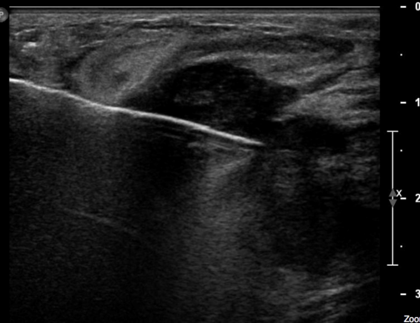
ODIN Link for Breast Mass Needle Biopsy images (Ultrasound), Figure 13.1: mistr.usask.ca/odin/?caseID=20151202232017924
Imaging Assessment
Findings:
Both mammography and breast ultrasound were used to evaluate a mass in the right breast. It was considered to be of intermediate risk for malignancy and was classified as a BiRads 4c lesion. Biopsy was recommended.
Interpretation:
The ultrasound images demonstrated the large caliber, 14g, needle in the breast mass.
Diagnosis:
Adenocarcinoma of the breast.
Discussion:
Ultrasound guided biopsy is warranted for the diagnosis of malignancy or infection. The abnormality in question must be visible with ultrasound, can be safely accessed, and be anatomically accessible for biopsy. Ultrasound affords real-time visualization of the needle tip which facilitates accurate biopsy sample acquisition.
Case 2
CT Guided Biopsy
Clinical:
History – Weight loss, fatigue, cough, and hemoptysis.
Symptoms – The patient complained of a chronic cough with hemoptysis.
Physical – There was evidence of chronic obstructive lung disease. The patient was cachectic and pale.
DDx:
Tuberculosis
Lung Abscess
Cavitating Lung Malignancy
Imaging Recommendation
CT Guided Lung Biopsy
ODIN Link for Left Upper Lobe Lung Mass Biopsy images (CT), Figure 13.2: mistr.usask.ca/odin/?caseID=20170118173043442

Imaging Assessment
Findings:
The patient had a cavitating mass in the left upper lobe. CT guidance was utilized to obtain a core needle biopsy specimen from the periphery of the mass where the tissue was more likely to be viable, not necrotic.
Interpretation:
Successful needle biopsy of the left upper lung mass.
Diagnosis:
Adenocarcinoma of the Lung, Non-small cell
Discussion:
CT guided biopsy is commonly used when the abnormality cannot be biopsied using ultrasound guidance. For the most part, CT biopsy is not a real-time imaging modality and multiple image acquisitions are required to position the needle for the biopsy.
Attributions
Figure 13.1 Ultrasound guided biopsy of a breast mass by Dr. Brent Burbridge MD, FRCPC, University Medical Imaging Consultants, College of Medicine, University of Saskatchewan is used under a CC-BY-NC-SA 4.0 license.
Figure 13.2 CT guided biopsy of a left lung mass by Dr. Brent Burbridge MD, FRCPC, University Medical Imaging Consultants, College of Medicine, University of Saskatchewan is used under a CC-BY-NC-SA 4.0 license.