
13.4: Percutaneous Fluid Drainage
- Page ID
- 14870

ACR – Interventional – Radiologic Management of Infected Fluid Collections
Case 1
Abscess Drain – CT Guided
Clinical:
History – 1 month in ICU after right hemi-colectomy for an obstructing colonic malignancy. The patient has had multiple intra-abdominal abscesses due to an anastomotic dehiscence.
Symptoms – Fever, on ventilator, septic.
Physical – The patient was intubated and septic. He was hemodynamically very fragile due to his advanced age of 86 year.
DDx:
Peri-hepatic abscess, deep.
ACR – Radiologic Management of Infected Fluid Collections
This fluid collection could not be seen well with ultrasound. CT guided, transhepatic abscess drain was requested due to his poor clinical status and ineligibility for surgical drain insertion.
CT Guided Abscess Drainage
ODIN Link for CT Abscess Drainage images, Figure 13.3A and B: mistr.usask.ca/odin/?caseID=20170404111333875


Imaging Assessment
Findings:
There was a low attenuation collection in the upper abdomen that was medial to the cranial liver. A route of needle access was planned via a transhepatic route. Needle insertion was CT guided and followed by insertion of a 12F drainage catheter.
Interpretation:
Successful insertion of a 12F, transhepatic, abscess drain resulted in drainage of thick, foul-smelling, green fluid.
Diagnosis:
Perihepatic Abscess
Discussion:
The elevated risk of this procedure was explained to the family and the patient. There are additional risks of hepatic venous or arterial bleeding due to the drain route through the liver and for the development of intrahepatic abscesses along the drain tract. The surgical team could not offer this man any treatment options that were safer and his potential morbidity and mortality from an open surgical drainage was felt to be greater than the risk of CT guided drainage.
Drainage of fluid can be performed utilizing needle aspiration and/or small or large caliber drainage tubes. The use of imaging guidance i.e. ultrasound, CT, and/or fluoroscopy facilitates accurate and safe insertion of the needle into the fluid collection. The drainage tube follows the positioning of a guidewire into the fluid via the puncture needle. The tract in the soft tissues can then be sequentially dilated to accommodate the small or large draining catheter based upon the location of the tube and the viscosity of the draining fluid. The process of needle, guidewire, dilation, drainage tube, is called the Seldinger Technique. It is widely used for interventional radiology procedures.
Case 2
Infected Polycystic Kidney Cyst, Drained, Ultrasound Guided
Clinical:
History – Autosomal Dominant Polycystic Kidney Disease
Symptoms – Left Flank and upper abdominal pain. Fever.
Physical – The kidneys were massively enlarged and palpable on physical examination. The patient was tender in the left upper quadrant. The patient’s temperature was 39.5C.
Laboratory – The white blood cell count was elevated.
DDx:
Renal Cyst Hemorrhage
Renal Cyst Infection
ACR – Radiologic Management of Infected Fluid Collections
Ultrasound Guided Fluid Drainage
ODIN Link for Kidney Cyst Drainage images (CT and Ultrasound), Figure 13.4A and B: mistr.usask.ca/odin/?caseID=20161107172446656

Imaging Assessment
Findings:
CT
The patient had polycystic kidneys. There was a massive cyst in the left upper kidney. No fluid/fluid levels or unusual internal attenuation of this large cyst.
Given the CT findings of the massive cyst, aspiration with Ultrasound guidance was suggested as draining this cyst may improve the patient’s condition and samples could be sent for culture and sensitivity.
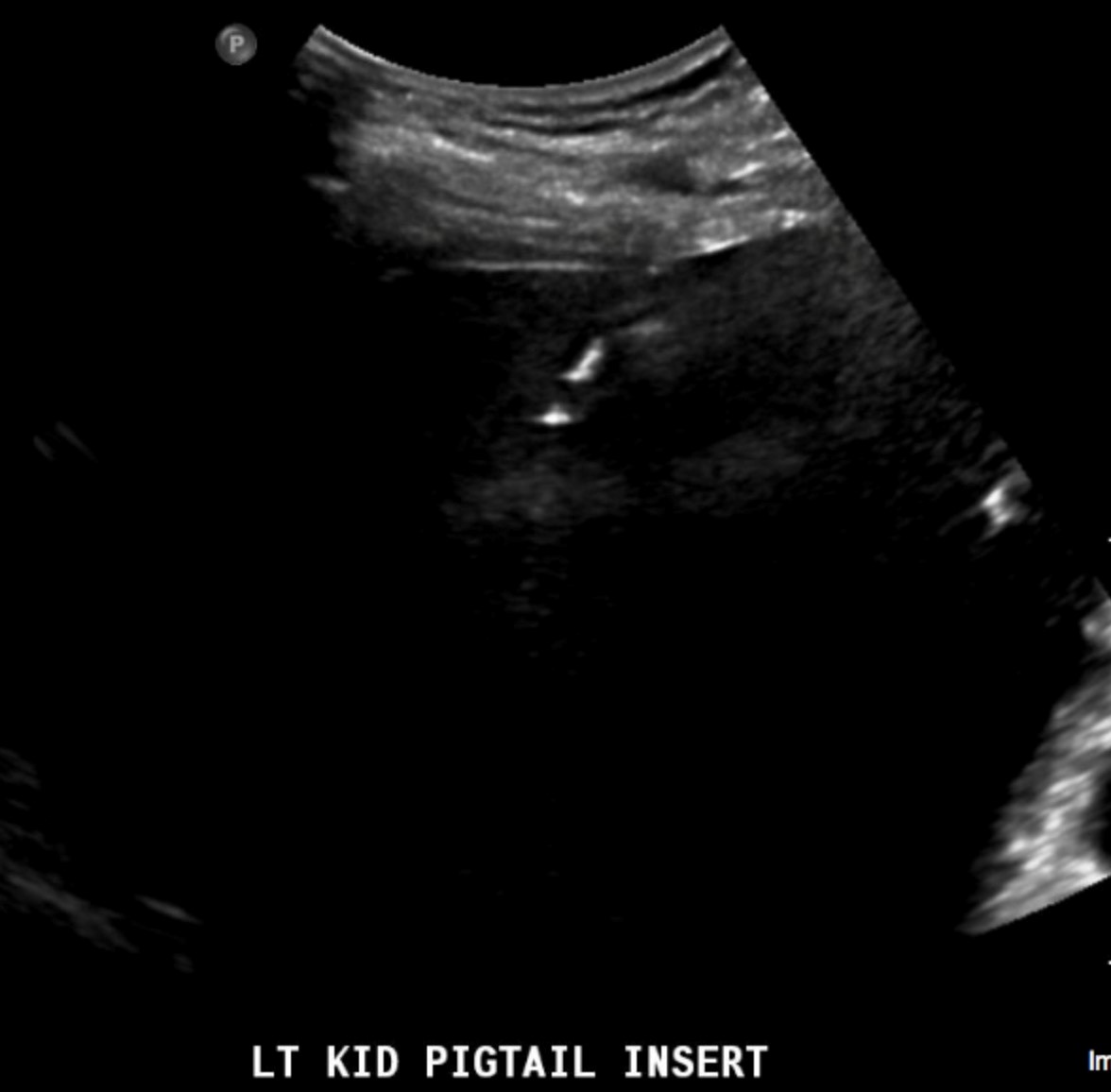
Ultrasound
Ultrasound was used to aspirate some fluid from the largest cyst on the left. It was frankly purulent and a 10F drain was inserted.
Interpretation:
Polycystic kidney with an infected cyst in the left upper kidney.
Diagnosis:
Secondary infection of a left renal cyst.
Attributions
Figure 13.3A CT scan of abdomen with a grid and measurements to plan drainage by Dr. Brent Burbridge MD, FRCPC, University Medical Imaging Consultants, College of Medicine, University of Saskatchewan is used under a CC-BY-NC-SA 4.0 license.
Figure 13.3B CT scan of a guided drainage of perihepatic abscess by Dr. Brent Burbridge MD, FRCPC, University Medical Imaging Consultants, College of Medicine, University of Saskatchewan is used under a CC-BY-NC-SA 4.0 license.
Figure 13.4A CT scan of polycystic kidneys with a very large cyst on the left by Dr. Brent Burbridge MD, FRCPC, University Medical Imaging Consultants, College of Medicine, University of Saskatchewan is used under a CC-BY-NC-SA 4.0 license.
Figure 13.4B Ultrasound guided drainage of the left kidney cyst. Needle visualized with ultrasound by Dr. Brent Burbridge MD, FRCPC, University Medical Imaging Consultants, College of Medicine, University of Saskatchewan is used under a CC-BY-NC-SA 4.0 license.