
3.2: Types of Tissues
- Page ID
- 63375
- Identify the four main tissue types
- Discuss the general functions of each tissue type
- Discuss the embryonic origin of tissue
- Identify the three major germ layers
- Identify the main types of tissue membranes
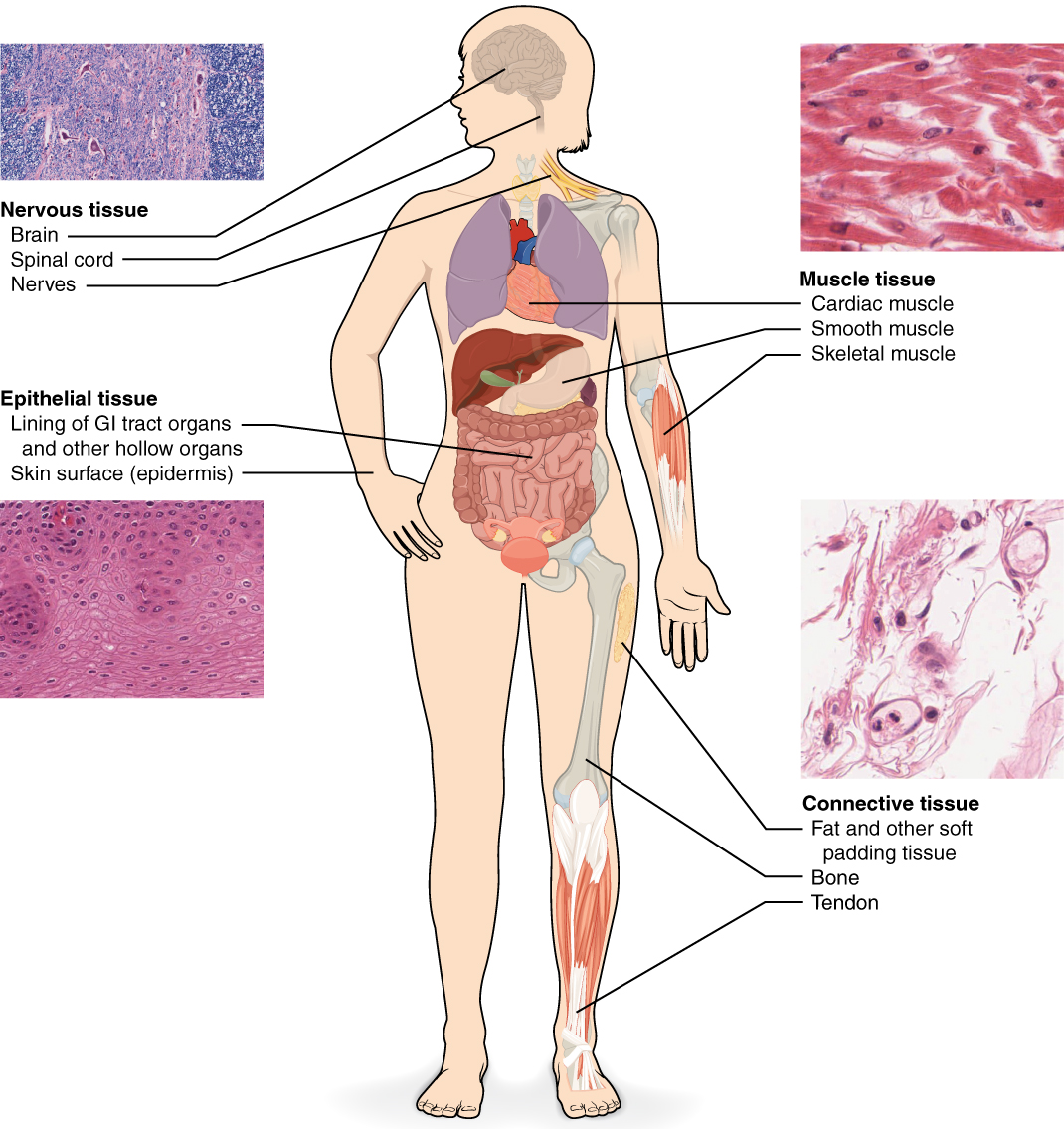
Although there are hundreds of types of cells in the human body, they are organized into four broad categories of tissues: epithelial, connective, muscle, and nervous (Figure \(\PageIndex{1}\)). Each of these categories is characterized by specific functions that contribute to the overall health and maintenance of the body. A disruption of the structure is a sign of injury or disease. Such changes can be detected through histology, the microscopic study of tissue appearance, organization, and function.
The Four Types of Tissues
- Epithelial tissue, also referred to as epithelium (plural = epithelia), refers to the sheets of cells that cover exterior surfaces of the body (e.g. the skin surface), line internal body cavities (e.g. the pericardium), and form the lining of internal and many external surfaces of organs (e.g. the GI tract organs), and forms certain glands (e.g the adrenal glands).
- Connective tissue, as its name implies, binds the cells and organs of the body together and functions in the protection, support, and integration of all parts of the body. Some examples include fat, bones, and tendons.
- Muscle tissue is excitable, responding to stimulation and contracting to provide movement, and occurs as three major types: skeletal muscle, smooth muscle, and cardiac muscle. Skeletal muscle is under the voluntary control of the brain, but cardiac muscle and smooth muscle are involuntary so you cannot control their actions.
- Nervous tissue, which can be found in the brain, spinal cord, and nerves, is also excitable, allowing the propagation of electrochemical signals in the form of nerve impulses that communicate between different regions of the body.
The next level of organization is the organ, where several types of tissues come together to form a working unit. Just as knowing the structure and function of cells helps you in your study of tissues, knowledge of tissues will help you understand how organs function.
Embryonic Origin of Tissues
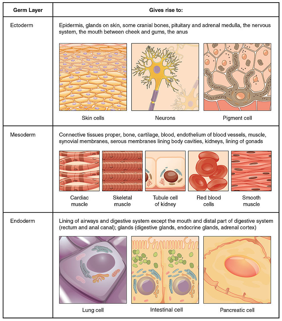
The zygote, or fertilized egg, is a single cell formed by the fusion of an egg and sperm. After fertilization the zygote gives rise to rapid mitotic cycles, generating many cells to form the embryo. The first embryonic cells generated have the ability to differentiate into any type of cell in the body and, as such, are called totipotent, meaning each has the capacity to divide, differentiate, and develop into a new organism. As cell proliferation progresses, three major cell lineages are established within the embryo. Each of these lineages of embryonic cells forms the distinct germ layers from which all the tissues and organs of the human body eventually form. Each germ layer is identified by its relative position: ectoderm (ecto- = “outer”), mesoderm (meso- = “middle”), and endoderm (endo- = “inner”).
Figure \(\PageIndex{2}\) shows the types of tissues and organs associated with the each of the three germ layers. The ectoderm gives rise to the epidermis, glands on the skin, some cranial bones, the pituitary gland and adrenal medulla, the nervous system, the mouth between cheek and gums, and the anus. The mesoderm gives rise to connective tissues proper, bone, cartilage, blood, endothelium of blood vessels, muscle, synovial membranes, serous membranes lining body cavities, kidneys, and the lining of the gonads. The endoderm gives rise to the lining of the airways and digestive system, (expect the mouth and distal portion (rectum and anal canal)), and some glands including digestive glands, endocrine glands, and the adrenal cortex. Note that epithelial tissue originates in all three layers, whereas nervous tissue derives primarily from the ectoderm and muscle tissue from mesoderm.
Stem Cells

View this slideshow video to learn more about stem cells. How do somatic stem cells differ from embryonic stem cells?
- Answer
-
Answer: Most somatic stem cells give rise to only a few cell types.
Tissue Membranes
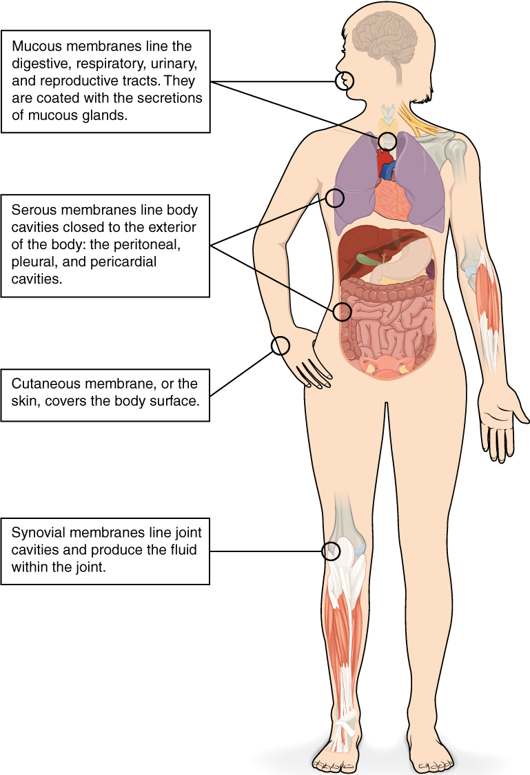
A tissue membrane is a thin layer or sheet of cells that covers the outside of the body (for example, skin), the organs (for example, pericardium), internal passageways that lead to the exterior of the body (for example, the digestive tract), and the lining of the moveable joint cavities. There are two basic types of tissue membranes: epithelial membranes and connective tissue membranes (Figure \(\PageIndex{3}\)).
Epithelial Membranes
The epithelial membrane is composed of epithelium attached to a layer of connective tissue, for example, your skin. The mucous membrane is also a composite of connective and epithelial tissues. Sometimes called mucosae (singular mucosa), these epithelial membranes line the body cavities and hollow passageways that open to the external environment, and include the digestive, respiratory, urinary, and reproductive tracts. Mucous, produced by the epithelial exocrine glands, covers the epithelial layer and protects it in a variety of specific ways. The underlying connective tissue, called the lamina propria (literally “own layer”), help support the fragile epithelial layer.
A serous membrane is an epithelial membrane composed of mesodermally derived epithelium called the mesothelium that is supported by connective tissue. These membranes line the coelomic cavities of the body, that is, those cavities that do not open to the outside, and they cover the organs located within those cavities. They are essentially membranous bags, with mesothelium lining the inside and connective tissue on the outside. Serous fluid secreted by the cells of the thin squamous mesothelium lubricates the membrane and reduces abrasion and friction between organs. Serous membranes are identified according to location. Three serous membranes line the thoracic cavity: the two pleura that cover the lungs and the pericardium that covers the heart. A fourth, the peritoneum, is the serous membrane in the abdominal cavity that covers abdominal organs and forms double sheets of mesenteries that suspend many of the digestive organs.
The skin is an epithelial membrane also called the cutaneous membrane. It is a stratified squamous epithelial membrane resting on top of connective tissue. The apical surface of this membrane is exposed to the external environment and is covered with dead, keratinized cells that help protect the body from desiccation (drying out) and pathogens.
Connective Tissue Membranes
The connective tissue membrane is formed solely from connective tissue. These membranes encapsulate organs, such as the kidneys, and line our movable joints, so each is typically referred to as a capsule. A synovial membrane is a type of connective tissue membrane that lines the cavity of a freely movable joint. For example, synovial membranes surround the joints of the shoulder, elbow, and knee. Fibroblasts in the inner layer of the synovial membrane release hyaluronan into the joint cavity. The hyaluronan effectively traps available water to form the synovial fluid, a natural lubricant that enables the bones of a joint to move freely against one another without much friction. This synovial fluid readily exchanges water and nutrients with blood, as do all body fluids.
Concept Review
The human body contains more than 200 types of cells that can all be classified into four types of tissues: epithelial, connective, muscle, and nervous. Epithelial tissues act as coverings. Connective tissue integrates the various parts of the body and provides support and protection to organs. Muscle tissue allows the body to move. Nervous tissues propagate information.
The study of the shape and arrangement of cells in tissue is called histology. All cells and tissues in the body derive from three germ layers in the embryo: the ectoderm, mesoderm, and endoderm.
Different types of tissues form membranes that enclose organs, provide a friction-free interaction between organs, and keep organs together. Synovial membranes are connective tissue membranes that protect and line the joints. Epithelial membranes are formed from epithelial tissue attached to a layer of connective tissue. There are three types of epithelial membranes: mucous, which contain glands; serous, which secrete fluid; and cutaneous which makes up the skin.
Review Questions
Q. Which of the following is not a type of tissue?
A. muscle
B. nervous
C. synovial
D. epithelial
- Answer
-
Answer: C
Q. The process by which a less specialized cell matures into a more specialized cell is called ________.
A. differentiation
B. maturation
C. modification
D. specialization
- Answer
-
Answer: A
Q. Differentiated cells in a developing embryo derive from ________.
A. endothelium, mesothelium, and epithelium
B. ectoderm, mesoderm, and endoderm
C. connective tissue, epithelial tissue, and muscle tissue
D. epidermis, mesoderm, and endothelium
- Answer
-
Answer: B
Q. Which of the following lines the body cavities exposed to the external environment?
A. mesothelium
B. lamina propria
C. mesenteries
D. mucosa
- Answer
-
Answer: D
Critical Thinking Questions
Q. Identify the four types of tissue in the body, and describe the major functions of each tissue.
- Answer
-
A. The four types of tissue in the body are epithelial, connective, muscle, and nervous. Epithelial tissue is made of layers of cells that cover the surfaces of the body that come into contact with the exterior world, line internal cavities, and form glands. Connective tissue binds the cells and organs of the body together and performs many functions, especially in the protection, support, and integration of the body. Muscle tissue, which responds to stimulation and contracts to provide movement, is divided into three major types: skeletal (voluntary) muscles, smooth muscles, and the cardiac muscle in the heart. Nervous tissue allows the body to receive signals and transmit information as electric impulses from one region of the body to another.
Q. The zygote is described as totipotent because it ultimately gives rise to all the cells in your body including the highly specialized cells of your nervous system. Describe this transition, discussing the steps and processes that lead to these specialized cells.
- Answer
-
A. The zygote divides into many cells. As these cells become specialized, they lose their ability to differentiate into all tissues. At first they form the three primary germ layers. Following the cells of the ectodermal germ layer, they too become more restricted in what they can form. Ultimately, some of these ectodermal cells become further restricted and differentiate in to nerve cells.
Q. What is the function of synovial membranes?
- Answer
-
A. Synovial membranes are a type of connective tissue membrane that supports mobility in joints. The membrane lines the joint cavity and contains fibroblasts that produce hyaluronan, which leads to the production of synovial fluid, a natural lubricant that enables the bones of a joint to move freely against one another.
Glossary
- connective tissue
- type of tissue that serves to hold in place, connect, and integrate the body’s organs and systems
- connective tissue membrane
- connective tissue that encapsulates organs and lines movable joints
- cutaneous membrane
- skin; epithelial tissue made up of a stratified squamous epithelial cells that cover the outside of the body
- ectoderm
- outermost embryonic germ layer from which the epidermis and the nervous tissue derive
- endoderm
- innermost embryonic germ layer from which most of the digestive system and lower respiratory system derive
- epithelial membrane
- epithelium attached to a layer of connective tissue
- epithelial tissue
- type of tissue that serves primarily as a covering or lining of body parts, protecting the body; it also functions in absorption, transport, and secretion
- histology
- microscopic study of tissue architecture, organization, and function
- lamina propria
- areolar connective tissue underlying a mucous membrane
- mesoderm
- middle embryonic germ layer from which connective tissue, muscle tissue, and some epithelial tissue derive
- mucous membrane
- tissue membrane that is covered by protective mucous and lines tissue exposed to the outside environment
- muscle tissue
- type of tissue that is capable of contracting and generating tension in response to stimulation; produces movement.
- nervous tissue
- type of tissue that is capable of sending and receiving impulses through electrochemical signals.
- serous membrane
- type of tissue membrane that lines body cavities and lubricates them with serous fluid
- synovial membrane
- connective tissue membrane that lines the cavities of freely movable joints, producing synovial fluid for lubrication
- tissue
- group of cells that are similar in form and perform related functions
- tissue membrane
- thin layer or sheet of cells that covers the outside of the body, organs, and internal cavities
- totipotent
- embryonic cells that have the ability to differentiate into any type of cell and organ in the body
Contributors and Attributions
OpenStax Anatomy & Physiology (CC BY 4.0). Access for free at https://openstax.org/books/anatomy-and-physiology