
5.5: Fractures and Bone Repair
- Page ID
- 63392
- Differentiate among the different types of fractures
- Describe the steps involved in bone repair
A fracture is a broken bone. It will heal whether or not a physician resets it in its anatomical position. If the bone is not reset correctly, the healing process will keep the bone in its deformed position. When a broken bone is manipulated and set into its natural position without surgery, the procedure is called a closed reduction. Open reduction requires surgery to expose the fracture and reset the bone. While some fractures can be minor, others are quite severe and result in grave complications. For example, a fractured diaphysis of the femur has the potential to release fat globules into the bloodstream. These can become lodged in the capillary beds of the lungs, leading to respiratory distress and if not treated quickly, death.
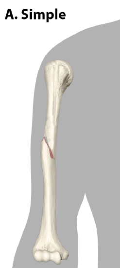
Types of Fractures
Fractures are classified by their complexity, location, and other features (Figure \(\PageIndex{1}\) and Figure \(\PageIndex{2}\)). Table \(\PageIndex{1}\) outlines common types of fractures. Some fractures may be described using more than one term because it may have the features of more than one type (e.g., an open transverse fracture).
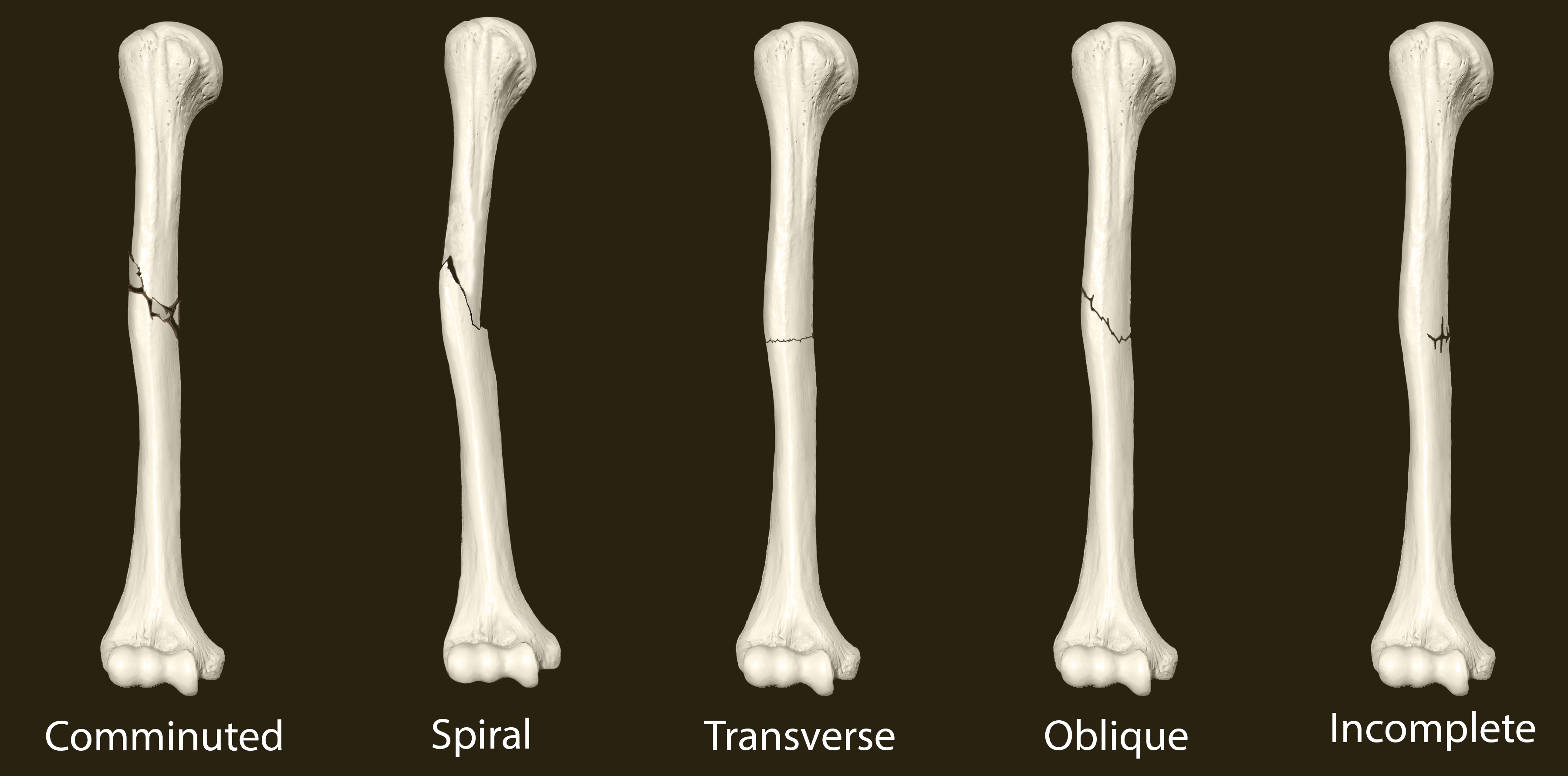
| Type of fracture | Description |
|---|---|
| Transverse | Occurs straight across the long axis of the bone |
| Oblique | Occurs at an angle that is not 90 degrees |
| Spiral | Bone segments are pulled apart as a result of a twisting motion |
| Comminuted | Several breaks result in many small pieces between two large segments |
| Impacted | One fragment is driven into the other, usually as a result of compression |
| Greenstick (children), Incomplete (adults) | A partial fracture in which only one side of the bone is broken |
| Compound (or Open) | A fracture in which at least one end of the broken bone tears through the skin; carries a high risk of infection |
| Simple (or Closed) | A fracture in which the skin remains intact |
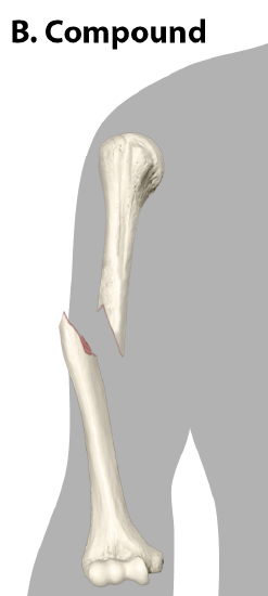
Bone Repair
When a bone breaks, blood flows from any vessel torn by the fracture. These vessels could be in the periosteum, osteons, and/or medullary cavity. The blood begins to clot, and about six to eight hours after the fracture, the clotting blood has formed a fracture hematoma (Figure \(\PageIndex{3.a}\)). The disruption of blood flow to the bone results in the death of bone cells around the fracture.
Within about 48 hours after the fracture, chondrocytes from the endosteum have created an internal callus (plural = calli) by secreting a fibrocartilaginous matrix between the two ends of the broken bone, while the periosteal chondrocytes and osteoblasts create an external callus of hyaline cartilage and bone, respectively, around the outside of the break (Figure \(\PageIndex{3.b}\)). This stabilizes the fracture.
Over the next several weeks, osteoclasts resorb the dead bone; osteogenic cells become active, divide, and differentiate into osteoblasts. The cartilage in the calli is replaced by trabecular bone via endochondral ossification (Figure \(\PageIndex{3.c}\)).
Eventually, the internal and external calli unite, compact bone replaces spongy bone at the outer margins of the fracture, and healing is complete. A slight swelling may remain on the outer surface of the bone, but quite often, that region undergoes remodeling (Figure \(\PageIndex{3.d}\)), and no external evidence of the fracture remains.
Concept Review
Fractured bones may be repaired by closed reduction or open reduction. Fractures are classified by their complexity, location, and other features. Common types of fractures are transverse, oblique, spiral, comminuted, impacted, greenstick, open (or compound), and closed (or simple). Healing of fractures begins with the formation of a hematoma, followed by internal and external calli. Osteoclasts resorb dead bone, while osteoblasts create new bone that replaces the cartilage in the calli. The calli eventually unite, remodeling occurs, and healing is complete.
Review Questions
Q. A fracture can be both ________.
A. open and closed
B. open and transverse
C. transverse and greenstick
D. greenstick and comminuted
- Answer
-
Answer: B
Q. How can a fractured diaphysis release fat globules into the bloodstream?
A. The bone pierces fat stores in the skin.
B. The yellow marrow in the diaphysis is exposed and damaged.
C. The injury triggers the body to release fat from healthy bones.
D. The red marrow in the fractured bone releases fat to heal the fracture.
- Answer
-
Answer: B
Q. In a compound fracture, ________.
A. the break occurs at an angle to the bone
B. the broken bone does not tear the skin
C. one fragment of broken bone is compressed into the other
D. broken bone pierces the skin
- Answer
-
Answer: D
Q. The internal and external calli are replaced by ________.
A. hyaline cartilage
B. trabecular bone
C. osteogenic cells
D. osteoclasts
- Answer
-
Answer: B
Q. The first type of bone to form during fracture repair is ________ bone.
A. compact
B. lamellar
C. spongy
D. dense
- Answer
-
Answer: C
Critical Thinking Questions
Q. What is the difference between closed reduction and open reduction? In what type of fracture would closed reduction most likely occur? In what type of fracture would open reduction most likely occur?
- Answer
-
A. In closed reduction, the broken ends of a fractured bone can be reset without surgery. Open reduction requires surgery to return the broken ends of the bone to their correct anatomical position. A partial fracture would likely require closed reduction. A compound fracture would require open reduction.
Q. In terms of origin and composition, what are the differences between an internal callus and an external callus?
- Answer
-
A. The internal callus is produced by cells in the endosteum and is composed of a fibrocartilaginous matrix. The external callus is produced by cells in the periosteum and consists of hyaline cartilage and bone.
Glossary
- closed reduction
- manual manipulation of a broken bone to set it into its natural position without surgery
- external callus
- collar of hyaline cartilage and bone that forms around the outside of a fracture
- fracture
- broken bone
- fracture hematoma
- blood clot that forms at the site of a broken bone
- internal callus
- fibrocartilaginous matrix, in the endosteal region, between the two ends of a broken bone
- open reduction
- surgical exposure of a bone to reset a fracture
Contributors and Attributions
OpenStax Anatomy & Physiology (CC BY 4.0). Access for free at https://openstax.org/books/anatomy-and-physiology