
6.10: Embryonic Development of the Skeleton
- Page ID
- 63401
\( \newcommand{\vecs}[1]{\overset { \scriptstyle \rightharpoonup} {\mathbf{#1}} } \)
\( \newcommand{\vecd}[1]{\overset{-\!-\!\rightharpoonup}{\vphantom{a}\smash {#1}}} \)
\( \newcommand{\id}{\mathrm{id}}\) \( \newcommand{\Span}{\mathrm{span}}\)
( \newcommand{\kernel}{\mathrm{null}\,}\) \( \newcommand{\range}{\mathrm{range}\,}\)
\( \newcommand{\RealPart}{\mathrm{Re}}\) \( \newcommand{\ImaginaryPart}{\mathrm{Im}}\)
\( \newcommand{\Argument}{\mathrm{Arg}}\) \( \newcommand{\norm}[1]{\| #1 \|}\)
\( \newcommand{\inner}[2]{\langle #1, #2 \rangle}\)
\( \newcommand{\Span}{\mathrm{span}}\)
\( \newcommand{\id}{\mathrm{id}}\)
\( \newcommand{\Span}{\mathrm{span}}\)
\( \newcommand{\kernel}{\mathrm{null}\,}\)
\( \newcommand{\range}{\mathrm{range}\,}\)
\( \newcommand{\RealPart}{\mathrm{Re}}\)
\( \newcommand{\ImaginaryPart}{\mathrm{Im}}\)
\( \newcommand{\Argument}{\mathrm{Arg}}\)
\( \newcommand{\norm}[1]{\| #1 \|}\)
\( \newcommand{\inner}[2]{\langle #1, #2 \rangle}\)
\( \newcommand{\Span}{\mathrm{span}}\) \( \newcommand{\AA}{\unicode[.8,0]{x212B}}\)
\( \newcommand{\vectorA}[1]{\vec{#1}} % arrow\)
\( \newcommand{\vectorAt}[1]{\vec{\text{#1}}} % arrow\)
\( \newcommand{\vectorB}[1]{\overset { \scriptstyle \rightharpoonup} {\mathbf{#1}} } \)
\( \newcommand{\vectorC}[1]{\textbf{#1}} \)
\( \newcommand{\vectorD}[1]{\overrightarrow{#1}} \)
\( \newcommand{\vectorDt}[1]{\overrightarrow{\text{#1}}} \)
\( \newcommand{\vectE}[1]{\overset{-\!-\!\rightharpoonup}{\vphantom{a}\smash{\mathbf {#1}}}} \)
\( \newcommand{\vecs}[1]{\overset { \scriptstyle \rightharpoonup} {\mathbf{#1}} } \)
\( \newcommand{\vecd}[1]{\overset{-\!-\!\rightharpoonup}{\vphantom{a}\smash {#1}}} \)
- Discuss the two types of embryonic bone development within the skull
- Describe the development of the vertebral column and thoracic cage
- Describe the growth and development of the embryonic limb buds
- Discuss the appearance of primary and secondary ossification centers
The axial skeleton begins to form during early embryonic development. However, growth, remodeling, and ossification (bone formation) continue for several decades after birth before the adult skeleton is fully formed. Knowledge of the developmental processes that give rise to the skeleton is important for understanding the abnormalities that may arise in skeletal structures.
Embryologically, the appendicular skeleton arises from mesenchyme, a type of embryonic tissue that can differentiate into many types of tissues, including bone or muscle tissue. Mesenchyme gives rise to the bones of the upper and lower limbs, as well as to the pectoral and pelvic girdles. Development of the limbs begins near the end of the fourth embryonic week, with the upper limbs appearing first. Thereafter, the development of the upper and lower limbs follows similar patterns, with the lower limbs lagging behind the upper limbs by a few days.
Development of the Skull
During the third week of embryonic development, a rod-like structure called the notochord develops dorsally along the length of the embryo. The tissue overlying the notochord enlarges and forms the neural tube, which will give rise to the brain and spinal cord. By the fourth week, mesoderm tissue located on either side of the notochord thickens and separates into a repeating series of block-like tissue structures, each of which is called a somite. As the somites enlarge, each one will split into several parts. The most medial of these parts is called a sclerotome. The sclerotomes consist of an embryonic tissue called mesenchyme, which will give rise to the fibrous connective tissues, cartilages, and bones of the body.
The bones of the skull arise from mesenchyme during embryonic development in two different ways. The first mechanism produces the bones that form the top and sides of the brain case. This involves the local accumulation of mesenchymal cells at the site of the future bone. These cells then differentiate directly into bone producing cells, which form the skull bones through the process of intramembranous ossification. As the brain case bones grow in the fetal skull, they remain separated from each other by large areas of dense connective tissue, each of which is called a fontanelle (Figure \(\PageIndex{1}\)). The fontanelles are the soft spots on an infant’s head. They are important during birth because these areas allow the skull to change shape as it squeezes through the birth canal. After birth, the fontanelles allow for continued growth and expansion of the skull as the brain enlarges. The largest fontanelle is located on the anterior head, at the junction of the frontal and parietal bones. The fontanelles decrease in size and disappear by age 2. However, the skull bones remain separated from each other at the sutures, which contain dense fibrous connective tissue that unites the adjacent bones. The connective tissue of the sutures allows for continued growth of the skull bones as the brain enlarges during childhood growth.
The second mechanism for bone development in the skull produces the facial bones and floor of the brain case. This also begins with the localized accumulation of mesenchymal cells. However, these cells differentiate into cartilage cells, which produce a hyaline cartilage model of the future bone. As this cartilage model grows, it is gradually converted into bone through the process of endochondral ossification. This is a slow process and the cartilage is not completely converted to bone until the skull achieves its full adult size.
At birth, the brain case and orbits of the skull are disproportionally large compared to the bones of the jaws and lower face. This reflects the relative underdevelopment of the maxilla and mandible, which lack teeth, and the small sizes of the paranasal sinuses and nasal cavity. During early childhood, the mastoid process enlarges, the two halves of the mandible and frontal bone fuse together to form single bones, and the paranasal sinuses enlarge. The jaws also expand as the teeth begin to appear. These changes all contribute to the rapid growth and enlargement of the face during childhood.


Use this 3D model to explore further the structure of the fetal skull:
Development of the Vertebral Column and Thoracic Cage
Development of the vertebrae begins with the accumulation of mesenchyme cells from each sclerotome around the notochord. These cells differentiate into a hyaline cartilage model for each vertebra, which then grow and eventually ossify into bone through the process of endochondral ossification. As the developing vertebrae grow, the notochord largely disappears. However, small areas of notochord tissue persist between the adjacent vertebrae and this contributes to the formation of each intervertebral disc.
The ribs and sternum also develop from mesenchyme. The ribs initially develop as part of the cartilage model for each vertebra, but in the thorax region, the rib portion separates from the vertebra by the eighth week. The cartilage model of the rib then ossifies, except for the anterior portion, which remains as the costal cartilage. The sternum initially forms as paired hyaline cartilage models on either side of the anterior midline, beginning during the fifth week of development. The cartilage models of the ribs become attached to the lateral sides of the developing sternum. Eventually, the two halves of the cartilaginous sternum fuse together along the midline and then ossify into bone. The manubrium and body of the sternum are converted into bone first, with the xiphoid process remaining as cartilage until late in life.
Craniosynostosis
The premature closure (fusion) of a suture line is a condition called craniosynostosis. This error in the normal developmental process results in abnormal growth of the skull and deformity of the head. It is produced either by defects in the ossification process of the skull bones or failure of the brain to properly enlarge. Genetic factors are involved, but the underlying cause is unknown. It is a relatively common condition, occurring in approximately 1:2000 births, with males being more commonly affected. Primary craniosynostosis involves the early fusion of one cranial suture, whereas complex craniosynostosis results from the premature fusion of several sutures.
The early fusion of a suture in primary craniosynostosis prevents any additional enlargement of the cranial bones and skull along this line. Continued growth of the brain and skull is therefore diverted to other areas of the head, causing an abnormal enlargement of these regions. For example, the early disappearance of the anterior fontanelle and premature closure of the sagittal suture prevents growth across the top of the head. This is compensated by upward growth by the bones of the lateral skull, resulting in a long, narrow, wedge-shaped head. This condition, known as scaphocephaly, accounts for approximately 50 percent of craniosynostosis abnormalities. Although the skull is misshapen, the brain still has adequate room to grow and thus there is no accompanying abnormal neurological development.
In cases of complex craniosynostosis, several sutures close prematurely. The amount and degree of skull deformity is determined by the location and extent of the sutures involved. This results in more severe constraints on skull growth, which can alter or impede proper brain growth and development.
Cases of craniosynostosis are usually treated with surgery. A team of physicians will open the skull along the fused suture, which will then allow the skull bones to resume their growth in this area. In some cases, parts of the skull will be removed and replaced with an artificial plate. The earlier after birth that surgery is performed, the better the outcome. After treatment, most children continue to grow and develop normally and do not exhibit any neurological problems.
Limb Growth
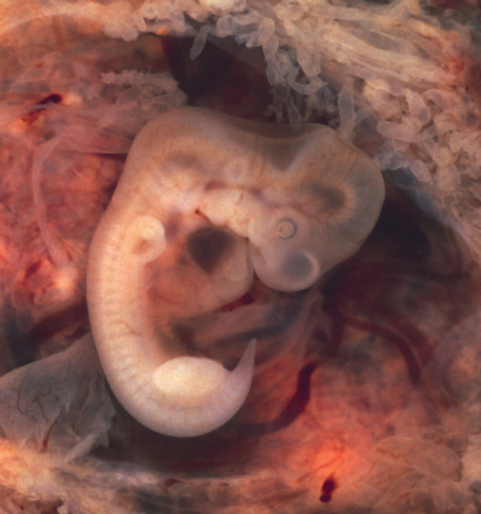
Each upper and lower limb initially develops as a small bulge called a limb bud, which appears on the lateral side of the early embryo. The upper limb bud appears near the end of the fourth week of development, with the lower limb bud appearing shortly after (Figure \(\PageIndex{2}\)) .
Initially, the limb buds consist of a core of mesenchyme covered by a layer of ectoderm. The ectoderm at the end of the limb bud thickens to form a narrow crest called the apical ectodermal ridge. This ridge stimulates the underlying mesenchyme to rapidly proliferate, producing the outgrowth of the developing limb. As the limb bud elongates, cells located farther from the apical ectodermal ridge slow their rates of cell division and begin to differentiate. In this way, the limb develops along a proximal-to-distal axis.
During the sixth week of development, the distal ends of the upper and lower limb buds expand and flatten into a paddle shape. This region will become the hand or foot. The wrist or ankle areas then appear as a constriction that develops at the base of the paddle. Shortly after this, a second constriction on the limb bud appears at the future site of the elbow or knee. Within the paddle, areas of tissue undergo cell death, producing separations between the growing fingers and toes. Also during the sixth week of development, mesenchyme within the limb buds begins to differentiate into hyaline cartilage that will form models of the future limb bones.
The early outgrowth of the upper and lower limb buds initially has the limbs positioned so that the regions that will become the palm of the hand or the bottom of the foot are facing medially toward the body, with the future thumb or big toe both oriented toward the head. During the seventh week of development, the upper limb rotates laterally by 90 degrees, so that the palm of the hand faces anteriorly and the thumb points laterally. In contrast, the lower limb undergoes a 90-degree medial rotation, thus bringing the big toe to the medial side of the foot.
Ossification of Appendicular Bones
All of the girdle and limb bones, except for the clavicle, develop by the process of endochondral ossification. This process begins as the mesenchyme within the limb bud differentiates into hyaline cartilage to form cartilage models for future bones. By the twelfth week, a primary ossification center will have appeared in the diaphysis (shaft) region of the long bones, initiating the process that converts the cartilage model into bone. A secondary ossification center will appear in each epiphysis (expanded end) of these bones at a later time, usually after birth. The primary and secondary ossification centers are separated by the epiphyseal plate, a layer of growing hyaline cartilage. This plate is located between the diaphysis and each epiphysis. It continues to grow and is responsible for the lengthening of the bone. The epiphyseal plate is retained for many years, until the bone reaches its final, adult size, at which time the epiphyseal plate disappears and the epiphysis fuses to the diaphysis. (Seek additional content on ossification in the chapter on bone tissue.)
Small bones, such as the phalanges, will develop only one secondary ossification center and will thus have only a single epiphyseal plate. Large bones, such as the femur, will develop several secondary ossification centers, with an epiphyseal plate associated with each secondary center. Thus, ossification of the femur begins at the end of the seventh week with the appearance of the primary ossification center in the diaphysis, which rapidly expands to ossify the shaft of the bone prior to birth. Secondary ossification centers develop at later times. Ossification of the distal end of the femur, to form the condyles and epicondyles, begins shortly before birth. Secondary ossification centers also appear in the femoral head late in the first year after birth, in the greater trochanter during the fourth year, and in the lesser trochanter between the ages of 9 and 10 years. Once these areas have ossified, their fusion to the diaphysis and the disappearance of each epiphyseal plate follow a reversed sequence. Thus, the lesser trochanter is the first to fuse, doing so at the onset of puberty (around 11 years of age), followed by the greater trochanter approximately 1 year later. The femoral head fuses between the ages of 14–17 years, whereas the distal condyles of the femur are the last to fuse, between the ages of 16–19 years. Knowledge of the age at which different epiphyseal plates disappear is important when interpreting radiographs taken of children. Since the cartilage of an epiphyseal plate is less dense than bone, the plate will appear dark in a radiograph image. Thus, a normal epiphyseal plate may be mistaken for a bone fracture.
The clavicle is the one appendicular skeleton bone that does not develop via endochondral ossification. Instead, the clavicle develops through the process of intramembranous ossification. During this process, mesenchymal cells differentiate directly into bone-producing cells, which produce the clavicle directly, without first making a cartilage model. Because of this early production of bone, the clavicle is the first bone of the body to begin ossification, with ossification centers appearing during the fifth week of development. However, ossification of the clavicle is not complete until age 25.
Appendicular System: Congenital Clubfoot
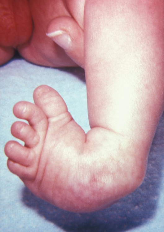
Clubfoot, also known as talipes, is a congenital (present at birth) disorder of unknown cause and is the most common deformity of the lower limb. It affects the foot and ankle, causing the foot to be twisted inward at a sharp angle, like the head of a golf club (Figure \(\PageIndex{2}\)) . Clubfoot has a frequency of about 1 out of every 1,000 births, and is twice as likely to occur in a male child as in a female child. In 50 percent of cases, both feet are affected.
At birth, children with a clubfoot have the heel turned inward and the anterior foot twisted so that the lateral side of the foot is facing inferiorly, commonly due to ligaments or leg muscles attached to the foot that are shortened or abnormally tight. These pull the foot into an abnormal position, resulting in bone deformities. Other symptoms may include bending of the ankle that lifts the heel of the foot and an extremely high foot arch. Due to the limited range of motion in the affected foot, it is difficult to place the foot into the correct position. Additionally, the affected foot may be shorter than normal, and the calf muscles are usually underdeveloped on the affected side. Despite the appearance, this is not a painful condition for newborns. However, it must be treated early to avoid future pain and impaired walking ability.
Although the cause of clubfoot is idiopathic (unknown), evidence indicates that fetal position within the uterus is not a contributing factor. Genetic factors are involved, because clubfoot tends to run within families. Cigarette smoking during pregnancy has been linked to the development of clubfoot, particularly in families with a history of clubfoot.
Previously, clubfoot required extensive surgery. Today, 90 percent of cases are successfully treated without surgery using new corrective casting techniques. The best chance for a full recovery requires that clubfoot treatment begin during the first 2 weeks after birth. Corrective casting gently stretches the foot, which is followed by the application of a holding cast to keep the foot in the proper position. This stretching and casting is repeated weekly for several weeks. In severe cases, surgery may also be required, after which the foot typically remains in a cast for 6 to 8 weeks. After the cast is removed following either surgical or nonsurgical treatment, the child will be required to wear a brace part-time (at night) for up to 4 years. In addition, special exercises will be prescribed, and the child must also wear special shoes. Close monitoring by the parents and adherence to postoperative instructions are imperative in minimizing the risk of relapse.
Despite these difficulties, treatment for clubfoot is usually successful, and the child will grow up to lead a normal, active life. Numerous examples of individuals born with a clubfoot who went on to successful careers include Dudley Moore (comedian and actor), Damon Wayans (comedian and actor), Troy Aikman (three-time Super Bowl-winning quarterback), Kristi Yamaguchi (Olympic gold medalist in figure skating), Mia Hamm (two-time Olympic gold medalist in soccer), and Charles Woodson (Heisman trophy and Super Bowl winner).
Concept Review
Formation of the axial skeleton begins during early embryonic development with the appearance of the rod-like notochord along the dorsal length of the early embryo. Repeating, paired blocks of tissue called somites then appear along either side of notochord. As the somites grow, they split into parts, one of which is called a sclerotome. This consists of mesenchyme, the embryonic tissue that will become the bones, cartilages, and connective tissues of the body.
Mesenchyme in the head region will produce the bones of the skull via two different mechanisms. The bones of the brain case arise via intramembranous ossification in which embryonic mesenchyme tissue converts directly into bone. At the time of birth, these bones are separated by fontanelles, wide areas of fibrous connective tissue. As the bones grow, the fontanelles are reduced to sutures, which allow for continued growth of the skull throughout childhood. In contrast, the cranial base and facial bones are produced by the process of endochondral ossification, in which mesenchyme tissue initially produces a hyaline cartilage model of the future bone. The cartilage model allows for growth of the bone and is gradually converted into bone over a period of many years.
The vertebrae, ribs, and sternum also develop via endochondral ossification. Mesenchyme accumulates around the notochord and produces hyaline cartilage models of the vertebrae. The notochord largely disappears, but remnants of the notochord contribute to formation of the intervertebral discs. In the thorax region, a portion of the vertebral cartilage model splits off to form the ribs. These then become attached anteriorly to the developing cartilage model of the sternum. Growth of the cartilage models for the vertebrae, ribs, and sternum allow for enlargement of the thoracic cage during childhood and adolescence. The cartilage models gradually undergo ossification and are converted into bone.
The bones of the appendicular skeleton arise from embryonic mesenchyme. Limb buds appear at the end of the fourth week. The apical ectodermal ridge, located at the end of the limb bud, stimulates growth and elongation of the limb. During the sixth week, the distal end of the limb bud becomes paddle-shaped, and selective cell death separates the developing fingers and toes. At the same time, mesenchyme within the limb bud begins to differentiate into hyaline cartilage, forming models for future bones. During the seventh week, the upper limbs rotate laterally and the lower limbs rotate medially, bringing the limbs into their final positions.
Endochondral ossification, the process that converts the hyaline cartilage model into bone, begins in most appendicular bones by the twelfth fetal week. This begins as a primary ossification center in the diaphysis, followed by the later appearance of one or more secondary ossifications centers in the regions of the epiphyses. Each secondary ossification center is separated from the primary ossification center by an epiphyseal plate. Continued growth of the epiphyseal plate cartilage provides for bone lengthening. Disappearance of the epiphyseal plate is followed by fusion of the bony components to form a single, adult bone.
The clavicle develops via intramembranous ossification, in which mesenchyme is converted directly into bone tissue. Ossification within the clavicle begins during the fifth week of development and continues until 25 years of age.
Review Questions
Q. Which event takes place during the seventh week of development?
A. appearance of the upper and lower limb buds
B. flattening of the distal limb bud into a paddle shape
C. the first appearance of hyaline cartilage models of future bones
D. the rotation of the limbs
- Answer
-
Answer: D
Q. During endochondral ossification of a long bone, ________.
A. a primary ossification center will develop within the epiphysis
B. mesenchyme will differentiate directly into bone tissue
C. growth of the epiphyseal plate will produce bone lengthening
D. all epiphyseal plates will disappear before birth
- Answer
-
Answer: C
Q. The clavicle ________.
A. develops via intramembranous ossification
B. develops via endochondral ossification
C. is the last bone of the body to begin ossification
D. is fully ossified at the time of birth
- Answer
-
Answer: A
Q. Embryonic development of the axial skeleton involves ________.
A. intramembranous ossification, which forms the facial bones.
B. endochondral ossification, which forms the ribs and sternum
C. the notochord, which produces the cartilage models for the vertebrae
D. the formation of hyaline cartilage models, which give rise to the flat bones of the skull
- Answer
-
Answer: B
Q. A fontanelle ________.
A. is the cartilage model for a vertebra that later is converted into bone
B. gives rise to the facial bones and vertebrae
C. is the rod-like structure that runs the length of the early embryo
D. is the area of fibrous connective tissue found at birth between the brain case bones
- Answer
-
Answer: D
Critical Thinking Questions
Q. How can a radiograph of a child’s femur be used to determine the approximate age of that child?
- Answer
-
A. A radiograph (X-ray image) of a child’s femur will show the epiphyseal plates associated with each secondary ossification center. These plates of hyaline cartilage will appear dark in comparison to the white imaging of the ossified bone. Since each epiphyseal plate appears and disappears at a different age, the presence or absence of these plates can be used to give an approximate age for the child. For example, the epiphyseal plate located at the base of the lesser trochanter of the femur appears at age 9–10 years and disappears at puberty (approximately 11 years of age). Thus, a child’s radiograph that shows the presence of the lesser trochanter epiphyseal plate indicates an approximate age of 10 years.
Q. How does the development of the clavicle differ from the development of other appendicular skeleton bones?
- Answer
-
A. Unlike other bones of the appendicular skeleton, the clavicle develops by the process of intramembranous ossification. In this process, embryonic mesenchyme accumulates at the site of the future bone and then differentiates directly into bone-producing tissue. Because of this direct and early production of bone, the clavicle is the first bone of the skeleton to begin to ossify. However, the growth and enlargement of the clavicle continues throughout childhood and adolescence, and thus, it is not fully ossified until 25 years of age.
Q. Discuss the processes by which the brain-case bones of the skull are formed and grow during skull enlargement.
- Answer
-
A. The brain-case bones that form the top and sides of the skull are produced by intramembranous ossification. In this, mesenchyme from the sclerotome portion of the somites accumulates at the site of the future bone and differentiates into bone-producing cells. These generate areas of bone that are initially separated by wide regions of fibrous connective tissue called fontanelles. After birth, as the bones enlarge, the fontanelles disappear. However, the bones remain separated by the sutures, where bone and skull growth can continue until the adult size is obtained.
Q. Discuss the process that gives rise to the base and facial bones of the skull.
- Answer
-
A. The facial bones and base of the skull arise via the process of endochondral ossification. This process begins with the localized accumulation of mesenchyme tissue at the sites of the future bones. The mesenchyme differentiates into hyaline cartilage, which forms a cartilage model of the future bone. The cartilage allows for growth and enlargement of the model. It is gradually converted into bone over time.
Q. Discuss the development of the vertebrae, ribs, and sternum.
- Answer
-
A. The vertebrae, ribs, and sternum all develop via the process of endochondral ossification. Mesenchyme tissue from the sclerotome portion of the somites accumulates on either side of the notochord and produces hyaline cartilage models for each vertebra. In the thorax region, a portion of this cartilage model splits off to form the ribs. Similarly, mesenchyme forms cartilage models for the right and left halves of the sternum. The ribs then become attached anteriorly to the developing sternum, and the two halves of sternum fuse together. Ossification of the cartilage model into bone occurs within these structures over time. This process continues until each is converted into bone, except for the sternal ends of the ribs, which remain as the costal cartilages.
Glossary
- apical ectodermal ridge
- enlarged ridge of ectoderm at the distal end of a limb bud that stimulates growth and elongation of the limb
- fontanelle
- expanded area of fibrous connective tissue that separates the brain case bones of the skull prior to birth and during the first year after birth
- limb bud
- small elevation that appears on the lateral side of the embryo during the fourth or fifth week of development, which gives rise to an upper or lower limb
- notochord
- rod-like structure along dorsal side of the early embryo; largely disappears during later development but does contribute to formation of the intervertebral discs
- sclerotome
- medial portion of a somite consisting of mesenchyme tissue that will give rise to bone, cartilage, and fibrous connective tissues
- somite
- one of the paired, repeating blocks of tissue located on either side of the notochord in the early embryo
OpenStax Anatomy & Physiology (CC BY 4.0). Access for free at https://openstax.org/books/anatomy-and-physiology