|

- Image Credit
-
"Anterior Skull -Bones Atlas" by John Polos is licensed under CC BY-NC-SA 4.0, modification of original by BlueLink is licensed under CC BY-NC 4.0 with notification of the original authors.
|
- Answer Key (click to show/hide answers)
-
A - frontal bone
B - nasal bone
C - lacrimal bone
D - sphenoid bone
E - zygomatic bone
F - maxilla (maxillary bone)
G - ethmoid bone
H - inferior nasal concha
I - vomer
J - mandible
|
|

- Image Credit
-
"Anterior Skull -Landmarks Atlas" by John Polos is licensed under CC BY-NC-SA 4.0, modification of original by BlueLink is licensed under CC BY-NC 4.0 with notification of the original authors.
|
- Answer Key (click to show/hide answers)
-
A - supraorbital foramen
B - optic canal
C - supraorbital fissure
D - infraorbital fissure
E - infraorbital foramen
F - alveolar process
G - perpendicular plate
H - inferior nasal concha
|
|

- Image Credit
-
"Skull Bones Posterolateral View - Atlas" by Jennifer Lange is licensed under CC BY-NC-SA 4.0, modification of image from Anatomy Standard.
|
- Answer Key (click to show/hide answers)
-
A - occipital bone
B - parietal bone
C - frontal bone
D - temporal bone
E - sphenoid bone
F - mandible
G - maxilla
H - zygomatic bone
I - nasal bone
|
|

- Image Credit
-
"Skull Landmarks Posterolateral View - Atlas" by Jennifer Lange is licensed under CC BY-NC-SA 4.0, modification of image from Anatomy Standard.
|
- Answer Key (click to show/hide answers)
-
A - lambdoid suture
B - mastoid process
C - external acoustic meatus
D - styloid process
E - squamousal suture
F - zygomatic process
G - mandibular condyle
H - coronal suture
I - temporal process
|
|

- Image Credit
-
"Skull Interior Landmarks - Atlas" by Jennifer Lange is licensed under CC BY-NC-SA 4.0.
|
- Answer Key (click to show/hide answers)
-
A - cribriform plate
B - optic groove
C - crista galli
D - hypophyseal fossa (part of sella turcica)
|
|

- Image Credit
-
"Skull Foramina Interior View - Atlas" by Jennifer Lange is licensed under CC BY-NC-SA 4.0.
|
- Answer Key (click to show/hide answers)
-
A - foramen ovale
B - carotid canal
C - foramen magnum
D - optic canal
E - foramen lacerum
|
|

- Image Credit
-
"Inferior Skull -Bones Atlas" by John Polos is licensed under CC BY-NC-SA 4.0, modification of original by BlueLink is licensed under CC BY-NC 4.0 with notification of the original authors.
|
- Answer Key (click to show/hide answers)
-
A - maxilla (maxillary bone)
B - palatine bone
C - zygomatic bone
D - sphenoid bone
E - temporal bone
F - occipital bone
H - vomer
|
|

- Image Credit
-
"Inferior Skull -Landmarks Atlas" by John Polos is licensed under CC BY-NC-SA 4.0, modification of original by BlueLink is licensed under CC BY-NC 4.0 with notification of the original authors.
|
- Answer Key (click to show/hide answers)
-
A - temporal process of zygomatic bone
B - zygomatic process of temporal bone
C - mastoid process
D - styloid process
E - carotid canal
F - occipital condyle
G - foramen magnum
H - foramen lacerum
I - foramen ovale
J - foramen ovale repeated
K - foramen spinosum
L - mandibular fossa
|
|

- Image Credit
-
"Cervical Vertebra C2 - image for atlas" by Jennifer Lange is licensed under CC BY-NC-SA 4.0, modification of image from Anatomy Standard.
|
- Answer Key (click to show/hide answers)
-
1 - anterior arch
2 - superior articular facet
3 - transverse foramen
4 - posterior arch
|
| |
|
|

- Image Credit
-
"Cervical Vertebra C2 - image for atlas" by Jennifer Lange is licensed under CC BY-NC-SA 4.0, modification of image from Anatomy Standard.
|
- Answer Key (click to show/hide answers)
-
A - superior articular facet
B - vertebral body
C - transverse process
D - transverse foramen
E - spinous process
F - dens (odontoid process)
|
| |
|
|

- Image Credit
-
"Cervical Vertebra C5 - image for atlas" by John Polos is licensed under CC BY-NC-SA 4.0, modification of image from Anatomy Standard.
|
- Answer Key (click to show/hide answers)
-
1 - vertebral body
2 - superior articular facet
3 - transverse foramen
4 - spinous process
5 - vertebral foramen
6 - lamina portion of arch
|
| |
|
|

- Image Credit
-
"Thoracic vertebra - image for atlas" by John Polos is licensed under CC BY-NC-SA 4.0, modification of image from Anatomy Standard.
|
- Answer Key (click to show/hide answers)
-
1 - vertebral body
2 - superior articular facet
3 - transverse foramen
4 - spinous process
5 - demifacets (superior and inferior)
6 - transverse (costal) facet
|
| |
|
|

- Image Credit
-
"Lumbar Vertebra - image for atlas" by John Polos is licensed under CC BY-NC-SA 4.0, modification of image from Anatomy Standard.
|
- Answer Key (click to show/hide answers)
-
1 - vertebral body
2 - superior articular facet
3 - transverse foramen
4 - spinous process
5 - pedicel of vertebral arch
6 - inferior articular process
|
| |
|
|
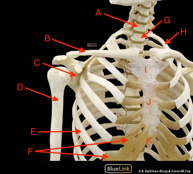
- Image Credit
-
"Thoracic Cage, Pectoral Girdle, and Humerus Anterior View - Atlas" by Jennifer Lange is licensed under CC BY-NC-SA 4.0, modification of original by BlueLink is licensed under CC BY-NC 4.0 with notification of the original authors.
|
- Answer Key (click to show/hide answers)
-
A - cervical vertebra #7
B - clavicle
C - scapula
D - humerus
E - rib #6, right
F - costal cartilages
G - thoracic vertebra #1
H - rib #1, left
I - manubrium of the sternum
J - body of the sternum
K - xiphoid process of the sternum
|
|

- Image Credit
-
"Thoracic Cage, Pectoral Girdle, and Humerus Posterior View - Atlas" by Jennifer Lange is licensed under CC BY-NC-SA 4.0, modification of original by BlueLink is licensed under CC BY-NC 4.0 with notification of the original authors.
|
- Answer Key (click to show/hide answers)
-
A - bifed spinous process
B - transverse process
C - tubercle of the rib
D - supraspinous fossa
E - spine of the scapula
F - acromial end of the clavicle
G - acromion process
H - head of the humerus
I - greater tubercle
J - glenoid fossa (cavity)
K - infraspinous fossa
L - head of the rib
|
|

- Image Credit
-
"Forearm and Hand Bones - Atlas" by Jennifer Lange is licensed under CC BY-NC-SA 4.0, modification of original by DrJanaOfficial.
|
- Answer Key (click to show/hide answers)
-
A - humerus
B - radius
C - ulna
D - carpals
E - metacarpals
F - phalanges
|
|

- Image Credit
-
"Anterior Scapula - Atlas" by John Polos is licensed under CC BY-NC-SA 4.0, modification of original by BlueLink is licensed under CC BY-NC 4.0 with notification of the original authors.
|
- Answer Key (click to show/hide answers)
-
1 - coracoid process
2 - glenoid fossa (cavity)
3 - acromion or acromial process
4 - lateral border
5 - inferior angle
6 - medial border
7 - superior angle
8 - subscapular fossa
|
|

- Image Credit
-
"Posterior Scapula - Atlas" by John Polos is licensed under CC BY-NC-SA 4.0, modification of original by BlueLink is licensed under CC BY-NC 4.0 with notification of the original authors.
|
- Answer Key (click to show/hide answers)
-
1 - coracoid process
2 - glenoid fossa (cavity)
3 - acromion or acromial process
4 - lateral border
5 - inferior angle
6 - medial border
7 - superior angle
8 - Spine (of scapula)
9 - supraspinous fossa
10 - infraspinous fossa
|
|

- Image Credit
-
"Lateral Scapula - Atlas" by John Polos is licensed under CC BY-NC-SA 4.0, modification of original by BlueLink is licensed under CC BY-NC 4.0 with notification of the original authors.
|
- Answer Key (click to show/hide answers)
-
1 - coracoid process
2 - glenoid fossa (cavity)
3 - acromion or acromial process
4 - lateral border
5 - supraspinous fossa
|
|

- Image Credit
-
"Proximal Humerus- Atlas" by John Polos is licensed under CC BY-NC-SA 4.0, modification of original by BlueLink is licensed under CC BY-NC 4.0 with notification of the original authors.
|
- Answer Key (click to show/hide answers)
-
1 - greater tubercle
2 - intertubercular groove (sulcus)
3 - lesser tubercle
4 - Head
|
|

- Image Credit
-
"Distal Humerus - Atlas" by John Polos is licensed under CC BY-NC-SA 4.0, modification of original by BlueLink is licensed under CC BY-NC 4.0 with notification of the original authors.
|
- Answer Key (click to show/hide answers)
-
1 - lateral epicondyle of humerus
2 - capitulum
3 - trochlea anterior view
4 - olecranon fossa
5 - medial epicondyle of humerus
6 - coronoid fossa
7 - radial fossa
8 - subscapular fossa
|
|
- Image Credit
-
"Anterior Radius - Atlas" by John Polos is licensed under CC BY-NC-SA 4.0, modification of original by BlueLink is licensed under CC BY-NC 4.0 with notification of the original authors.
|
- Answer Key (click to show/hide answers)
-
1 - Head
2 - Neck
3 - radial tuberosity
4 - interosseous border of the radius
5 - ulnar notch
6 - styloid process of radius
|
|

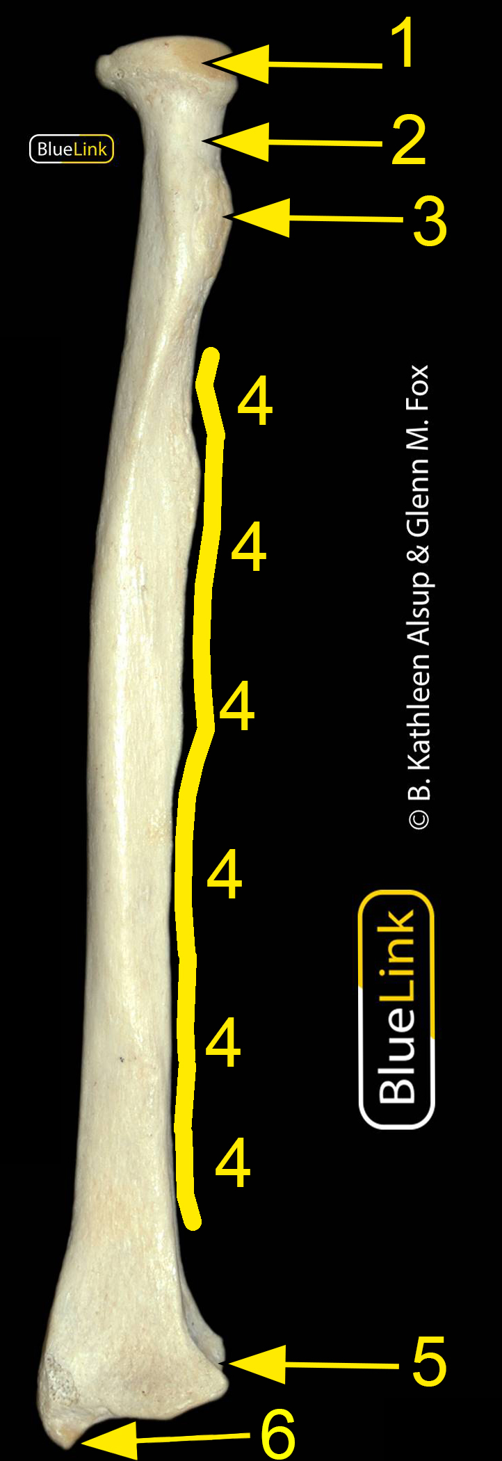
- Image Credit
-
"Ulna - Atlas" by John Polos is licensed under CC BY-NC-SA 4.0, modification of original by BlueLink is licensed under CC BY-NC 4.0 with notification of the original authors.
|
- Answer Key (click to show/hide answers)
-
1 - olecranon process
2 - trochlear notch
3 - coronoid process
4 - radial notch of ulna
5 - styloid process of ulna
6 - head of ulna
|
-
 - Image Credit
-
"Wrist and Hand Bones - Atlas" by Jennifer Lange is licensed under CC BY-NC-SA 4.0, modification of original by DrJanaOfficial.
|
- Answer Key (click to show/hide answers)
-
A - scaphoid
B - trapezius
C - trapezoid
D - first distal phalange, right hand
E - metacarpal #2, right hand
F - second middle phalange, right hand
G - third distal phalange, right hand
H - lunate
I - triquetrum
J - pisiform
K - hamate
L - metacarpal #5, right hand
M - fifth proximal phalange, right hand
|
|
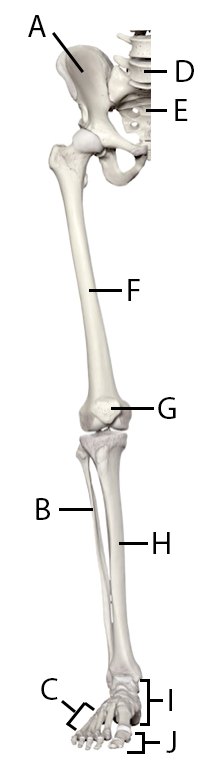
- Image Credit
-
"Lower Limb Bones - Atlas" by Jennifer Lange is licensed under CC BY-NC-SA 4.0, modification of image from Anatomy Standard.
|
- Answer Key (click to show/hide answers)
-
A - os coxa
B - fibula
C - metatarsals
D - lumbar vertebra #5
E - sacrum
F - femur
G - patella
H - tibia
I - tarsals
J - phalanges
|
| |
|
| |
|
|

- Image Credit
-
"Anterior hip - Atlas" by John Polos is licensed under CC BY-NC-SA 4.0, modification of original by BlueLink is licensed under CC BY-NC 4.0 with notification of the original authors.
|
- Answer Key (click to show/hide answers)
-
1 - anterior superior iliac spine
2 - anterior inferior iliac spine
3 - acetabulum
4 - pubic tubercle
5 - illiac crest
6 - Superior articulating facet/process
7 - obturator foramen
|
|

- Image Credit
-
"Posterior Femur - Atlas" by John Polos is licensed under CC BY-NC-SA 4.0, modification of original by BlueLink is licensed under CC BY-NC 4.0 with notification of the original authors.
|
- Answer Key (click to show/hide answers)
-
1 - head of femur
2 - neck of femur
3 - lesser trochanter
4 - greater trochanter
5 - linea aspera
6 - medial epicondyle
7 - medial condyle
8 - lateral condyle
9 - lateral epicondyle
|
|

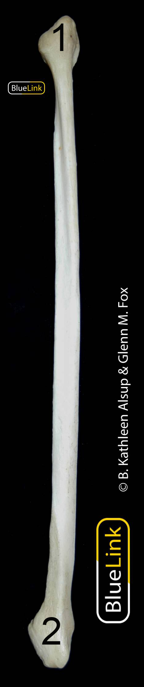
- Image Credit
-
"Anterior Tibia - Atlas" by John Polos is licensed under CC BY-NC-SA 4.0, modification of original by BlueLink is licensed under CC BY-NC 4.0 with notification of the original authors.
|
- Answer Key (click to show/hide answers)
-
1 - lateral condyle
2 - tibial tuberosity
3 - medial condyle
4 - medial malleolus
|
|
- Image Credit
-
"Fibula- Atlas" by John Polos is licensed under CC BY-NC-SA 4.0, modification of original by BlueLink is licensed under CC BY-NC 4.0 with notification of the original authors.
|
- Answer Key (click to show/hide answers)
-
1 - head of fibula
2 - lateral malleolus
|
|

- Image Credit
-
"Foot - Atlas" by John Polos is licensed under CC BY-NC-SA 4.0, modification of original by BlueLink is licensed under CC BY-NC 4.0 with notification of the original authors.
|
- Answer Key (click to show/hide answers)
-
1 - calcaneus
2 - talus
3 - navicular
4 - medial cuneiform
5 - intermediate cuneiform
6 - lateral cuneiform
7 - cuboid
8 - metatarsal 1 (I)
9 - proximal phalange
10 - distal phalange
11 - intermediate phalange
|
