
13.3: Hearing and Balance
- Page ID
- 63449
\( \newcommand{\vecs}[1]{\overset { \scriptstyle \rightharpoonup} {\mathbf{#1}} } \)
\( \newcommand{\vecd}[1]{\overset{-\!-\!\rightharpoonup}{\vphantom{a}\smash {#1}}} \)
\( \newcommand{\id}{\mathrm{id}}\) \( \newcommand{\Span}{\mathrm{span}}\)
( \newcommand{\kernel}{\mathrm{null}\,}\) \( \newcommand{\range}{\mathrm{range}\,}\)
\( \newcommand{\RealPart}{\mathrm{Re}}\) \( \newcommand{\ImaginaryPart}{\mathrm{Im}}\)
\( \newcommand{\Argument}{\mathrm{Arg}}\) \( \newcommand{\norm}[1]{\| #1 \|}\)
\( \newcommand{\inner}[2]{\langle #1, #2 \rangle}\)
\( \newcommand{\Span}{\mathrm{span}}\)
\( \newcommand{\id}{\mathrm{id}}\)
\( \newcommand{\Span}{\mathrm{span}}\)
\( \newcommand{\kernel}{\mathrm{null}\,}\)
\( \newcommand{\range}{\mathrm{range}\,}\)
\( \newcommand{\RealPart}{\mathrm{Re}}\)
\( \newcommand{\ImaginaryPart}{\mathrm{Im}}\)
\( \newcommand{\Argument}{\mathrm{Arg}}\)
\( \newcommand{\norm}[1]{\| #1 \|}\)
\( \newcommand{\inner}[2]{\langle #1, #2 \rangle}\)
\( \newcommand{\Span}{\mathrm{span}}\) \( \newcommand{\AA}{\unicode[.8,0]{x212B}}\)
\( \newcommand{\vectorA}[1]{\vec{#1}} % arrow\)
\( \newcommand{\vectorAt}[1]{\vec{\text{#1}}} % arrow\)
\( \newcommand{\vectorB}[1]{\overset { \scriptstyle \rightharpoonup} {\mathbf{#1}} } \)
\( \newcommand{\vectorC}[1]{\textbf{#1}} \)
\( \newcommand{\vectorD}[1]{\overrightarrow{#1}} \)
\( \newcommand{\vectorDt}[1]{\overrightarrow{\text{#1}}} \)
\( \newcommand{\vectE}[1]{\overset{-\!-\!\rightharpoonup}{\vphantom{a}\smash{\mathbf {#1}}}} \)
\( \newcommand{\vecs}[1]{\overset { \scriptstyle \rightharpoonup} {\mathbf{#1}} } \)
\( \newcommand{\vecd}[1]{\overset{-\!-\!\rightharpoonup}{\vphantom{a}\smash {#1}}} \)
- Describe the gross and microscopic structures responsible for the special senses of hearing and balance
- Trace the pathway of auditory and equilibrium information from the inner ear to the brain
Audition (Hearing)
Hearing, or audition, is the transduction of sound waves into a neural signal that is made possible by the structures of the ear (Figure \(\PageIndex{1}\)).
External Ear
The large, fleshy structure on the lateral aspect of the head is known as the auricle. Some sources will also refer to this structure as the pinna, though that term is more appropriate for a structure that can be moved, such as the external ear of a cat. The C-shaped curves of the auricle direct sound waves toward the auditory canal. The canal enters the skull through the external auditory meatus of the temporal bone. At the end of the auditory canal is the tympanic membrane, or ear drum, which vibrates after it is struck by sound waves. The auricle, ear canal, and tympanic membrane are often referred to as the external ear.
Middle Ear
The middle ear consists of a space, the tympanic cavity, spanned by three small bones called the ossicles. The three ossicles are the malleus, incus, and stapes, which are Latin names that roughly translate to hammer, anvil, and stirrup. The malleus is attached to the tympanic membrane and articulates with the incus. The incus, in turn, articulates with the stapes. The stapes is then attached to the oval window, which is the entrance to the inner ear, where the sound waves will be transduced into a neural signal. The tympanic cavity is connected to the pharynx through the auditory (or Eustachian) tube, which helps equilibrate air pressure across the tympanic membrane by providing and entrance/exit point for air. The tube is normally closed but will pop open when the muscles of the pharynx contract during swallowing or yawning. This helps to equalize the pressure on both sides of the ear drum with change in altitude, such as driving up the mountain or in a plane.

Inner Ear
The inner ear is often described as a bony labyrinth, as it is composed of a series of canals embedded within the temporal bone (Figure \(\PageIndex{2}\)). Within the bony labyrinth are membranes that separate tubes and spaces filled with liquid. This is called the membranous labyrinth. The inner ear has three separate regions, the cochlea, the vestibule, and the semicircular canals. The cochlea is responsible for hearing while the vestibule and semicircular canals are important for balance. The neural signals from these regions are relayed to the brainstem through separate fiber bundles called the cochlear nerve and the vestibular nerve. However, these two distinct bundles travel together from the inner ear to the brainstem as the vestibulocochlear nerve (CN VIII).

Sound is transduced into neural signals within the cochlear region of the inner ear, which contains the sensory neurons of the spiral ganglia. These ganglia are located within the spiral-shaped cochlea of the inner ear. The cochlea is attached to the stapes through the oval window. The oval window is located at the beginning of a fluid-filled tube within the cochlea called the scala vestibuli. The scala vestibuli extends from the oval window, traveling above the cochlear duct, which is the central cavity of the cochlea that contains the sound-transducing neurons. At the uppermost tip of the cochlea, the scala vestibuli curves over the top of the cochlear duct. The fluid-filled tube, now called the scala tympani, returns to the base of the cochlea, this time traveling under the cochlear duct. The scala tympani ends at the round window, which is covered by a membrane that contains the fluid within the scala.
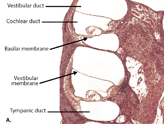
A cross-sectional view of the cochlea shows that the scala vestibuli and scala tympani run along both sides of the cochlear duct (Figure \(\PageIndex{2}\) and Figure \(\PageIndex{3}\)). The cochlear duct contains the spiral organ (organs of Corti), which transduce the wave motion of the two scala into neural signals. The spiral organ lies on top of the basilar membrane, which separates the cochlear duct and the scala tympani. Another membrane called the vestibular membrane separates the cochlear duct from the scala vestibuli.


The spiral organ contains hair cells, which are named for the hair-like stereocilia extending from the cell’s apical surface (Figure \(\PageIndex{5}\)). The stereocilia are an array of microvilli-like structures arranged from tallest to shortest. The stereocilia extend up from the hair cells to the overlying tectorial membrane, which is attached medially to the spiral organ. Protein fibers tether adjacent stereocilia together within each array, such that relative shifts between the basilar membrane and the tectorial membrane bend the stereocilia, causing ion channels to open. This converts the mechanical force of the fluid wave into an electrical signal.

The transmission and transduction of sound require the participation of the majority of the structures of the ear. Each sound wave has a specific frequency, which depends on its pitch, and amplitude, which depends on its loudness. Sound waves are funneled into the ear canal by the auricle and reach the tympanic membrane (Figure \(\PageIndex{6}\)). The vibration of the tympanic membrane is amplified across the ossicles. As vibrations of the ossicles travel through the oval window, the fluid of the scala vestibuli and scala tympani moves in a wave-like motion. The frequency of the fluid waves match the frequencies of the sound waves. The membrane covering the round window will bulge out or pucker in with the movement of the fluid within the scala tympani. As the fluid waves move through the scala vestibuli and scala tympani, the basilar membrane moves at a specific spot of the cochlea, depending on the frequency of the waves. Higher frequency waves move the region of the basilar membrane that is close to the base of the cochlea. Lower frequency waves move the region of the basilar membrane that is near the tip of the cochlea. When the fluid waves from the scala move the basilar membrane, the tectorial membrane slides across the stereocilia. This bends the stereocilia either toward or away from the tallest member of each array of stereocilia. When the stereocilia bend toward the tallest member of their array, tension in the protein tethers opens ion channels in the hair cell membrane. This will electrically change the hair cell membrane, triggering nerve impulses that travel down the afferent nerve fibers attached to the hair cells. When the stereocilia bend toward the shortest member of their array, the tension on the tethers slackens and the ion channels close. The relative movement of different arrays of stereocilia along the length of the basilar membrane allows the brain to perceive frequency, or pitch. The greater force of louder sounds causes an increase in the range of movement of the stereocilia of the hair cells, determining the loudness of a sound.

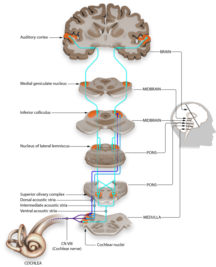
The initial afferent fibers are bipolar neurons. The dendrites of these neurons receive the signal from the hair cell then carry the signal to the cell cell body located in the spiral ganglion. Axons emerging from the spiral ganglion then carry neural signals through the cochlear nerve which travels within the vestibulocochlear nerve (CN VIII), terminating in the cochlear nuclei in the pons(Figure \(\PageIndex{7}\)). From the cochlear nuclei the auditory information travels along multiple pathways, but the primary auditory pathway goes through the inferior colliculus (auditory reflexes) and then through the thalamus and into the primary auditory cortex of the temporal lobe.
Equilibrium (Balance)
Along with audition, the inner ear is responsible for encoding information about equilibrium, the sense of balance. A similar mechanoreceptor—a hair cell with stereocilia—senses head position, head movement, and whether our bodies are in motion. These cells are located in the vestibule and semicircular canals within the inner ear. Vestibule and semicircular canals compose the vestibular system. The vestibule is the central part of the bony labyrinth, and is located posterior to the cochlea and anterior to the semicircular canals. Within the vestibule, the membranous labyrinth forms two chambers, the utricle and saccule, which contain endolymph. The utricle and saccule are interconnected by a narrow endolymphatic duct and their endolymph is confluent with that of the semicircular ducts and cochlear duct. Head position is sensed by the utricle and saccule, whereas head movement is sensed by the semicircular canals.
The utricle and saccule are both largely composed of macula tissue (plural = maculae). The macula is composed of hair cells surrounded by supporting cells. The stereocilia of the hair cells extend into a viscous gel called the otolithic membrane (Figure \(\PageIndex{8}\)). On top of the otolithic membrane is a layer of calcium carbonate crystals, called otoliths. The otoliths essentially make the otolithic membrane top-heavy. The otolithic membrane moves separately from the macula in response to head movements. Tilting the head causes the otolithic membrane to slide over the macula in the direction of gravity. The moving otolithic membrane, in turn, bends the stereocilia, causing the hair cells to elicit electrical changes. The exact position of the head is interpreted by the brain based on the pattern of hair-cell activation.

The semicircular canals are three ring-like extensions of the vestibule. One is oriented in the horizontal plane, whereas the other two are oriented in the vertical plane. The anterior and posterior vertical canals are oriented at approximately 45 degrees relative to the sagittal plane (Figure \(\PageIndex{9}\)). The base of each semicircular canal, where it meets with the vestibule, connects to an enlarged region known as the ampulla. The ampulla contains the hair cells that respond to rotational movement, such as turning the head while saying “no.” The stereocilia of these hair cells extend into the cupula, a gelatinous membrane that attaches to the top of the ampulla. As the head rotates in a plane parallel to the semicircular canal, the fluid lags, deflecting the cupula in the direction opposite to the head movement. The semicircular canals contain several ampullae, with some oriented horizontally and others oriented vertically. By comparing the relative movements of both the horizontal and vertical ampullae, the vestibular system can detect the direction of most head movements within three-dimensional (3-D) space.

The neural signals generated in the vestibule and semicircular canals are transmitted through the vestibular nerve which travels within the vestibulocochlear nerve (CN VIII). The nerve carries the equilibrium information into the brain, through the thalamus, and to cerebral nuclei, brainstem and cerebellum.
Explore the structure of the inner ear in the 3D rendering of an inner ear model.
Concept Review
Hearing and balance belong to the special senses with their receptors present in the inner ear.
The ear is divided in three regions: external, middle and inner ear. In the external ear, the auricle is the fleshy structure that convey sounds into the auditory canal. At the end of the canal is the tympanic membrane that vibrates and transforms sound waves into mechanical waves. The middle ear consists of a space spanned by three small bones called ossicles (malleus, incus, and stapes) that conduct the mechanical waves. The middle ear is connected to the pharynx through the Eustachian (or auditory) tube, which helps equilibrate air pressure across the tympanic membrane. The inner ear is made of a bony labyrinth lined with a membranous labyrinth that separate tubes and spaces. The space between the two labyrinths is filled with a fluid called perilymph. Inside the membranous labyrinth the space is filled with endolymph. The inner ear is responsible for transforming mechanical waves into electrical signals, which are then sent to the brain through the vestibulocochlear nerve (CN VIII).
The cochlea is a spiral-shaped tube, divided into three compartments: the scala vestibuli, scala tympani and cochlear duct. All compartments are filled with endolymph. The scala vestibuli starts at the oval window, curves over the top of the cochlear duct and becomes the scala tympani, that returns to the base of the cochlea, traveling under the cochlear duct and ends at the round window. As vibrations of the ossicles travel through the oval window, the fluid of the scala vestibuli and scala tympani moves in a wave-like motion. The cochlear duct contains several organs of Corti, which transduce the wave motion of the two scala into neural signals. The organs of Corti lie on top of the basilar membrane, which is the side of the cochlear duct located between the organs of Corti and the scala tympani. The organs of Corti contain hair cells, which are named for the hair-like stereocilia extending from the cell’s apical surfaces. The stereocilia extend up from the hair cells to the overlying tectorial membrane, which is attached medially to the organ of Corti. When the pressure waves from the scala move the basilar membrane, the tectorial membrane slides across the stereocilia. This bends the stereocilia either toward or away from the tallest member of each array, which causes an electrical signal to be generated.
The vestibule and the semicircular canals are responsible for the sense of equilibrium. The vestibule is composed of the utricle and saccule. Hair cells are located in maculae of the utricle and saccule. The stereocilia of the hair cells extend into a viscous gel called the otolithic membrane, on top of which is a layer of calcium carbonate crystals, called otoliths. The vestibule senses the linear acceleration of the head and gravity. When the head moves, the otoliths move and the otolithic membrane bends the stereocilia of the hair cells. Hair cells are also located in ampullae at the base of the three semicircular canals. One is oriented in the horizontal plane, whereas the other two are oriented in the vertical plane. The stereocilia of these hair cells extend into the cupula, a membrane that attaches to the top of the ampulla. The semicircular canals sense the head rotation. As the head rotates in a plane parallel to the semicircular canal, the fluid lags, deflecting the cupula in the direction opposite to the head movement, and bending the stereocilia of the hair cells.
Review Questions
Q. What type of receptor cell is involved in the sensations of sound and balance?
A. photoreceptor
B. chemoreceptor
C. mechanoreceptor
D. nociceptor
- Answer
-
C
Glossary
- ampulla
- in the ear, the structure at the base of a semicircular canal that contains the hair cells and cupula for transduction of rotational movement of the head
- audition
- sense of hearing
- auditory canal
- passageway of the external ear that leads to the tympanic membrane; also known as ear canal
- auditory tube
- tube that connects the nasopharynx to the middle ear; also known as Eustachian tube
- auricle
- fleshy external structure of the ear
- basilar membrane
- in the ear, the floor of the cochlear duct on which the organ of Corti sits
- bony labyrinth
- complex cavities in the inner ear made by bones
- brainstem
- region of the adult brain that includes the midbrain, pons, and medulla oblongata and develops from the mesencephalon, metencephalon, and myelencephalon of the embryonic brain
- cerebellum
- region of the adult brain connected primarily to the pons that developed from the metencephalon (along with the pons) and is largely responsible for comparing information from the cerebrum with sensory feedback from the periphery through the spinal cord
- cerebral nuclei
- deep gray matter of the cerebrum
- cochlea
- auditory portion of the inner ear containing structures to transduce sound stimuli
- cochlear duct
- space within the auditory portion of the inner ear that contains the spiral organ and is adjacent to the scala tympani and scala vestibuli on either side
- cochlear nerve
- branch of the vestibulocochlear nerve projecting from the cochlea
- cupula
- specialized structure within the base of a semicircular canal that bends the stereocilia of hair cells when the head rotates by way of the relative movement of the enclosed fluid
- ear canal
- passageway of the external ear that leads to the tympanic membrane; also known as auditory canal
- endolymph
- fluid in the membranous labyrinth of the ear
- equilibrium
- sense of balance that includes sensations of position and movement of the head
- Eustachian tube
- tube that connects the nasopharynx to the middle ear; also known as auditory tube
- external ear
- structures on the lateral surface of the head, including the auricle and the ear canal back to the tympanic membrane
- hair cells
- mechanoreceptor cells found in the inner ear that transduce stimuli for the senses of hearing and balance
- incus
- (also, anvil) ossicle of the middle ear that connects the malleus to the stapes
- inner ear
- structure within the temporal bone that contains the sensory apparati of hearing and balance
- macula
- enlargement at the base of a semicircular canal at which transduction of equilibrium stimuli takes place within the ampulla
- malleus
- (also, hammer) ossicle that is directly attached to the tympanic membrane
- membranous labyrinth
- membranes of the the inner ear that line the bony labyrinth
- middle ear
- space within the temporal bone between the ear canal and bony labyrinth where the ossicles amplify sound waves from the tympanic membrane to the oval window
- organ of Corti
- structure in the cochlea in which hair cells transduce movements from sound waves into electrochemical signals (also spiral organ)
- ossicles
- three small bones in the middle ear
- otolith
- layer of calcium carbonate crystals located on top of the otolithic membrane
- otolithic membrane
- gelatinous substance in the utricle and saccule of the inner ear that contains calcium carbonate crystals and into which the stereocilia of hair cells are embedded
- oval window
- membrane at the base of the cochlea where the stapes attaches, marking the beginning of the scala vestibuli
- perilymph
- fluid between the membranous labyrinth of the ear and the bony labyrinth
- primary auditory cortex
- region of the cerebral cortex within the temporal lobe responsible for the perception of sounds
- round window
- membrane that marks the end of the scala tympani
- saccule
- structure of the inner ear responsible for transducing linear acceleration in the vertical plane
- scala tympani
- portion of the cochlea that extends from the apex to the round window
- scala vestibuli
- portion of the cochlea that extends from the oval window to the apex
- semicircular canals
- structures within the inner ear responsible for transducing rotational movement information
- spiral ganglion
- location of neuronal cell bodies that transmit auditory information along the eighth cranial nerve
- spiral organ
- structure in the cochlea in which hair cells transduce movements from sound waves into electrochemical signals (also organ of Corti)
- stapes
- (also, stirrup) ossicle of the middle ear that is attached to the inner ear
- stereocilia
- array of apical membrane extensions in a hair cell that transduce movements when they are bent
- tectorial membrane
- component of the organ of Corti that lays over the hair cells, into which the stereocilia are embedded
- thalamus
- major region of the diencephalon that is responsible for relaying information between the cerebrum and the hindbrain, spinal cord, and periphery
- tympanic membrane
- ear drum
- utricle
- structure of the inner ear responsible for transducing linear acceleration in the horizontal plane
- vestibular membrane
- membrane separating the cochlear duct and scala vestibuli
- vestibular nerve
- branch of the vestibulocochlear nerve projecting from the vestibule
- vestibular apparatus
- system composed of the vestibule and semicircular canals to sense equilibrium
- vestibule
- in the ear, the portion of the inner ear responsible for the sense of equilibrium
- vestibulocochlear nerve
- eighth cranial nerve; responsible for the sensations of hearing and balance
Contributors and Attributions
OpenStax Anatomy & Physiology (CC BY 4.0). Access for free at https://openstax.org/books/anatomy-and-physiology