
15.4: Thyroid Gland and Parathyroid Glands
- Page ID
- 63461
\( \newcommand{\vecs}[1]{\overset { \scriptstyle \rightharpoonup} {\mathbf{#1}} } \)
\( \newcommand{\vecd}[1]{\overset{-\!-\!\rightharpoonup}{\vphantom{a}\smash {#1}}} \)
\( \newcommand{\id}{\mathrm{id}}\) \( \newcommand{\Span}{\mathrm{span}}\)
( \newcommand{\kernel}{\mathrm{null}\,}\) \( \newcommand{\range}{\mathrm{range}\,}\)
\( \newcommand{\RealPart}{\mathrm{Re}}\) \( \newcommand{\ImaginaryPart}{\mathrm{Im}}\)
\( \newcommand{\Argument}{\mathrm{Arg}}\) \( \newcommand{\norm}[1]{\| #1 \|}\)
\( \newcommand{\inner}[2]{\langle #1, #2 \rangle}\)
\( \newcommand{\Span}{\mathrm{span}}\)
\( \newcommand{\id}{\mathrm{id}}\)
\( \newcommand{\Span}{\mathrm{span}}\)
\( \newcommand{\kernel}{\mathrm{null}\,}\)
\( \newcommand{\range}{\mathrm{range}\,}\)
\( \newcommand{\RealPart}{\mathrm{Re}}\)
\( \newcommand{\ImaginaryPart}{\mathrm{Im}}\)
\( \newcommand{\Argument}{\mathrm{Arg}}\)
\( \newcommand{\norm}[1]{\| #1 \|}\)
\( \newcommand{\inner}[2]{\langle #1, #2 \rangle}\)
\( \newcommand{\Span}{\mathrm{span}}\) \( \newcommand{\AA}{\unicode[.8,0]{x212B}}\)
\( \newcommand{\vectorA}[1]{\vec{#1}} % arrow\)
\( \newcommand{\vectorAt}[1]{\vec{\text{#1}}} % arrow\)
\( \newcommand{\vectorB}[1]{\overset { \scriptstyle \rightharpoonup} {\mathbf{#1}} } \)
\( \newcommand{\vectorC}[1]{\textbf{#1}} \)
\( \newcommand{\vectorD}[1]{\overrightarrow{#1}} \)
\( \newcommand{\vectorDt}[1]{\overrightarrow{\text{#1}}} \)
\( \newcommand{\vectE}[1]{\overset{-\!-\!\rightharpoonup}{\vphantom{a}\smash{\mathbf {#1}}}} \)
\( \newcommand{\vecs}[1]{\overset { \scriptstyle \rightharpoonup} {\mathbf{#1}} } \)
\( \newcommand{\vecd}[1]{\overset{-\!-\!\rightharpoonup}{\vphantom{a}\smash {#1}}} \)
- Describe the location and anatomy of the thyroid gland
- Explain the role of thyroid hormones in the regulation of basal metabolism
- Identify the hormone produced by the parafollicular cells of the thyroid
- Describe the location and structure of the parathyroid glands
- Describe the hormonal control of blood calcium levels
Thyroid Gland
The thyroid gland, a butterfly-shaped organ, is located anterior to the trachea, just inferior to the larynx (Figure \(\PageIndex{1}\)). The medial region, called the isthmus, is flanked by wing-shaped left and right lobes. Each of the thyroid lobes has a pair of parathyroid glands embedded on its posterior surface. The tissue of the thyroid gland is composed mostly of thyroid follicles lined with simple cuboidal epithelium. The follicles are made up of a central cavity filled with a sticky fluid called colloid. Surrounded by a wall of epithelial follicle cells, the colloid is the center of thyroid hormone production, and that production is dependent on the essential and unique component of thyroid hormones: iodine.

Regulation of Thyroid Hormone Synthesis
Thyroglobulin is the precursor to two thyroid hormones: triiodothyronine (T3) with three iodines and thyroxine (T4) with four iodines. Thyroglobulin is produced by the follicle cells and secreted into the colloid where the iodines are attached to form T3 and T4. Ninety-nine percent of circulating T3 and T4 is bound to specialized transport proteins called thyroxine-binding globulins (TBGs), to albumin, or to other plasma proteins. This “packaging” prevents their free diffusion into body cells. When blood levels of T3 and T4 begin to decline, bound T3 and T4 are released from these plasma proteins, now referred to as "unbound", and may readily cross the membrane of target cells. T3 is more potent than T4, and many cells convert T4 to T3 through the removal of an iodine atom.
The release of T3 and T4 from the thyroid gland is regulated by thyroid-stimulating hormone (TSH). As shown in Figure \(\PageIndex{2}\), low blood levels of T3 and T4 stimulate the release of thyrotropin-releasing hormone (TRH) from the hypothalamus, which triggers secretion of TSH from the anterior pituitary. In turn, TSH stimulates the thyroid gland to secrete T3 and T4. The levels of TRH, TSH, T3, and T4 are regulated by a negative feedback system in which increasing levels of T3 and T4 decrease the production and secretion of TSH.

Functions of Thyroid Hormones
The thyroid hormones, T3 and T4, are often referred to as metabolic hormones because their levels influence the body’s basal metabolic rate, the amount of energy used by the body at rest. When T3 and T4 bind to intracellular receptors located on the mitochondria, they cause an increase in nutrient breakdown and the use of oxygen to produce ATP. In addition, T3 and T4 initiate the transcription of genes involved in glucose oxidation. Although these mechanisms prompt cells to produce more ATP, the process is inefficient, and an abnormally increased level of heat is released as a byproduct of these reactions. This so-called calorigenic effect (calor- = “heat”) raises body temperature.
Adequate levels of thyroid hormones are also required for protein synthesis and for fetal and childhood tissue development and growth. They are especially critical for normal development of the nervous system both in utero and in early childhood, and they continue to support neurological function in adults. As noted earlier, these thyroid hormones have a complex interrelationship with reproductive hormones, and deficiencies can influence libido, fertility, and other aspects of reproductive function. Finally, thyroid hormones increase the body’s sensitivity to catecholamines (epinephrine and norepinephrine) from the adrenal medulla by upregulation of receptors in the blood vessels. When levels of T3 and T4 hormones are excessive, this effect accelerates the heart rate, strengthens the heartbeat, and increases blood pressure. Because thyroid hormones regulate metabolism, heat production, protein synthesis, and many other body functions, thyroid disorders can have severe and widespread consequences.
Endocrine System: Iodine Deficiency, Hypothyroidism, and Hyperthyroidism
As discussed above, dietary iodine is required for the synthesis of T3 and T4. But for much of the world’s population, foods do not provide adequate levels of this mineral, because the amount varies according to the level in the soil in which the food was grown, as well as the irrigation and fertilizers used. Marine fish and shrimp tend to have high levels because they concentrate iodine from seawater, but many people in landlocked regions lack access to seafood. Thus, the primary source of dietary iodine in many countries is iodized salt. Fortification of salt with iodine began in the United States in 1924, and international efforts to iodize salt in the world’s poorest nations continue today.
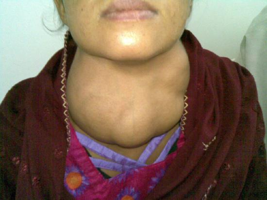
Dietary iodine deficiency can result in the impaired ability to synthesize T3 and T4, leading to a variety of severe disorders. When T3 and T4 cannot be produced, TSH is secreted in increasing amounts. As a result of this hyperstimulation, thyroglobulin accumulates in the thyroid gland follicles, increasing their deposits of colloid. The accumulation of colloid increases the overall size of the thyroid gland, a condition called a goiter (Figure \(\PageIndex{3}\)). A goiter is only a visible indication of the deficiency. Other iodine deficiency disorders include impaired growth and development, decreased fertility, and prenatal and infant death. Moreover, iodine deficiency is the primary cause of preventable intellectual disability worldwide. Neonatal hypothyroidism (cretinism) is characterized by cognitive deficits, short stature, and sometimes deafness and muteness in children and adults born to mothers who were iodine-deficient during pregnancy.
In the absence of iodine deficiency, inflammation of the thyroid gland is the more common cause of low blood levels of thyroid hormones. Called hypothyroidism, the condition is characterized by a low metabolic rate, weight gain, cold extremities, constipation, reduced libido, menstrual irregularities, and intellectual disability.
In contrast, hyperthyroidism—an abnormally elevated blood level of thyroid hormones—is often caused by a pituitary or thyroid tumor. In Graves’ disease, the hyperthyroid state results from an autoimmune reaction in which antibodies overstimulate the follicle cells of the thyroid gland. Hyperthyroidism can lead to an increased metabolic rate, excessive body heat and sweating, diarrhea, weight loss, tremors, and increased heart rate. The person’s eyes may bulge (called exophthalmos) as antibodies produce inflammation in the soft tissues of the orbits.
Calcitonin
The thyroid gland also secretes a hormone called calcitonin that is produced by the parafollicular cells (also called clear cells or C thyrocytes) that stud the tissue between distinct follicles. Calcitonin is released in response to a rise in blood calcium levels. It appears to function in decreasing blood calcium concentrations by:
- Inhibiting the activity of osteoclasts, bone cells that release calcium into the circulation by degrading bone matrix
- Increasing osteoblastic activity
- Decreasing calcium absorption in the intestines
- Decreasing calcium reabsorption in the kidneys, which increases calcium loss in the urine
The hormones secreted by the thyroid gland are summarized in Table \(\PageIndex{1}\).
Parathyroid Glands
The parathyroid glands are tiny, round structures usually found embedded in the posterior surface of the thyroid gland (Figure \(\PageIndex{4}\)). A thick connective tissue capsule separates the glands from the thyroid tissue. Most people have four parathyroid glands, but occasionally there are more in tissues of the neck or chest.

The primary functional cells of the parathyroid glands are the chief cells. These epithelial cells produce and secrete parathyroid hormone (PTH), a major hormone involved in the regulation of blood calcium levels (Figure \(\PageIndex{5}\)). When blood calcium levels drop too low, PTH is released from the parathyroid glands into the bloodstream. Parathyroid hormone encourages the release of stored calcium from the bones and stimulates the kidneys to reabsorb calcium. In bones, PTH inhibits osteoblast activity while stimulating osteoclast activity, resulting in calcium being released from bone matrix into the bloodstream. In the kidneys, PTH stimulates an increase in calcium reabsorption into the bloodstream and also stimulates the release of calcitriol. Calcitriol targets the intestines to stimulate increased absorption of calcium from food. The hormone secreted by the parathyroid glands is summarized in Table \(\PageIndex{1}\).
Calcium Regulation
Calcium is critical for many biological processes. It is a second messenger in many signaling pathways, and is essential for muscle contraction, nerve impulse transmission, and blood clotting. Given these roles, it is not surprising that blood calcium levels are tightly regulated by the endocrine system. When blood calcium levels are high, calcitonin is produced and secreted by the parafollicular cells of the thyroid gland. Calcitonin inhibits the activity of osteoclasts, reduces the absorption of dietary calcium in the intestine, and signals the kidneys to reabsorb less calcium, resulting in larger amounts of calcium excreted in the urine. When blood calcium levels are low, parathyroid hormone is secreted by the parathyroid glands. Targeting the same organs, parathyroid hormone has the opposite effects as calcitonin. Thus, calcitonin and parathyroid hormone have antagonistic functions.

Endocrine System: Hyperparathyroidism and Hypoparathyroidism
Abnormally high activity of the parathyroid gland can cause hyperparathyroidism, a disorder caused by an overproduction of PTH that results in excessive calcium reabsorption from bone. Hyperparathyroidism can significantly decrease bone density, leading to spontaneous fractures or deformities. As blood calcium levels rise, cell membrane permeability to sodium is decreased, and the responsiveness of the nervous system is reduced. At the same time, calcium deposits may collect in the body’s tissues and organs, impairing their functioning.
In contrast, abnormally low blood calcium levels may be caused by parathyroid hormone deficiency, called hypoparathyroidism, which may develop following injury or surgery involving the thyroid gland. Low blood calcium increases membrane permeability to sodium, resulting in muscle twitching, cramping, spasms, or convulsions. Severe deficits can paralyze muscles, including those involved in breathing, and can be fatal.
Concept Review
The thyroid gland is a butterfly-shaped organ located in the neck anterior to the trachea. The parathyroid glands are four small nodules on the posterior surface of the thyroid gland.
Calcium is required for a variety of important physiologic processes, including neuromuscular functioning; thus, blood calcium levels are closely regulated. The parathyroid glands are small structures located on the posterior of the thyroid gland that produce parathyroid hormone (PTH), which regulates blood calcium levels. Low blood calcium levels cause the production and secretion of PTH. In contrast, elevated blood calcium levels trigger secretion of the thyroid hormone calcitonin.
Thyroid hormones regulate basal metabolism, oxygen use, nutrient metabolism, the production of ATP, and calcium homeostasis. The thyroid hormones triiodothyronine (T3) and thyroxine (T4) are produced and secreted by the follicular cells in response to thyroid-stimulating hormone (TSH) from the anterior pituitary. Synthesis of the amino acid–derived T3 and T4 hormones requires iodine. Insufficient amounts of iodine in the diet can lead to goiter and many other disorders.
| Associated hormones | Target(s) | Effect(s) | Associated Disorders |
|---|---|---|---|
| Thyroid Hormone: Thyroxine (T4), triiodothyronine (T3) | Most body cells | Stimulate basal metabolic rate |
Hypothyroidism: goiter Hyperthyroidism: Graves disease |
| Calcitonin | Bones, kidneys | Reduces blood Ca+2 levels; increases Ca+2 deposition in bone | Hypersecretion: increased risk of some cancers; renal failure |
| Parathyroid Hormone | Bones, kidneys | Increases blood Ca+2 levels; decreases Ca+2 deposition in bone | Hypersecretion: increased risk of fractures, kidney stones, constipation; decreased mental activity and coma. |
Review Questions
Q. When blood calcium levels are low, PTH stimulates ________.
A. urinary excretion of calcium by the kidneys
B. a reduction in calcium absorption from the intestines
C. the activity of osteoblasts
D. the activity of osteoclasts
- Answer
-
Answer: D
Q. Which of the following can result from hyperparathyroidism?
A. increased bone deposition
B. fractures
C. convulsions
D. all of the above
- Answer
-
Answer: B
Q. Which of the following statements about the thyroid gland is true?
A. It is located anterior to the trachea and inferior to the larynx.
B. The parathyroid glands are embedded within it.
C. It manufactures three hormones.
D. all of the above
- Answer
-
Answer: D
Q. The secretion of thyroid hormones is controlled by ________.
A. TSH from the hypothalamus
B. TSH from the anterior pituitary
C. thyroxine from the anterior pituitary
D. thyroglobulin from the thyroid’s parafollicular cells
- Answer
-
Answer: B
Q. The development of a goiter indicates that ________.
A. the anterior pituitary is abnormally enlarged
B. there is hypertrophy of the thyroid’s follicle cells
C. there is an excessive accumulation of colloid in the thyroid follicles
D. the anterior pituitary is secreting excessive growth hormone
- Answer
-
Answer: C
Critical Thinking Questions
Q. Explain why maternal iodine deficiency might lead to neurological impairment in the fetus.
- Answer
-
A. Iodine deficiency in a pregnant woman would also deprive the fetus. Iodine is required for the synthesis of thyroid hormones, which contribute to fetal growth and development, including maturation of the nervous system. Insufficient amounts would impair these functions.
Q. Define hyperthyroidism and explain why one of its symptoms is weight loss.
- Answer
-
A. Hyperthyroidism is an abnormally elevated blood level of thyroid hormones due to an overproduction of T3 and T4. An individual with hyperthyroidism is likely to lose weight because one of the primary roles of thyroid hormones is to increase the body’s basal metabolic rate, increasing the breakdown of nutrients and the production of ATP.
Q. Describe the role of negative feedback in the function of the parathyroid gland.
- Answer
-
A. The production and secretion of PTH is regulated by a negative feedback loop. Low blood calcium levels initiate the production and secretion of PTH. PTH increases bone resorption, calcium absorption from the intestines, and calcium reabsorption by the kidneys. As a result, blood calcium levels begin to rise. This, in turn, inhibits the further production and secretion of PTH.
Q. Explain why someone with a parathyroid gland tumor might develop kidney stones.
- Answer
-
A. A parathyroid gland tumor can prompt hypersecretion of PTH. This can raise blood calcium levels so excessively that calcium deposits begin to accumulate throughout the body, including in the kidney tubules, where they are referred to as kidney stones.
Glossary
- calcitonin
- peptide hormone produced and secreted by the parafollicular cells (C cells) of the thyroid gland that functions to decrease blood calcium levels
- calcitriol
- hormone released by kidney tubule cells in response to parathyroid hormone that stimulates increased absorption of calcium from digested food in the intestines
- colloid
- viscous fluid in the central cavity of thyroid follicles, containing the glycoprotein thyroglobulin
- goiter
- enlargement of the thyroid gland either as a result of iodine deficiency or hyperthyroidism
- hyperparathyroidism
- disorder caused by overproduction of PTH that results in abnormally elevated blood calcium
- hypoparathyroidism
- disorder caused by underproduction of PTH that results in abnormally low blood calcium
- hyperthyroidism
- clinically abnormal, elevated level of thyroid hormone in the blood; characterized by an increased metabolic rate, excess body heat, sweating, diarrhea, weight loss, and increased heart rate
- hypothyroidism
- clinically abnormal, low level of thyroid hormone in the blood; characterized by low metabolic rate, weight gain, cold extremities, constipation, and reduced mental activity
- neonatal hypothyroidism
- condition characterized by cognitive deficits, short stature, and other signs and symptoms in people born to women who were iodine-deficient during pregnancy
- parathyroid glands
- small, round glands embedded in the posterior thyroid gland that produce parathyroid hormone (PTH)
- parathyroid hormone (PTH)
- peptide hormone produced and secreted by the parathyroid glands that targets bone and the kidneys to raise blood calcium levels and targets the kidneys to release calcitriol
- thyroid gland
- large endocrine gland responsible for the synthesis of thyroid hormones
- thyroxine
- (also, tetraiodothyronine, T4) amino acid–derived thyroid hormone that is more abundant but less potent than T3 and often converted to T3 by target cells
- triiodothyronine
- (also, T3) amino acid–derived thyroid hormone that is less abundant but more potent than T4
Contributors and Attributions
OpenStax Anatomy & Physiology (CC BY 4.0). Access for free at https://openstax.org/books/anatomy-and-physiology