
3.1: Examining epithelial tissue under the microscope
- Page ID
- 52725
Information
Epithelial tissue serves two main functions in the body.
- It provides linings for external and internal surfaces that face harsh environments. The outer layer of the skin is epithelial tissue, as are the innermost layers of the digestive tract, the respiratory tract, and blood vessels.
- It forms glands that secrete materials onto epithelial surfaces or into the blood. Sweat glands, salivary glands, mammary glands, adrenal glands, and pituitary glands are examples of glands made of epithelial tissue.
Epithelial tissue is often classified according to numbers of layers of cells present, and by the shape of the cells. See Figure 3.1.
A simple epithelium is only one layer of cells thick. A stratified epithelium is more than one layer of cells thick. A pseudostratified epithelium is really a specialized form of a simple epithelium in which there appears at first glance to be more than one layer of epithelial cells, but a closer inspection reveals that each cell in the layer actually extends to the basolateral surface of the epithelium.
There are three basic shapes used to classify epithelial cells. A squamous epithelial cell looks flat under a microscope. A cuboidal epithelial cell looks close to a square.
A columnar epithelial cell looks like a column or a tall rectangle. A few epithelial layers are constructed from cells that are said to have a transitional shape. Transitional epithelial cells are epithelial cells specialized to change shape if they are stretched laterally. They can transition from columnar- and cuboidal-looking shapes in their unstretched state to more squamous-looking shapes in their stretched state.
When classifying a stratified epithelial sheet, the sheet is named for the shape of the cells in its most superficial layers. So, a stratified squamous epithelium only necessarily has squamous-shaped cells in its highest layers and might have a different-shaped cell in its lower layers.
Under a microscope, epithelial cells are readily distinguished by the following features:
- The cells will usually be one of the three basic cell shapes – squamous, cuboidal, or columnar.
- The cells will be closely attached to one another, in either a single layer or in multiple layers, and usually will not have room for extracellular material between the attached cells.
The epithelial layer on one side will face an empty space (or, in some organs, it will face a secreted substance like mucus) and on the other side will usually be attached to connective tissue proper.
Usually, a slide will have a section of tissue cut out of a larger organ. Slides with epithelial tissues usually have some of the underlying tissue found beneath the epithelial tissue with them.
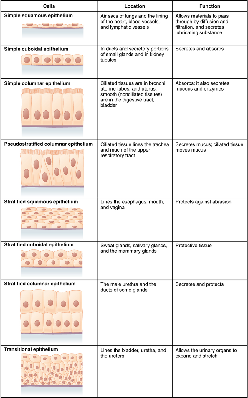
Figure \(\PageIndex{1}\): The different ways sheets of epithelial cells are categorized. (CC-BY, OpenStax, Human Anatomy)
A layer of epithelial cells always serves as an outer layer for some structure, but, when looking at a tissue preparation on a slide, do not assume that just because you have found one end of the tissue sample you are automatically looking at epithelial tissue. Look for the cell characteristics listed above to be sure you are on the epithelial side of a tissue slice.
In Figure \(\PageIndex{2}\), only one edge of the tissue slice has epithelial cells. In Figure \(\PageIndex{2}\)A that edge is indicated with an arrow, but when looking at a specimen under a microscope, you have to figure out for yourself where the edge with the epithelial cells is.
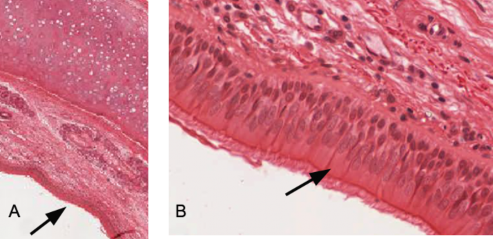
Figure \(\PageIndex{2}\): A slice of a trachea. A. Magnified 1.8x. The arrow indicates which edge in this slice contains the epithelial cells B. Magnified 20x. The arrow indicates an individual columnar epithelial cell. (CC-BY-SA-NC, University of Michigan Histology and Virtual Microscopy Learning Resources)
In the tissue slice in Figure \(\PageIndex{2}\), there are three edges that are not epithelial cells. If you just mindlessly started viewing the first edge you find, you have a good chance of looking as something other than the epithelial cells in the preparation. Be sure what you are looking at has the three visual characteristics of epithelial tissue:
- The cells will usually be one of the three basic cell shapes – squamous, cuboidal, or columnar.
- The cells will be closely attached to one another, in either a single layer or in multiple layers, and usually will not have room for extracellular material between the attached cells.
- The epithelial layer on one side will face an empty space (or, in some organs, it will face a secreted substance like mucus) and on the other side will usually be attached to connective tissue proper.
In the following figures are some more epithelial layers. Note that whether the epithelial layer of the specimen is on the top, bottom, right, or left of the slice varies with how the specimen slice was positioned on the slide. In every case, you have to find which edge has the epithelial layer.
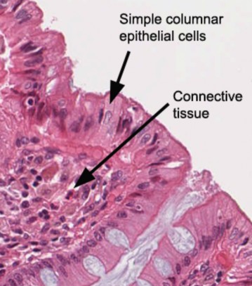
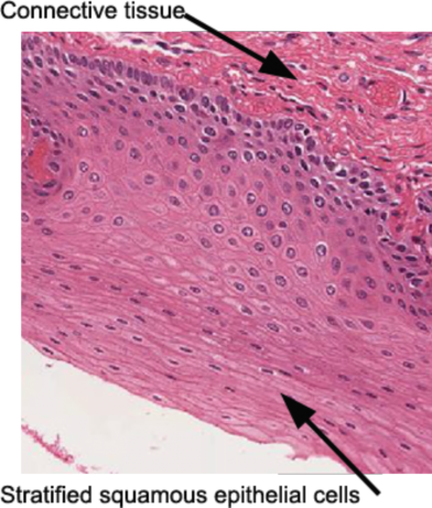
Figure \(\PageIndex{3}\): Left a slice of the colon, 20x. Right Figure 5-4. A slice of the esophagus, 10x. (CC-BY-SA-NC, University of Michigan Histology and Virtual Microscopy Learning Resources)
-------------------------------------------------------------------------------------------------------------------
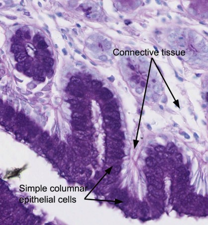
Figure \(\PageIndex{4}\): (CC-BY-SA-NC, University of Michigan Histology and Virtual Microscopy Learning Resources)
-----------------------------------------------------------------------------------------------------------
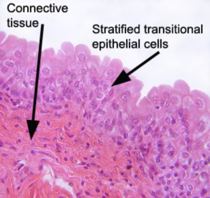
Figure \(\PageIndex{5}\): slice of the urinary bladder, 10x. (CC-BY-SA-NC, University of Michigan Histology and Virtual Microscopy Learning Resources)
Setting up a microscope
Lab 1 Exercise 1
![]() 1. Plug in the microscope & turn on light source.
1. Plug in the microscope & turn on light source.
![]() 2. Pick up microscope by carrying arm, position it so it is accessible to your seat, with open side of the stage facing you
2. Pick up microscope by carrying arm, position it so it is accessible to your seat, with open side of the stage facing you
![]() 3. Rotate the objectives so that the lowest power objective (smallest in size) clicks into place.
3. Rotate the objectives so that the lowest power objective (smallest in size) clicks into place.
![]() 4. Look at the slide with your naked eye and find the location of the specimen.
4. Look at the slide with your naked eye and find the location of the specimen.
![]() 5. Clip the slide into place with the stage clips. The cover slip on the slide must face up. Find the stage controls and make sure that, when they are turned, the slide moves smoothly left and right or up and down, depending on the knob.
5. Clip the slide into place with the stage clips. The cover slip on the slide must face up. Find the stage controls and make sure that, when they are turned, the slide moves smoothly left and right or up and down, depending on the knob.
![]() 6. Use the stage controls to move the slide so that the light source is shining directly on to the specimen to be magnified.
6. Use the stage controls to move the slide so that the light source is shining directly on to the specimen to be magnified.
![]() 7. Find the coarse and fine focus knobs. Watching the stage and objective, use the coarse focus knob to bring the low power objective as close to the slide as it will go.
7. Find the coarse and fine focus knobs. Watching the stage and objective, use the coarse focus knob to bring the low power objective as close to the slide as it will go.
![]() 8. Put your eye to the eyepiece (or eyepieces, if the microscope is binocular) and rotate the coarse focus knob in the lowering direction until some aspect of the specimen comes into focus.
8. Put your eye to the eyepiece (or eyepieces, if the microscope is binocular) and rotate the coarse focus knob in the lowering direction until some aspect of the specimen comes into focus.
![]() 9. Move your hand to the fine focus knob and get the specimen into perfect focus for your eyes. Do NOT touch the coarse focus knob again.
9. Move your hand to the fine focus knob and get the specimen into perfect focus for your eyes. Do NOT touch the coarse focus knob again.
![]() 10. Use the stage control knobs to move you specimen to close to the exact center of your field of view
10. Use the stage control knobs to move you specimen to close to the exact center of your field of view
![]() 11. Move to the next highest power objective (do not skip the individual objectives) and use only the fine focus to get your image into perfect focus for your eyes.
11. Move to the next highest power objective (do not skip the individual objectives) and use only the fine focus to get your image into perfect focus for your eyes.
![]() 12. If you need further magnification, move to the next highest power objective and use only the fine focus to get your image into perfect focus for your eyes.
12. If you need further magnification, move to the next highest power objective and use only the fine focus to get your image into perfect focus for your eyes.
![]() 13. Do not use the 100x objective (if you have one) in this course. It must be used with immersion oil and we won’t have students doing that.
13. Do not use the 100x objective (if you have one) in this course. It must be used with immersion oil and we won’t have students doing that.
Lab 3 Exercise \(\PageIndex{1}\)
Simple columnar
Simple cuboidal
 |
Total Magnification: _________________
Type of eptihelium: _________________
Source of tissue: ____________________
Function of this phase: _______________
__________________________________ |
Simple squamous
Stratified squamous
Pseudostratified columnar
|
Total Magnification: _________________
Type of eptihelium: _________________
Source of tissue: ____________________
Function of this phase: _______________
__________________________________ |
Transitional
|
Total Magnification: _________________
Type of eptihelium: _________________
Source of tissue: ____________________
Function of this phase: _______________
__________________________________ |
- Obtain a slide of one of the tissues listed below from the slide box at your table.
- Follow the checklist above to set up your slide for viewing.
- View the slide on the objective which provides the best view. Find the representative object.
- In the circle below the name, draw a representative sample of the tissue, taking care to correctly and clearly draw their true shape in the slide. If it is a stratified epithelium draw all the layers. Draw your structures proportionately to their size in your microscope’s field of view.
- Fill in the blanks next to your drawing.
A) Prophase
|
Total Magnification: _________________
Type of eptihelium: _________________
Source of tissue: ____________________
Function of this phase: _______________
__________________________________ |
|
Total Magnification: _________________
Type of eptihelium: _________________
Source of tissue: ____________________
Function of this phase: _______________
__________________________________ |
Repeat this for each of the tissue types seen below.
References
A&P Labs. Authored by: Ross Whitwam. Provided by: Mississippi University for Women. Located at: http://www.muw.edu/. License: CC BY-SA: Attribution-ShareAlike
CC LICENSED CONTENT, SHARED PREVIOUSLY
Figure \(\PageIndex{1}\). The different ways sheets of epithelial cells are categorized.. Provided by: OpenStax College. Located at: https://commons.wikimedia.org/wiki/F...sue_CellsN.jpg. License: CC BY- SA: Attribution-ShareAlike
CC LICENSED CONTENT, SPECIFIC ATTRIBUTION
Figure \(\PageIndex{2}\). A slice of a trachea. A. Magnified 1.8x. The arrow indicates which edge in this slice contains the epithelial cells B. Magnified 20x. The arrow indicates an individual columnar epithelial cell.. Authored by: Kent Christensen, Ph.D., J. Matthew Velkey, Ph.D., Lloyd M. Stoolman, M.D., Laura Hessler, and Diedra Mosley-Brower. Provided by: University of Michigan Histology and Virtual Microscopy Learning Resources. Located at: http://141.214.65.171/Histology/Basic%20Tissues/Epithelium%20and%20CT/020_HISTO_20X.svs/view.apml. License: CC BY-NC-SA: Attribution-NonCommercial-ShareAlike
Figure \(\PageIndex{3L}\). A slice of the colon, 20x.. Authored by: Kent Christensen, Ph.D., J. Matthew Velkey, Ph.D., Lloyd M. Stoolman, M.D., Laura Hessler, and Diedra Mosley-Brower. Provided by: University of Michigan Histology and Virtual Microscopy Learning Resources. Located at: http://141.214.65.171/Histology/Basic%20Tissues/Epithelium%20and%20CT/176_HISTO_20X.svs/view.apml. License: CC BY-NC-SA: Attribution-NonCommercial-ShareAlike
Figure \(\PageIndex{3R}\). A slice of the esophagus, 10x.. Authored by: Kent Christensen, Ph.D., J. Matthew Velkey, Ph.D., Lloyd M. Stoolman, M.D., Laura Hessler, and Diedra Mosley-Brower. Provided by: University of Michigan Histology and Virtual Microscopy Learning Resources. Located at: http://141.214.65.171/Histology/Basic%20Tissues/Epithelium%20and%20CT/153_HISTO_20X.svs/view.apml. License: CC BY-NC-SA: Attribution-NonCommercial-ShareAlike
Figure \(\PageIndex{4}\) A slice of the stomach, 20x.. Authored by: Kent Christensen, Ph.D., J. Matthew Velkey, Ph.D., Lloyd M. Stoolman, M.D., Laura Hessler, and Diedra Mosley-Brower. Provided by: University of Michigan Histology and Virtual Microscopy Learning Resources. Located at: http://141.214.65.171/Histology/Basic%20Tissues/Epithelium%20and%20CT/160_HISTO_40X.svs/view.apml. License: CC BY-NC-SA: Attribution-NonCommercial-ShareAlike
Figure \(\PageIndex{5}\). A slice of the urinary bladder, 10x.. Authored by: Kent Christensen, Ph.D., J. Matthew Velkey, Ph.D., Lloyd M. Stoolman, M.D., Laura Hessler, and Diedra Mosley-Brower. Provided by: University of Michigan Histology and Virtual Microscopy Learning Resources. Located at: http://141.214.65.171/Histology/Urinary%20System/212_HISTO_40X.svs/view.apml. License: CC BY- NC-SA: Attribution-NonCommercial-ShareAlike