
3.2: Examining Connective Tissue
- Page ID
- 52726
Information
Connective tissue is found throughout the body, usually in association with other tissues. As its name indicates, it often serves to connect different tissues together, but it also can serve as a wrapper (in locations where a tough epithelial wrapping is not required), a structural support, cushioning, a storage repository, a protective layer, or a transport medium.
Connective tissue has the most types of subcategories and the most varied functions of all the four major tissue types (epithelial, muscular, nervous, and connective tissues.) Bone and cartilage are connective tissues, as are blood and lymph, fat, ligaments, and tendons. Epimysium, the connective tissue wrapping around skeletal muscles, and periosteum, the connective tissue wrapping around bones, are both connective tissues.
The different types of connective tissue are so diverse, there is no one set of characteristics that encompasses all the different types. However, there are three characteristics that we consider diagnostic of most connective tissue types.
- The cells are dispersed. Connective tissues generally have cells that are not tightly connected to each other, the way the cells in epithelial and muscular tissues usually are. There is usually a fair amount of space between the connective tissue cells. An exception to this is adipose tissue (also known as fat), the rare type of connective tissue in which the cells are packed tightly together.
- The tissue has more extracellular material than cells. Most connective tissues are solid (blood and lymph are the exceptions) because all the volume between the dispersed cells is filled with an extracellular matrix of viscous ground substance and protein fibers.
- An extensive network of protein fibers is found in the extracellular matrix. Protein fibers are complexes of millions of individual proteins threaded into long fibrous structures that provide strength and elasticity to the tissue as a whole. The protein fibers are so large, they are longer than the cells they surround and enmesh. Blood and lymph, being liquid connective tissues, do not have these enmeshing protein fibers, but they still have an extensive liquid extracellular matrix.
A common way of classifying the many different types of connective tissue is to subdivide it into three main sub-categories, and further divide those subcategories into specific types of connective tissue. The three main sub-categories of connective tissue are:
- Connective tissue proper. These are the types of connective tissue that typically have all three of the defining characteristics listed above. It is further subdivided into dense connective tissue proper, in which the extracellular protein fibers predominate, and loose connective tissue proper, in which the extracellular protein fibers are not so densely woven.
- Supporting connective tissue. Bones and cartilage are the two types of connective tissue in this sub-category. They both have all three of the defining characteristics listed above, but their extracellular matrix is tougher, denser, and more solid than the various types of connective tissue proper.
- Fluid connective tissue.
Blood and lymph are the two types of connective tissue in this sub-category. Both are fluid, rather than solid, and both lack the network of extracellular protein fibers found in the other types of connective tissue.
A&P Labs. Authored by: Ross Whitwam. Provided by: Mississippi University for Women. Located at: http://www.muw.edu. License: CC BY-SA: Attribution-ShareAlike
Connective Tissue Proper
Information
Connective tissue proper encompasses the types of connective tissue that usually show all three of the defining cellular characteristics of connective tissue with the fewest deviations from those characteristics.
Dispersed cells
More extracellular material than cells.
Extensive protein fibers in the extracellular matrix.
Nonetheless there is still a great variety among the subcategories of connective tissue proper. Some are classified as dense connective tissue proper and have a dense arrangement of extracellular protein fibers that give the tissue strength and toughness. Tendons connecting muscles to bone and ligaments connecting bone to bone are examples of dense connective tissue proper. Other tissues are classified loose connective tissue proper and have fewer extracellular protein fibers and more ground substance (the extracellular material surrounding the protein fibers), making the tissues spongier but more fragile. Areolar tissue, found in the hypodermis of the skin and below the epithelial layers of the digestive, respiratory, and urinary tracts, is a loose connective tissue proper, as is adipose tissue, also known as fat.
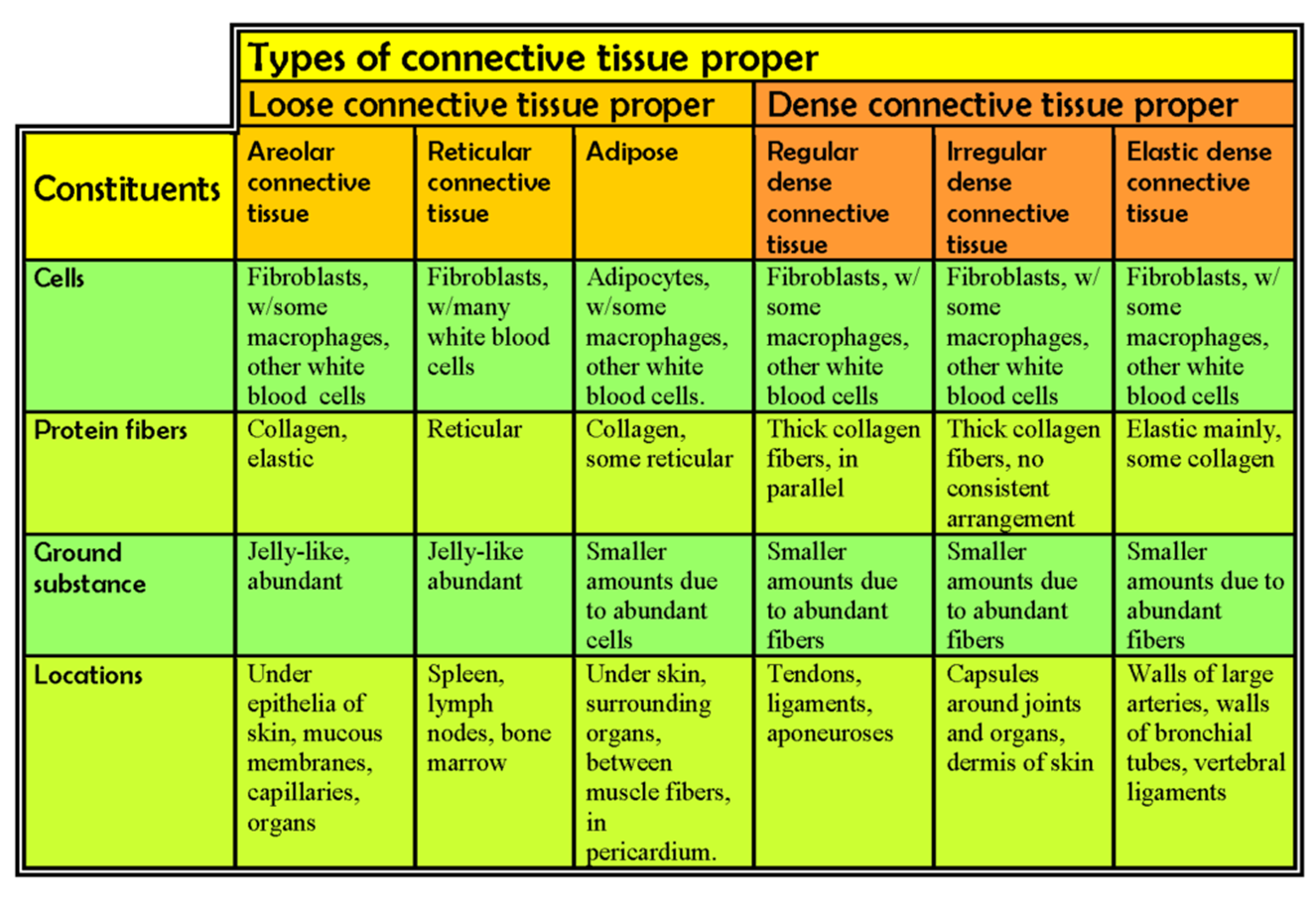
Table \(\PageIndex{1}\) lists some of the subcategories of connective tissue proper, along with some of their characteristics and properties.
Table \(\PageIndex{1}\): Summary of the properties of the major types of connective tissue proper.
In drawing images of connective tissue proper preparations seen under the microscope, it is important to simplify the visuals. Connective tissue preparations are often messy with a number of blotches and shapes irrelevant to the main components of the tissue, which are the cells and the extracellular protein fibers. Especially with connective tissue slides, it is important to make sure you know what you are looking for, find those components, and draw only them in as simple a form as possible, usually with just lines and with minimal shadings or hatchings. Leave out the unnecessary and irrelevant stuff on the slide.
For instance, Figure 3.7 A shows a section of connective tissue taken from just below (deep to) the epithelial layer of the stomach, magnified 40x. Figure 3.7 B illustrates how you should represent that view as a line drawing.
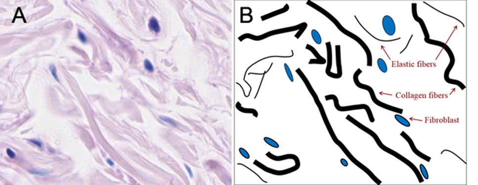
Figure \(\PageIndex{2}\): A photomicrograph, A, and a drawing from the photomicrograph, B, of the connective tissue in the wall of the stomach, just below the epithelial layer. (CC-BY-SA-NC, University of Michigan Histology and Virtual Microscopy Learning Resources)
Notice that in the line drawing not every single thick collagen fiber, nor every single thin elastic fiber, not every single fibroblast was drawn. Also notice that some of the out-of-focus, blurry fibers were not drawn at all, rather than draw fuzzy blotches. The key is to get the important structures (once you know what those are) and leave out distracting, non-essential messiness.
LAB 3 EXERCISE \(\PageIndex{2}\)
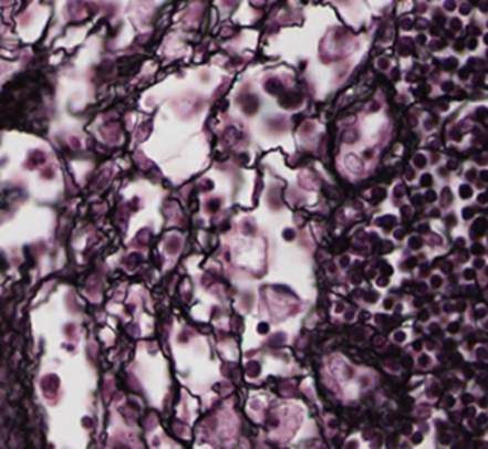
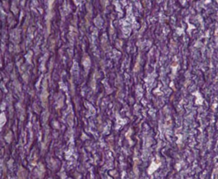
- Table 3.1 lists six categories of connective tissue proper. Use the information in that table to identify which category each of the following samples belong to.
- Under each picture, list the evidence that points to the category you choose.
- The key identifying features to look for in each picture are: the type(s) of protein fibers present, the type of cells present, and whether there are significant amounts of ground substance present.
- Collagen protein fibers are thick. Elastic protein fibers are thin. Reticular protein fibers are thin but form a web-like arrangement.
- If there is abundant space between protein fibers, the tissue is likely one of the loose connective tissues. If there is little space between protein fibers, the tissue is likely one of the dense connective tissues.
- Adipose is mainly large adipocyte cells containing a large droplet of lipids and nucleus and cytoplasm crammed into one corner of the cell. There are more adipocytes than extracellular material in adipose.
- The protein fibers in regular dense connective tissue proper will largely parallel each other, but they are often undulate in a wave-like arrangement while being parallel.
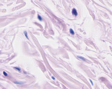 |
1. Source: Stomach wall
Category:
Evidence: |
 |
1. Joint Capsule
Category:
Evidence: |
 |
1. Source: Lymph node
Category:
Evidence: |
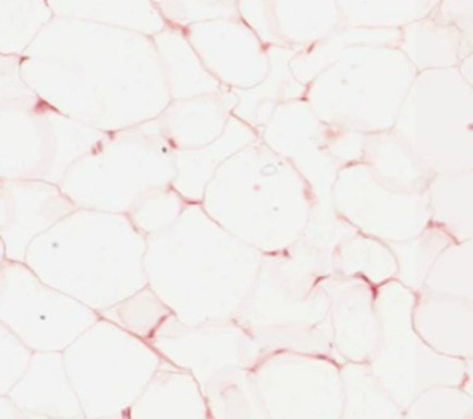 |
1. Source: Fat
Category:
Evidence: |
 |
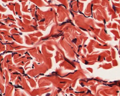
1. Source: Aorta Wall
Category:
Evidence: |
 |
1. Source: Tendon
Category:
Evidence: |
LAB 3 EXERCISE \(\PageIndex{3}\)
- Obtain a slide of connective tissue proper/areolar CT from the slide box.
- View the slide on an appropriate objective.
- Fill out the blanks next to your drawing.
- In the circle below, draw a representative sample of key features you identified, taking care to correctly and clearly draw their true shapes and directions. Draw your structures proportionately to their size in your microscope’s field of view.
  |
Total Magnification: _________________
Type of eptihelium: _________________
Source of tissue: ____________________
Function of this phase: _______________
__________________________________ |
Slide \(\PageIndex{2}\):connective tissue proper/areolar CT
- Obtain a slide of dense irregular connective tissue from the slide box.
- View the slide on an appropriate objective.
- Fill out the blanks next to your drawing.
- In the circle below, draw a representative sample of key features you identified, taking care to correctly and clearly draw their true shapes and directions. Draw your structures proportionately to their size in your microscope’s field of view.
|
Total Magnification: _________________
Type of eptihelium: _________________
Source of tissue: ____________________
Function of this phase: _______________
__________________________________ |
Slide \(\PageIndex{3}\): irregular connective tissue
- Obtain a slide of a spleen or lymph node with reticular connective tissue from the slide box.
- View the slide on an appropriate objective.
- Fill out the blanks next to your drawing.
- In the circle below, draw a representative sample of key features you identified, taking care to correctly and clearly draw their true shapes and directions. Draw your structures proportionately to their size in your microscope’s field of view.
|
Total Magnification: _________________
Type of eptihelium: _________________
Source of tissue: ____________________
Function of this phase: _______________
__________________________________ |
Slide \(\PageIndex{4}\): spleen or lymph node with reticular connective tissue
- Obtain a slide of adipose connective tissue from the slide box.
- View the slide on an appropriate objective.
- Fill out the blanks next to your drawing.
- In the circle below, draw a representative sample of key features you identified, taking care to correctly and clearly draw their true shapes and directions. Draw your structures proportionately to their size in your microscope’s field of view.
|
Total Magnification: _________________
Type of eptihelium: _________________
Source of tissue: ____________________
Function of this phase: _______________
__________________________________ |
Slide \(\PageIndex{5}\): adipose connective tissue
- Obtain a slide of a large artery with elastic connective tissue from the slide box.
- View the slide on an appropriate objective.
- Fill out the blanks next to your drawing.
- In the circle below, draw a representative sample of key features you identified, taking care to correctly and clearly draw their true shapes and directions. Draw your structures proportionately to their size in your microscope’s field of view.
|
Total Magnification: _________________
Type of eptihelium: _________________
Source of tissue: ____________________
Function of this phase: _______________
__________________________________ |
Figure \(\PageIndex{6}\): adipose connective tissue
- Obtain a slide of a tendon with dense regular connective tissue from the slide box.
- View the slide on an appropriate objective.
- Fill out the blanks next to your drawing.
- In the circle below, draw a representative sample of key features you identified, taking care to correctly and clearly draw their true shapes and directions. Draw your structures proportionately to their size in your microscope’s field of view.
|
Total Magnification: _________________
Type of eptihelium: _________________
Source of tissue: ____________________
Function of this phase: _______________
__________________________________ |
Slide \(\PageIndex{7}\): elastic connective tissue
LICENSES AND ATTRIBUTIONS
CC LICENSED CONTENT, ORIGINAL
- A&P Labs. Authored by: Ross Whitwam. Provided by: Mississippi University for Women. Located at: http://www.muw.edu. License: CC BY-SA: Attribution-ShareAlike
CC LICENSED CONTENT, SPECIFIC ATTRIBUTION
- Figure 3-1A photomicrograph, A, and a drawing from the photomicrograph, B, of the connective tissue in the wall of the stomach, just below the epithelial layer.. Authored by: Kent Christensen, Ph.D., J. Matthew Velkey, Ph.D., Lloyd M. Stoolman, M.D., Laura Hessler, and Diedra Mosley-Brower. Provided by: University of Michigan Histology and Virtual Microscopy Learning Resources. Located
at: 141.214.65.171/Histology/Basic%20Tissues/Epithelium%20and%20CT/160_HISTO_40X.svs/view.apml. License: CC BY-NC-SA: Attribution-NonCommercial-ShareAlike
-
Exercise 3.1 A. Source: Stomach wall. Authored by: Kent Christensen, Ph.D., J. Matthew Velkey, Ph.D., Lloyd M. Stoolman, M.D., Laura Hessler, and Diedra Mosley-Brower. Provided by: University of Michigan Histology and Virtual Microscopy Learning Resources. Located
-
at: 141.214.65.171/Histology/Basic%20Tissues/Epithelium%20and%20CT/160_HISTO_40X.svs/view.apml. License: CC BY-NC-SA: Attribution-NonCommercial-ShareAlike
- Exercise 3.1 B. Joint Capsule. Authored by: Kent Christensen, Ph.D., J. Matthew Velkey, Ph.D., Lloyd M. Stoolman, M.D., Laura Hessler, and Diedra Mosley-Brower. Provided by: University of Michigan Histology and Virtual Microscopy Learning
Resources. Located
at: 141.214.65.171/Histology/Basic%20Tissues/Epithelium%20and%20CT/033_HISTO_20X.svs/view.apml. License: CC BY-NC-SA: Attribution-NonCommercial-ShareAlike
- Exercise 3.1 C. Source: Lymph node. Authored by: Kent Christensen, Ph.D., J. Matthew Velkey, Ph.D., Lloyd M. Stoolman, M.D., Laura Hessler, and Diedra Mosley-Brower. Provided by: University of Michigan Histology and Virtual Microscopy Learning Resources. Located at: 141.214.65.171/Histology/Basi...0and%20CT/028- 2_HISTO_40X.svs/view.apml. License: CC BY-NC-SA: Attribution-NonCommercial-ShareAlike
- Exercise 3.1 D. Source: Fat. Authored by: Kent Christensen, Ph.D., J. Matthew Velkey, Ph.D., Lloyd M. Stoolman, M.D., Laura Hessler, and Diedra Mosley-Brower. Provided by: University of Michigan Histology and Virtual Microscopy Learning Resources. Located at: 141.214.65.171/Histology/Basi...0and%20CT/019- 2_HISTO_20X.svs/view.apml. License: CC BY-NC-SA: Attribution-NonCommercial-ShareAlike
- Exercise 3.1 E. Source: Aorta Wall. Authored by: Kent Christensen, Ph.D., J. Matthew Velkey, Ph.D., Lloyd M. Stoolman, M.D., Laura Hessler, and Diedra Mosley-Brower. Provided by: University of Michigan Histology and Virtual Microscopy Learning Resources. Located
at: 141.214.65.171/Histology/Cardiovascular%20System/036_HISTO_20X.svs/view.apml%20%20. License: CC BY-NC- SA: Attribution-NonCommercial-ShareAlike
- Exercise 3.1 F. Source: Tendon. Authored by: Kent Christensen, Ph.D., J. Matthew Velkey, Ph.D., Lloyd M. Stoolman, M.D., Laura Hessler, and Diedra Mosley-Brower. Provided by: University of Michigan Histology and Virtual Microscopy Learning
Resources. Located at: 141.214.65.171/Histology/Basic%20Tissues/Epithelium%20and%20CT/74.svs/view.apml. License: CC BY-NC-SA: Attribution-NonCommercial-ShareAlike