
3.3: Examining Connective Tissue
- Page ID
- 52727
Information
Supporting connective tissue comprises bone and cartilage. We will examine those tissues in greater detail in Lab 5 The Appendicular Skeleton & Lab 6 The Axial Skeleton.
In both bone and cartilage, as in the different types of connective tissue proper, there are extracellular protein fibers embedded in a viscous ground substance. However, in bone and cartilage, the ground substance is so viscous as to be very hard and tough solids. Both bone and cartilage use mainly collagen and elastic protein fibers in their extracellular matrix, but cartilage uses a ground substance rich in the carbohydrate hyaluronan and bone uses a ground substance rich in a mineralized calcium phosphate compound known as hydroxyapatite.
The carbohydrate hyaluronan (sometimes known as hyaluronic acid or hyaluronate) binds up huge numbers of water molecules in the extracellular matrix of cartilage. This helps solidify the ground substance around the collagen and elastic fibers of cartilage. As a result, it is often difficult to see the protein fibers in cartilage when viewing preparations under the microscope.
They hydroxyapatite that surrounds the mostly collagen protein fibers in the ground substance of bone is not soluble in water and forms a mineral solid in which both the bone cells and the collagen fibers are embedded. As with cartilage, it is usually difficult to see the collagen fibers in the extracellular matrix of bone due to the density of the ground substance that surrounds them.
There is only one type of cell in cartilage, chondrocytes. They secrete and maintain the extracellular matrix of the tissue. Chondrocytes arise from mesenchymal stem cells, just like the fibroblasts of connective tissue proper do, but chondrocytes are specialized to produce just cartilage. The extracellular matrix produced by the chondrocytes is so tough and durable, the chondrocytes are in danger of being crushed by it. This is why chondrocytes always leave a region around themselves free of the cartilaginous extracellular matrix that makes up the rest of the tissue. These non-cartilaginous pockets around each chondrocyte are called lacunae and are clearly visible when examining cartilage under the microscope.
There are four types of bone cells, osteoprogenitor cells, osteoblasts, osteoclasts, and osteocytes, but the osteocytes are the most abundant and the only ones found throughout the bone. Osteocytes are found in concentric circles of mineralized extracellular matrix. Each circle is called a lamella (plural: lamellae) and the osteocytes are found along the edges of each lamellae. In compact bone, groups of lamellae and osteocytes are arranged into individual osteons, the cylindrical arrangement of material that makes up the fundamental building block of the compact bone. Each osteon has a hollow central canal in its center that blood vessels and nerves can travel through. In spongy bone, groups of lamellae are arranged into trabeculae (singular: trabecula), which are the individual projections of spongy bone. Trabeculae do not have central canals.
Osteocytes, like chondrocytes, are protected from the extracellular matrix that surrounds them by being housed in lacunae, which are spaces free of mineralized extracellular matrix. Osteocytes, unlike chondrocytes, have numerous cytoplasmic extensions that project off of the main cell body. These extension connect up with the extensions from other near-by osteocytes. These projections, like the osteocyte cell body, are in tiny spaces free of the mineralized extracellular matrix. These spaces (but not the cytoplasmic projections themselves) are called canaliculi (singular: canaliculum) because, under the microscope, they look like tiny little canals.
LAB 3 EXERCISE \(\PageIndex{1}\)
- In the photomicrograph below of cartilage tissue, find and label the indicated structures.
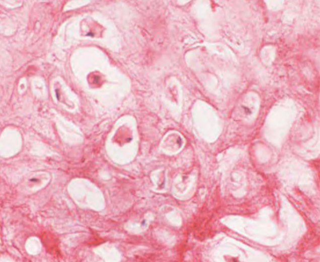 |
Lacuna: _________________
Chondrocyle: _________________
Elastic protein fibers: ____________________
Extracellular matix: _______________
|
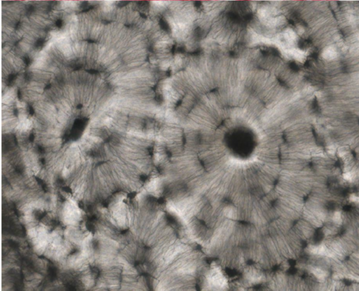 |
Osteon: _________________
Lanella: _________________
Lacuna: ____________________
Osteocyte: _______________
Canaliculi: _________________
Central canal: _______________ |
2. In the photomicrograph below of compact bone tissue, find and label the indicated structures.
- Obtain a slide of hyaline cartilage connective tissue from the slide box.
- View the slide on an appropriate objective.
- Fill out the blanks next to your drawing.
- In the circle below, draw a representative sample of key features you identified, taking care to correctly and clearly draw their true shapes and directions. Draw your structures proportionately to their size in your microscope’s field of view.
 |
Total Magnification: _________________
Type of eptihelium: _________________
Source of tissue: ____________________
Function tissue: _______________
__________________________________ |
Slide \(\PageIndex{1}\): hyaline cartilage connective tissue
- Obtain a slide of ground compact bone connective tissue from the slide box.
- View the slide on an appropriate objective.
- Fill out the blanks next to your drawing.
- In the circle below, draw a representative sample of key features you identified, taking care to correctly and clearly draw their true shapes and directions. Draw your structures proportionately to their size in your microscope’s field of view.
|
Total Magnification: _________________
Type of connective tissue: _________________
Source of tissue: ____________________
Function of this tissue: _______________
__________________________________
Key features to find and draw: ________________________________ |
Slide \(\PageIndex{2}\): ground compact bone connective tissue
LICENSES AND ATTRIBUTIONS
CC LICENSED CONTENT, ORIGINAL
A&P Labs. Authored by: Ross Whitwam. Provided by: Mississippi University for Women. Located at: http:www.muw.edu. License: CC BY-SA: Attribution-ShareAlike
CC LICENSED CONTENT, SPECIFIC ATTRIBUTION
Exercise 3.3. 1. In the photomicrograph below of cartilage tissue, find and label the indicated structures.. Authored by: Kent Christensen, Ph.D., J. Matthew Velkey, Ph.D., Lloyd M. Stoolman, M.D., Laura Hessler, and Diedra Mosley-Brower. Provided by: University of Michigan Histology and Virtual Microscopy Learning Resources. Located at: 141.214.65.171/Histology/Basic%20Tissues/Cartilage%20and%20Bone/044H_HISTO_20X.svs/view.apml. License: C C BY-NC-SA: Attribution-NonCommercial-ShareAlike
Exercise 3.3. 2. In the photomicrograph below of compact bone tissue, find and label the indicated structures.. Authored by: Kent Christensen, Ph.D., J. Matthew Velkey, Ph.D., Lloyd M. Stoolman, M.D., Laura Hessler, and Diedra Mosley-Brower. Provided by: University of Michigan Histology and Virtual Microscopy Learning Resources. Located at: 141.214.65.171/Histology/Basic%20Tissues/Cartilage%20and%20Bone/051xc_HISTO_40X.svs/view.apml. License: C C BY-NC-SA: Attribution-NonCommercial-ShareAlike