
3.4: Distinguishing Between The Three Types of Muscle Tissue
- Page ID
- 52728
Information
Muscular tissue is the third of the four major categories of animal tissue. Muscle tissue is subdivided into three broad categories: skeletal muscle, cardiac muscle, and smooth muscle. The three types of muscle can be distinguished by both their locations and their microscopic features.
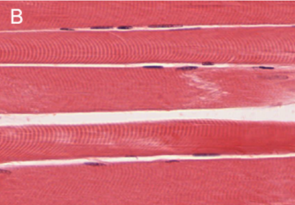
Skeletal muscle is found attached to bones. It consists of long multinucleate fibers. The fibers run the entire length of the muscle they come from and so are usually too long to have their ends visible when viewed under the microscope. The fibers are relatively wide and very long, but unbranched. Fibers are not individual cells, but are formed from the fusion of thousands of precursor cells. This is why they are so long and why individual fibers are multinucleate (a single fiber has many nuclei). The nuclei are usually up against the edge of the fiber. There are striations in skeletal muscle. These are alternating dark and light bands perpendicular to the edge of the fiber that are present all along the fiber.
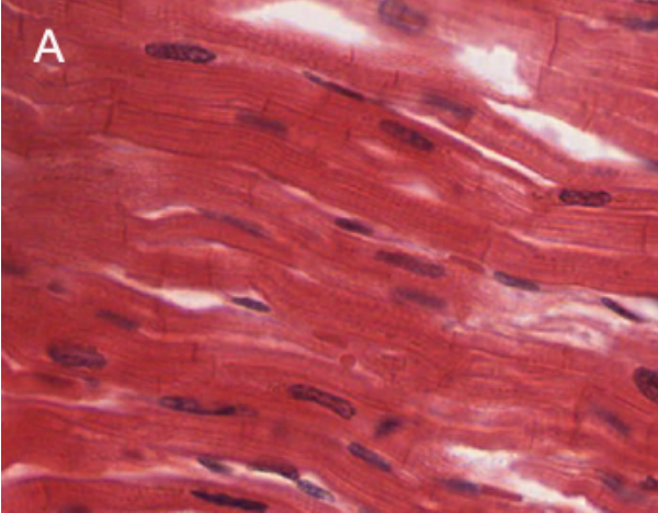
Cardiac muscle is only found in the heart. Its fibers are longer than they are wide, and they are striated, like skeletal muscle fibers. But, unlike skeletal muscle fibers, cardiac muscle
fibers have distinct ends to them, called intercalated discs. These are dark lines that run from one side of the fiber to the other. The intercalated discs are not much thicker than the striations, but they are usually darker and so distinct for that reason. One cardiac muscle fiber is the material between two intercalated discs. Cardiac muscle fibers are mononucleate, with only one nucleus per fiber, and they can sometimes be branched.
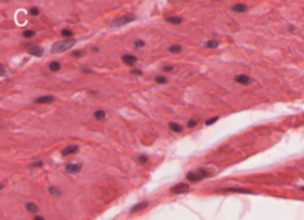
Smooth muscle is found in the walls of internal organs, such as the organs of the digestive tract, blood vessels, and others. It consists of mononucleate fibers with tapered edges. No striations are visible in smooth muscle under the microscope. Because smooth muscle often is wrapping around the organ it is associated with, it can be hard to find an entire smooth muscle fiber in profile in a tissue slice on a microscope slide. Most of the fibers will be sectioned at angles or will be difficult to get into a single plane of focus, but a little bit of searching can usually turn up some with all of the defining characteristics visible.
LAB 3 EXERCISE \(\PageIndex{1}\)
In each of the three photomicrographs below, identify which type of muscle is present. List the defining visual characteristics of that type of muscle, and draw arrows to features on the photograph that illustrate each characteristic.
 |
Muscle type: _________________ Visual characteristics 1. 2. 3. 4. |
 |
Muscle type: _________________ Visual characteristics 1. 2. 3. 4. |
 |
Muscle type: _________________ Visual characteristics 1. 2. 3. 4. |
-
- Obtain a slide of cardiac muscle tissue from the slide box.
- View the slide on an appropriate objective.
- Fill out the blanks next to your drawing.
- In the circle below, draw a representative sample of key features you identified, taking care to correctly and clearly draw their true shapes and directions. Draw your structures proportionately to their size in your microscope’s field of view.
- Obtain a slide of skeletal muscle tissue from the slide box.
- View the slide on an appropriate objective.
- Fill out the blanks next to your drawing.
- In the circle below, draw a representative sample of key features you identified, taking care to correctly and clearly draw their true shapes and directions. Draw your structures proportionately to their size in your microscope’s field of view.
- Obtain a slide of smooth muscle tissue from the slide box.
- View the slide on an appropriate objective.
- Fill out the blanks next to your drawing.
- In the circle below, draw a representative sample of key features you identified, taking care to correctly and clearly draw their true shapes and directions. Draw your structures proportionately to their size in your microscope’s field of view.
LICENSES AND ATTRIBUTIONS
A&P Labs. Authored by: Ross Whitwam. Provided by: Mississippi University for Women. Located at: http://www.muw.edu. License: CC BY-SA: Attribution-ShareAlike
CC LICENSED CONTENT, SPECIFIC ATTRIBUTION
Exercise \(\PageIndex{1}\)A. Authored by: Kent Christensen, Ph.D., J. Matthew Velkey, Ph.D., Lloyd M. Stoolman, M.D., Laura Hessler, and Diedra Mosley-Brower. Provided by: University of Michigan Histology and Virtual Microscopy Learning Resources. Located at: 141.214.65.171/Histology/Cardiovascular%20System/098HE_HISTO_40X.svs/view.apml. License: CC BY-NC-SA: Attribution-NonCommercial-ShareAlike
Exercise \(\PageIndex{1}\)B. Authored by: Kent Christensen, Ph.D., J. Matthew Velkey, Ph.D., Lloyd M. Stoolman, M.D., Laura Hessler, and Diedra Mosley-Brower. Provided by: University of Michigan Histology and Virtual Microscopy Learning Resources. Located at: 141.214.65.171/Histology/Basic%20Tissues/Muscle/058L_HISTO_40X.svs/view.apml. License: CC BY-NC-SA: Attribution-NonCommercial-ShareAlike
Exercise \(\PageIndex{1}\)C. Authored by: Kent Christensen, Ph.D., J. Matthew Velkey, Ph.D., Lloyd M. Stoolman, M.D., Laura Hessler, and Diedra Mosley-Brower. Provided by: University of Michigan Histology and Virtual Microscopy Learning Resources. Located at: 141.214.65.171/Histology/Basic%20Tissues/Muscle/169_HISTO_40X.svs/view.apml. License: CC BY-NC-SA: Attribution-NonCommercial-ShareAlike