
2.2: Body Chemicals - Types, Structures, and Uses
- Page ID
- 83966
\( \newcommand{\vecs}[1]{\overset { \scriptstyle \rightharpoonup} {\mathbf{#1}} } \)
\( \newcommand{\vecd}[1]{\overset{-\!-\!\rightharpoonup}{\vphantom{a}\smash {#1}}} \)
\( \newcommand{\id}{\mathrm{id}}\) \( \newcommand{\Span}{\mathrm{span}}\)
( \newcommand{\kernel}{\mathrm{null}\,}\) \( \newcommand{\range}{\mathrm{range}\,}\)
\( \newcommand{\RealPart}{\mathrm{Re}}\) \( \newcommand{\ImaginaryPart}{\mathrm{Im}}\)
\( \newcommand{\Argument}{\mathrm{Arg}}\) \( \newcommand{\norm}[1]{\| #1 \|}\)
\( \newcommand{\inner}[2]{\langle #1, #2 \rangle}\)
\( \newcommand{\Span}{\mathrm{span}}\)
\( \newcommand{\id}{\mathrm{id}}\)
\( \newcommand{\Span}{\mathrm{span}}\)
\( \newcommand{\kernel}{\mathrm{null}\,}\)
\( \newcommand{\range}{\mathrm{range}\,}\)
\( \newcommand{\RealPart}{\mathrm{Re}}\)
\( \newcommand{\ImaginaryPart}{\mathrm{Im}}\)
\( \newcommand{\Argument}{\mathrm{Arg}}\)
\( \newcommand{\norm}[1]{\| #1 \|}\)
\( \newcommand{\inner}[2]{\langle #1, #2 \rangle}\)
\( \newcommand{\Span}{\mathrm{span}}\) \( \newcommand{\AA}{\unicode[.8,0]{x212B}}\)
\( \newcommand{\vectorA}[1]{\vec{#1}} % arrow\)
\( \newcommand{\vectorAt}[1]{\vec{\text{#1}}} % arrow\)
\( \newcommand{\vectorB}[1]{\overset { \scriptstyle \rightharpoonup} {\mathbf{#1}} } \)
\( \newcommand{\vectorC}[1]{\textbf{#1}} \)
\( \newcommand{\vectorD}[1]{\overrightarrow{#1}} \)
\( \newcommand{\vectorDt}[1]{\overrightarrow{\text{#1}}} \)
\( \newcommand{\vectE}[1]{\overset{-\!-\!\rightharpoonup}{\vphantom{a}\smash{\mathbf {#1}}}} \)
\( \newcommand{\vecs}[1]{\overset { \scriptstyle \rightharpoonup} {\mathbf{#1}} } \)
\( \newcommand{\vecd}[1]{\overset{-\!-\!\rightharpoonup}{\vphantom{a}\smash {#1}}} \)
\(\newcommand{\avec}{\mathbf a}\) \(\newcommand{\bvec}{\mathbf b}\) \(\newcommand{\cvec}{\mathbf c}\) \(\newcommand{\dvec}{\mathbf d}\) \(\newcommand{\dtil}{\widetilde{\mathbf d}}\) \(\newcommand{\evec}{\mathbf e}\) \(\newcommand{\fvec}{\mathbf f}\) \(\newcommand{\nvec}{\mathbf n}\) \(\newcommand{\pvec}{\mathbf p}\) \(\newcommand{\qvec}{\mathbf q}\) \(\newcommand{\svec}{\mathbf s}\) \(\newcommand{\tvec}{\mathbf t}\) \(\newcommand{\uvec}{\mathbf u}\) \(\newcommand{\vvec}{\mathbf v}\) \(\newcommand{\wvec}{\mathbf w}\) \(\newcommand{\xvec}{\mathbf x}\) \(\newcommand{\yvec}{\mathbf y}\) \(\newcommand{\zvec}{\mathbf z}\) \(\newcommand{\rvec}{\mathbf r}\) \(\newcommand{\mvec}{\mathbf m}\) \(\newcommand{\zerovec}{\mathbf 0}\) \(\newcommand{\onevec}{\mathbf 1}\) \(\newcommand{\real}{\mathbb R}\) \(\newcommand{\twovec}[2]{\left[\begin{array}{r}#1 \\ #2 \end{array}\right]}\) \(\newcommand{\ctwovec}[2]{\left[\begin{array}{c}#1 \\ #2 \end{array}\right]}\) \(\newcommand{\threevec}[3]{\left[\begin{array}{r}#1 \\ #2 \\ #3 \end{array}\right]}\) \(\newcommand{\cthreevec}[3]{\left[\begin{array}{c}#1 \\ #2 \\ #3 \end{array}\right]}\) \(\newcommand{\fourvec}[4]{\left[\begin{array}{r}#1 \\ #2 \\ #3 \\ #4 \end{array}\right]}\) \(\newcommand{\cfourvec}[4]{\left[\begin{array}{c}#1 \\ #2 \\ #3 \\ #4 \end{array}\right]}\) \(\newcommand{\fivevec}[5]{\left[\begin{array}{r}#1 \\ #2 \\ #3 \\ #4 \\ #5 \\ \end{array}\right]}\) \(\newcommand{\cfivevec}[5]{\left[\begin{array}{c}#1 \\ #2 \\ #3 \\ #4 \\ #5 \\ \end{array}\right]}\) \(\newcommand{\mattwo}[4]{\left[\begin{array}{rr}#1 \amp #2 \\ #3 \amp #4 \\ \end{array}\right]}\) \(\newcommand{\laspan}[1]{\text{Span}\{#1\}}\) \(\newcommand{\bcal}{\cal B}\) \(\newcommand{\ccal}{\cal C}\) \(\newcommand{\scal}{\cal S}\) \(\newcommand{\wcal}{\cal W}\) \(\newcommand{\ecal}{\cal E}\) \(\newcommand{\coords}[2]{\left\{#1\right\}_{#2}}\) \(\newcommand{\gray}[1]{\color{gray}{#1}}\) \(\newcommand{\lgray}[1]{\color{lightgray}{#1}}\) \(\newcommand{\rank}{\operatorname{rank}}\) \(\newcommand{\row}{\text{Row}}\) \(\newcommand{\col}{\text{Col}}\) \(\renewcommand{\row}{\text{Row}}\) \(\newcommand{\nul}{\text{Nul}}\) \(\newcommand{\var}{\text{Var}}\) \(\newcommand{\corr}{\text{corr}}\) \(\newcommand{\len}[1]{\left|#1\right|}\) \(\newcommand{\bbar}{\overline{\bvec}}\) \(\newcommand{\bhat}{\widehat{\bvec}}\) \(\newcommand{\bperp}{\bvec^\perp}\) \(\newcommand{\xhat}{\widehat{\xvec}}\) \(\newcommand{\vhat}{\widehat{\vvec}}\) \(\newcommand{\uhat}{\widehat{\uvec}}\) \(\newcommand{\what}{\widehat{\wvec}}\) \(\newcommand{\Sighat}{\widehat{\Sigma}}\) \(\newcommand{\lt}{<}\) \(\newcommand{\gt}{>}\) \(\newcommand{\amp}{&}\) \(\definecolor{fillinmathshade}{gray}{0.9}\)The atoms, ions, and molecules contained in the body or making up its parts are derived from atoms, ions, and molecules in the diet. However, many dietary substances are modified before entering the body and becoming integral parts of it. The following discussion focuses on body chemicals, which are chemicals that are already internal or compose parts of the body. Dietary chemicals and their relationships to body chemicals are discussed in Chapter 11.
Approximately 70 percent of the body consists of water molecules (Figure 2.6). Water dissolves and transports materials, lubricates and cushions structures, regulates temperature, and modulates osmotic pressure and acid/base balance. The other body chemicals are of myriad variety and complexity. Some of them are minerals that are present as ions (e.g., sodium ions). The bulk of the remaining body chemicals are molecules, many of which share common features and therefore can be grouped into broad categories. These categories are carbohydrates, lipids, proteins, and nucleic acids.

Carbohydrates
The smallest and simplest carbohydrate molecules are simple sugar molecules called monosaccharides. One of the most abundant monosaccharides is glucose (Figure 2.7).

Other carbohydrate molecules consist of monosaccharide molecules linked together. A carbohydrate containing two monosaccharides is called a disaccharide, and carbohydrates consisting of many monosaccharides are called polysaccharides. Polysaccharides may contain from dozens to thousands of monosaccharide molecules, which are arranged to form straight or branched chains (Figure 2.8). A common polysaccharide is glycogen, which is made of glucose. Carbohydrates are used for storing and supplying energy and for building materials and receptor molecules for communication.

Nucleic Acids
Nucleic acid molecules contain dozens to thousands of small molecules linked to form chains (Figure 2.9).

The small molecular units in nucleic acids are called nucleotides. Each nucleotide contains a sugar molecule with five carbon atoms, a molecular fragment containing phosphorus (a phosphate group), and a molecular fragment containing nitrogen (a nitrogen base). There are five types of nitrogen bases, which can be designated by the initials A, G, C, U, and T (adenine, guanine, cytosine, uracil, and thymine). Though many nucleotides are linked to form nucleic acids, others remain separate. These individual nucleotides often have additional phosphate groups or other modifications and are used to transfer energy from chemical reactions to other activities. Two of the most abundant and widely used nucleotides are adenosine diphosphate (ADP) and adenosine triphosphate (ATP) (Figure 2.10) .

The nucleic acids are divided into two main classes based on the type of sugar used in their nucleotides. The sugars in nucleic acids called ribonucleic acid (RNA) have one more oxygen atom than do the sugars in nucleic acids called deoxyribonucleic acid (DNA). A second difference between RNA and DNA is that nucleotides in RNA contain A, G, C, and U, while nucleotides in DNA contain A, G, C, and T. Most DNA molecules contain two chains of DNA linked side by side by matching nitrogen bases in complementary pairs. The double strand of DNA is twisted in the form of a double helix, which resembles a twisted ladder (Figure 2.9). Other combinations of DNA, RNA, and individual nucleotides are also made by matching complementary nitrogen bases.
Though there are only four different nucleotides in RNA and DNA, nucleic acid molecules in both classes have enormous variety. The reasons are the same as those permitting the writing of an essentially limitless variety of sentences using the limited number of letters in the alphabet. That is, nucleic acid molecules may be of different lengths, and more importantly, the nucleotides may be arranged in a virtually infinite number of ratios and sequences. As with the letters in a sentence, the nucleotide sequence in each nucleic acid molecule contains a message. Also like letters in sentences, changes in either the nitrogen bases employed or the sequence of the bases can change the message or make it meaningless (e.g., dog_dig_god_dgo). When complementary nitrogen bases are matched, the encoded message can be deciphered in cells and the information it contains can be used as instructions. Many of these instructions direct the formation of protein molecules by a process called translation.
Proteins
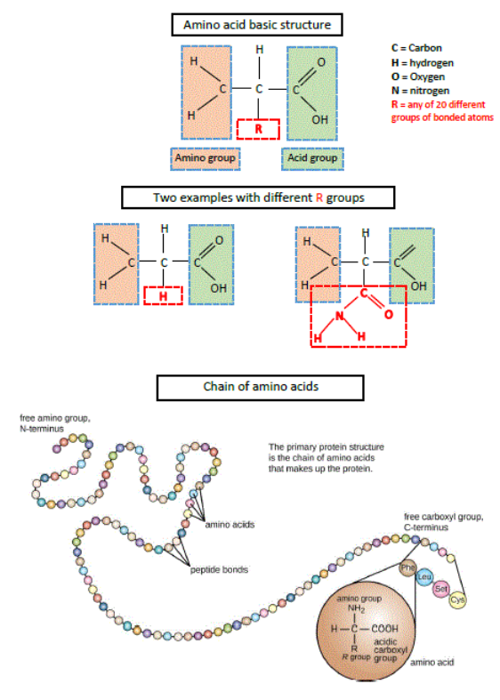
Like carbohydrate and nucleic acid molecules, protein molecules consist of many small molecules linked to form chains (Figure 2.11) (a) Two amino acids, each with a short segment of -N-C-C- and side groups of hydrogen (H), oxygen (O), and R-groups. (b) A chain of amino acids (i.e., a polypeptide). The small molecular units in proteins are called amino acids, of which there are 20 types.
Compared with many polysaccharides and nucleic acid molecules, protein molecules show much more variety in structure and functioning. This variety exists for five reasons. First, the different amino acids may be combined in a virtually infinite variety of lengths, ratios, and sequences. Second, polysaccharides and nucleic acids contain few different types of units, while each protein molecule may contain all 20 types of amino acids. Third, the shapes of different amino acids cause protein chains to form various twists and bends. Fourth, some amino acids link to others in a protein, causing it to take on and maintain other twists and bends (Figure 2.12). Fifth, some amino acids link to amino acids in adjacent protein molecules, causing additional changes in configurations and positions.

(Animated view: https://upload.wikimedia.org/Wikipedia/commons/9/91/Hemoglobin_1hho.gif)
Many twists, bends, and links, and therefore the shape and position of each protein, are determined by the conditions surrounding it (e.g., temperature, amount of acid present). When these conditions change, the shape and position of a protein may shift, and slight changes in these conditions cause dramatic shifts in some proteins. Furthermore, as with any tool or device, the shape and position of each protein determine what functions it can perform and how well it can perform them (Figure 2.13). This is a major reason body structures retain their normal shapes and proper functioning only under homeostatic conditions. Proteins serve as building materials; receptor molecules and hormones for communication; enzymes for regulating reactions; and antibodies for defense.


Lipids
Lipid molecules are placed into the same category because at least a large portion of each molecule does not dissolve well in water. We will mention only a few types of body lipid molecules.

Among the most water‑repellent lipid molecules are the triglycerides, also called fat. Triglycerides contain a backbone made of a glycerol molecule with three fatty acid arms protruding from it (Figure 2.14) . Fatty acids contain up to 20 carbon atoms in a row (Figure 2.15). The body also contains glycerol combined with only one or two fatty acid molecules, forming monoglycerides and diglycerides, respectively. Glycerides store and supply energy.

The carbon atoms in some fatty acids are linked to the maximum number of hydrogen atoms; such fatty acids are called saturated fatty acids (Figure 2.15). In other fatty acids, additional hydrogen atoms can be linked to the carbon atoms, and these fatty acids are called unsaturated fatty acids. Monounsaturated fatty acids have only one location that permits the addition of hydrogen atoms (Figure 2.15), while polyunsaturated fatty acids have more than one such location (Figure 2.15c, Figure 2.15d). Similar terms are applied to triglycerides with fatty acids able to hold zero, one, or more than one additional hydrogen atom (i.e., saturated fat, monounsaturated fat, and polyunsaturated fat).
Often, glycerol linked to two fatty acids is also linked to a molecular fragment containing phosphorus, forming phospholipids (Figure 2.14b) . While the regions of phospholipids containing fatty acids repel water, the region containing phosphorus attracts water. These properties cause phospholipids to align and form double‑layered membranes in the watery internal environment of the body.
A third group of lipid molecules are called steroids. Their carbon atoms are linked to form rings (Figure 2.16) . Well‑known examples of steroids include cholesterol, which is used as a building material, and sex steroids such as testosterone, estrogen, and progesterone, which serve as hormones for communication.

Molecular Complexes
Though many carbohydrate, nucleic acid, protein, and lipid molecules are not joined to any others in these categories, many molecules link together and form molecular complexes. Combinations of carbohydrate and protein are called glycoproteins or mucopolysaccharides, depending on whether carbohydrate or protein predominates. Combinations of nucleic acids and proteins are called nucleoproteins, and combinations of lipids and proteins are called lipoproteins. The formation of molecular complexes can modify the physical properties (e.g., flexibility) and activities (e.g., accessibility) of the molecules involved.
Free radicals
A free radical (*FR) is an atom or molecule with an unpaired electron (* = an unpaired electron). For example, an ordinary oxygen molecule is made of two oxygen atoms, and it contains 16 electrons. (Figure 2.3). If another electron is added to the molecule, one electron would be unpaired. The resulting molecule would be a free radical called a superoxide radical. Some free radicals are made from highly reactive substances that contain oxygen, and some free radicals produce such highly reactive substances. These substances, which are not free radicals themselves, are called reactive oxygen species (ROS).
Small *FRs in the body include the superoxide radical (*O2-), the hydroxyl radical (*OH), and the nitric oxide radical (*NO). Larger free radicals contain an organic molecule, such as a fatty acid, combined with extra oxygen. Examples include the alkoxyl radical (*RO) and the peroxyl radical (*ROO), where the R represents the original organic molecule. Peroxyl radicals containing a fatty acid are also called lipid peroxides (*LPs).
Reactive oxygen species in the body include hydrogen peroxide (H2O2), peroxynitrite anion (ONOO-), organic hydroperoxide (ROOH), plus certain amino acids (e.g., tryptophan) and other substances produced during cell metabolism. An organic hydroperoxide containing a fatty acid is also called a lipid hydroperoxide.
These *FRs and ROS are not equally important. Superoxide radicals and H2O2 result in damage only when they fuel reactions that produce hydroxyl radicals (*OH) or ONOO-, because these latter two substances are among the fastest acting and most toxic to the body. Free radicals containing fatty acids react much slower than these.
Importance in aging
Free radicals seem to be implicated in aging. Reasons include the apparent negative correlations between the following; mean longevities (MLs) and maximum longevities (XLs) of species and their rates of forming *FRs and ROS; MLs and XLs and their rates of developing damage from *FRs; age and the level of *FR defenses in some species; and age and the rate of repairing damage from *FRs.
Other reasons include the apparent positive correlations between the following; MLs and XLs of some species and their levels of *FR defenses; MLs and anti-*FR supplements (i.e., antioxidants); age and the rate of *FR formation; age and the amount of damage from *FRs; age-related diseases and *FRs (e.g., atherosclerosis, heart attacks, strokes, Alzheimer's diseases, parkinsonism, cataracts, kidney failure, cancers).
Formation of *FRs and ROS
Some *FRs and ROS produced by the body are useful. Examples include; *NO for signals among neurons; *NO to cause blood vessel dilation for blood pressure regulation; H2O2 to destroy bacteria; and other defense *FRs and ROS produced during defense processes such as inflammation and immune responses. Many *FRs and ROS are produced as by-products from other useful reactions. Examples include cells producing *O2-, H2O2, and *OH and other *FRs and ROS when cells obtain energy from nutrients; detoxifying certain plant materials; break down of dopamine (DOPA) and fatty acids; using iron or copper in reactions; and performing reactions peculiar to their special functions. Finally, *FRs and ROS result from unwanted conditions including exposure to ultraviolet light, internal bleeding, reversing unduly restricted blood flow, and reactions between glucose and proteins (see Glycation below).
Conditions that increase *FR production include elevated amounts of O2 in the body; high blood LDLs; high blood glucose levels; excess vitamin C; very high level of exercise; skin photosensitizers including certain cosmetics, medications, and air pollutants; menopause; and smoking; and increasing age. Conditions that decrease *FR production include reduced intake of polyunsaturated lipids; moderate exercise; increased intake of cruciferous vegetables (e.g., broccoli, cauliflower, cabbage); reduced intake of copper, iron, or magnesium; and reduced intake of certain amino acids (e.g., histidine, lysine).
Common chemical reactions by which *FRs and ROS are formed include the following, where e- represents an electron, H+ represents a hydrogen ion, and H- represents a hydrogen atom sharing electrons with another atom or molecule.
\(\begin{aligned}
& \mathrm{O}_2+\mathrm{e}^{-} \rightarrow{ }^* \mathrm{O}_2^{-} \\
& * \mathrm{O}_2^{-}+\mathrm{e}^{-}+2 \mathrm{H}^{+} \rightarrow \mathrm{H}_2 \mathrm{O}_2 \\
& \mathrm{H}_2 \mathrm{O}_2+\mathrm{e}^{-} \rightarrow \mathrm{OH}^{-}+{ }^* \mathrm{OH}
\end{aligned}\)
These reactions are common when cells use oxygen to derive energy from nutrients (see Mitochondria below); where iron or copper ions exist; in skin struck by ultraviolet light; and when enzymes in the brain use monoamine oxidases (MAOs).
Common reactions with *FRs involving fatty acids are shown next, where PUFA represents a polyunsaturated fatty acid. These reactions occur in three processes called initiation, propagation, and termination. The reactions are chain reactions because propagation or reinitiation may occur repeatedly before termination occurs. Each time propagation or reinitiation occurs, another damaged fatty acid is produced and a new *FR is formed, leaving a wake of oxidized damaged molecules. Oxidation damage from free radicals spreads though the cell like oxidation damage from a fire spreads from one house to the next along a street in a neighborhood. Since free radicals form in many areas in a cell, many molecular “fires” can be spreading at once through a molecular “neighborhood” such as a cell membrane (Figure 2.18).


initiation
H-PUFA1 + *FR → *PUFA1 + H-R
*PUFA1 molecule rearranges itself
rearranged *PUFA1 + O2 → *PUFA1-O-O (peroxyl radical = *LP)
propagation
*PUFA1-O-O + H-PUFA2 → H-PUFA1-O-O (a damaged fatty acid {lipid hydroperoxide}) + *PUFA2 (new *FR)
repeated propagations
*PUFA2 + O2 → *PUFA2-O-O
*PUFA2-O-O + H-PUFA3 → H-PUFA2-O-O (another damaged fatty acid) + *PUFA3 (new *FR)
etc., etc. → many H-PUFAx-O-O (many damaged fatty acids) + another *PUFAn
(another new *FR)
termination
*PUFAn + H-R → H-PUFAn + R
or
*PUFAn + *PUFAn → R-O-O-O-O-R → (toxic substances) + O2
reinitiation
R-O-O-O-O-R + iron or copper → *PUFA-O or *PUFA-O-O
Effects from *FRs
Free radicals damage body molecules by taking electrons from molecules, a process called oxidation. This process alters the shapes and functions of the molecules, causing the body to sustain structural damage and malfunctions.
The main types of molecules affected are nucleic acids, proteins, and lipids. The consequences from even a small alteration in DNA can be devastating because the effect is multiplied during protein production. Also, damaged DNA may be unable to be replicated, preventing cells from reproducing by mitosis. Alternatively, some types of DNA damage promote cancer.
Damage to proteins and lipids causes abnormal cell and body structures and operations. Protein molecules such as those in tendons and ligaments become excessively joined together, and the functions of enzymes and cell membranes become abnormal. Damaged mitochondria are often unable to produce adequate energy for maximum cell activity. *FR damage can initiate inflammation, cause excess blood clotting, and promote several diseases, especially cataracts and atherosclerosis. Some *FR effects on proteins and lipids compound problems by increasing the rate of *FR production, reducing the body's ability to eliminate *FRs, and decreasing the ability to repair or remove damaged molecules.
Clearly, free radicals damage a variety of essential bodily components and alter body functions. To defend against *FR damage, the body has mechanisms to eliminate free radicals and to remove and repair molecules damaged by them. Substances called antioxidants destroy them by helping to create pairs of electrons. Examples of dietary antioxidants include vitamin E, vitamin C, beta-carotene (a vitamin A precursor), and substances in fruits and vegetables. The body also makes many antioxidants (e.g., melatonin, glutathione, albumin, uric acid), and can even recycle some of these antioxidants after they have neutralized *FRs.
The body also makes enzymes to divert *FR production and to speed up *FR elimination. Extremely important examples remove superoxide radicals (superoxide dismutase) and H2O2 (glutathione peroxidase, catalase). These enzymes are especially important because they prevent the formation of *OH, the most reactive and harmful free radical. Selenium is a vital dietary constituent because it helps an enzyme (glutathione peroxidase) remove H2O2.
Glycation
Glucose joins chemically with certain amino acids in proteins. No enzymes are needed for these reaction, and they usually occur with the side groups of arginine and lysine (Figure 2.11) see R-). The products are altered amino acids attached to glucose (i.e., Amadori products). The amino acid/glucose portion may break down to form a distorted protein plus an ROS (e.g., H2O2), and the ROS may form *FRs. Distorting a protein in this way is like bending and twisting a wire clothes hanger until it is useless.
Alternatively, the amino acid/glucose portion may join with others on the same or different protein molecules. This is called a Maillard reaction. The results are cross-links among the protein strands, and they then resemble strips of tape that have become stuck and tangled together. The cross-links are extremely stable and long-lasting, and common ones are called pentosidine.
The reactions forming glucose cross-links between proteins are called nonenzymatic glycation because glucose reacts without the use of enzymes. They are also called glycation, or glycoxydation because *FRs help as oxidizing substances during the process. The glycated proteins are damaged and distorted, turn a darker color, and are called advanced glycation end-products (AGEs). Other sugars in the body perform similar reactions that produce altered proteins, and AGEs may be formed by other chemical pathways. Most types of protein in the body are subject to glycation.
Effects Studies on AGEs began in the 1970s, and studies on their relationship to aging began in the 1980s. To date, little is known about the exact identity and characteristics of AGEs formed by glycation. Though they do not seem to cause aging, they accumulate with aging and seem to contribute to aging and age-related diseases. For example, the rate of glycation is inversely correlated with the XL of animals, though the amount of AGEs formed is not related to XLs. Also, *FRs and glycation are related in at least four ways; *FRs increase glycation; glycation increases *FRs; both processes increase the effects from *FRs by damaging defense and repair enzymes.
There is no known benefit from glycation or from AGEs. However, glycation adversely affects all body parts and functions directly by distorting the proteins involved. Other adverse effects from glycation include indirect damage to DNA and lipids though the *FRs produced from glycation and by the abnormal operations of damaged proteins; body stiffening; poor movement of materials between cells, and distorted signaling of cells (see Intercellular Materials below); reduced ability to control blood vessels and blood pressure; damage to blood vessel linings and atherosclerosis; increased blood clotting; increased development of Alzheimer's disease; kidney injury and eye damage from AGEs in blood vessel walls; amplification of most effects from diabetes mellitus; and tissue damage and inflammation by activating defense cells. For example, macrophages and monocytes are activated when AGEs attach to their cell membrane receptors for AGEs. These receptors are called receptors for AGES (RAGEs).
Keeping blood glucose levels within normal levels seems to be a main way to minimize glycation and its adverse effects.