
6.5: Small Intestine
- Page ID
- 63659

\( \newcommand{\vecs}[1]{\overset { \scriptstyle \rightharpoonup} {\mathbf{#1}} } \)
\( \newcommand{\vecd}[1]{\overset{-\!-\!\rightharpoonup}{\vphantom{a}\smash {#1}}} \)
\( \newcommand{\id}{\mathrm{id}}\) \( \newcommand{\Span}{\mathrm{span}}\)
( \newcommand{\kernel}{\mathrm{null}\,}\) \( \newcommand{\range}{\mathrm{range}\,}\)
\( \newcommand{\RealPart}{\mathrm{Re}}\) \( \newcommand{\ImaginaryPart}{\mathrm{Im}}\)
\( \newcommand{\Argument}{\mathrm{Arg}}\) \( \newcommand{\norm}[1]{\| #1 \|}\)
\( \newcommand{\inner}[2]{\langle #1, #2 \rangle}\)
\( \newcommand{\Span}{\mathrm{span}}\)
\( \newcommand{\id}{\mathrm{id}}\)
\( \newcommand{\Span}{\mathrm{span}}\)
\( \newcommand{\kernel}{\mathrm{null}\,}\)
\( \newcommand{\range}{\mathrm{range}\,}\)
\( \newcommand{\RealPart}{\mathrm{Re}}\)
\( \newcommand{\ImaginaryPart}{\mathrm{Im}}\)
\( \newcommand{\Argument}{\mathrm{Arg}}\)
\( \newcommand{\norm}[1]{\| #1 \|}\)
\( \newcommand{\inner}[2]{\langle #1, #2 \rangle}\)
\( \newcommand{\Span}{\mathrm{span}}\) \( \newcommand{\AA}{\unicode[.8,0]{x212B}}\)
\( \newcommand{\vectorA}[1]{\vec{#1}} % arrow\)
\( \newcommand{\vectorAt}[1]{\vec{\text{#1}}} % arrow\)
\( \newcommand{\vectorB}[1]{\overset { \scriptstyle \rightharpoonup} {\mathbf{#1}} } \)
\( \newcommand{\vectorC}[1]{\textbf{#1}} \)
\( \newcommand{\vectorD}[1]{\overrightarrow{#1}} \)
\( \newcommand{\vectorDt}[1]{\overrightarrow{\text{#1}}} \)
\( \newcommand{\vectE}[1]{\overset{-\!-\!\rightharpoonup}{\vphantom{a}\smash{\mathbf {#1}}}} \)
\( \newcommand{\vecs}[1]{\overset { \scriptstyle \rightharpoonup} {\mathbf{#1}} } \)
\( \newcommand{\vecd}[1]{\overset{-\!-\!\rightharpoonup}{\vphantom{a}\smash {#1}}} \)
Partially digested food leaves the stomach through a sphincter located at the juncture between the stomach and small intestine. The sphincter controls the passage of food into the small intestine, and contracts to help prevent backtracking.
The adult small intestine is about 1½ inches wide and about 20 feet long when it relaxes, and about 10 feet when it contracts—it’s not very small! It’s the main site of digestion and absorption. The first 12 inches or so is called the duodenum (because so much happens here, this uppermost section has its own name-—derived from duodeni, Latin for twelve each). Except for the duodenum, the small intestine is unattached, which allows such a long tube to be stuffed into such a small space (Figure 6.1).
The lining of the small intestine is truly remarkable. Like a wadded terry cloth towel (that quickly absorbs water), its surface is expanded by folds and loops to quickly absorb food (Figure 6.2). The folds are densely covered with villi (finger-like projections), each of which is covered with microvilli (the “ruffled” membrane of intestinal cells).

Many proteins are embedded in these microvilli (see cross section of cell membrane in Figure 6.2), and are involved in digestion as well as absorption. Examples include sucrase, maltase, and lactase—digestive enzymes anchored in the membrane and protruding into the intestinal passageway. Although these enzymes are named for the double sugars (sucrose, maltose, lactose) that they digest, sucrase and maltase also can digest starch. This gives the digestive tract a large and versatile starch-digesting capacity: sucrase and maltase, and the starch-digesting enzymes of the saliva (salivary amylase) and pancreas (pancreatic amylase). Starch is the predominant source of calories for the world’s population.
“Starch blocker” pills (amylase blockers) were a popular, short-term money-maker: popular, because the sales pitch was irresistible (eat all the pasta and potatoes you want without gaining weight); short-term, because the pills didn’t block starch digestion. The pills blocked only a small amount of amylase (we make a lot). Also, maltase together with sucrase can digest starch.
The surface area of our small-intestinal lining is about 1800 square feet, about the floor space of a three-bedroom house. No wonder our food’s absorbed so efficiently! The lining is renewed continually; it’s completely replaced about every 3 days.
The cells that make up this lining are made at the base of the villi (in the crypts, as shown in Figure 6.2) and then migrate to the tips of the villi where, at an “old age” of 3 days, they come off and become a part of the food that’s digested and absorbed there.
About 20-50 million cells per minute are shed into the adult intestine. In fact, dietary protein represents only about half of the protein that we digest and absorb. About 25% comes from shed intestinal cells and another 25% from protein in digestive secretions (remember, enzymes are proteins).
Digestion and absorption take place mainly in the first half of the small intestine (protein, fat, carbohydrate, vitamins, and minerals are mostly absorbed here). Pancreatic and liver secretions enter the small intestine through the bile duct (Figure 6.1).
The pancreas secretes an alkaline solution of sodium bicarbonate (baking soda) that neutralizes the acidity of the material coming from the stomach. The pancreas also secretes digestive enzymes that break apart starch, protein, and fat. (As food, pancreas and thymus are called sweetbread; tripe is part of the stomach of a ruminant. Intestine can be used as casing for sausage.)
The liver’s job in digestion is to make and secrete bile. Bile emulsifies fat, making it easier to digest. As discussed earlier, emulsification finely divides the fat and suspends it in a water-based liquid. It’s like homogenizing whole milk, finely dividing the fat (cream) so that it stays suspended rather than rising to the top. In homogenization, globs of fat are made smaller by forcing the milk through a small nozzle, whereas an emulsifier such as bile has chemical properties that finely divide the fat.
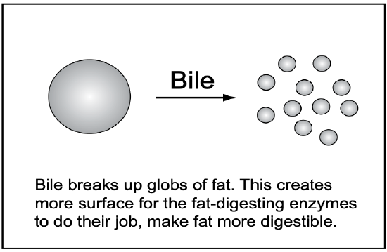
Finely dividing the fat increases its exposed surface. This is crucial. The fat-digesting enzymes are in a watery liquid and can’t penetrate the fat. The enzymes can reach and digest only the exposed surface. Bile is stored and concentrated in the gallbladder, which contracts and releases bile when food enters the small intestine.
A gallbladder isn’t essential. Many people have it removed without serious consequences. Without a gallbladder, bile enters the duodenum (the top of the small intestine) directly from the liver in steady and less concentrated amounts. Most fatty foods are comfortably digested, unless a lot is eaten in a short time.
By the time the intestinal contents reach the lower half of the small intestine, most of the nutrients have been digested and absorbed. But the lower part of the small intestine is the site of vitamin B12 absorption. Intrinsic factor (secreted in the stomach) bound to B12 is absorbed here.
About 90% of the bile acids secreted earlier into the duodenum are also absorbed in the lower small intestine and recycled via the blood to the liver to be made again into bile. Bile acids are recycled about twice during a meal. Without this efficient recycling, the liver wouldn’t have enough bile in time to digest the fat in a typical meal.
People who tend to put on weight bemoan the fact that digestion and absorption are so efficient. In the 1970s, some morbidly obese patients underwent a desperate procedure that speaks to the efficiency of the small intestine. All but about two or three feet of the small intestine was surgically bypassed so that they could overeat and steadily lose weight. As you might expect, an undesirable side effect was massive amounts of stool. Some patients also developed more serious side effects (e.g., liver failure).
Duodenal Ulcers
The acidic liquid from the stomach enters the small intestine at the duodenum, and this was thought to be why the duodenum is a common site of ulcers. A common treatment was to cut out part of the stomach, so less acid would be made. Drugs (e.g., cimetidine/Tagamet) that reduce the stomach’s acid production have now replaced surgery as the standard treatment.
To treat ulcers, physicians used to prescribe a diet that avoided certain acid foods. Studies since have shown this was mostly unwarranted. Because the stomach is already very acid, acid food (e.g., citrus fruits) don’t appreciably increase the acidity. Today, physicians commonly limit the “forbidden” list to coffee (caffeinated and decaffeinated) and alcohol, both of which stimulate acid production in the stomach.
Ulcers can bleed or cause pain, but pain isn’t a dependable sign. About half the people diagnosed with duodenal ulcers don’t report pain as a symptom. Sometimes, an ulcer’s discomfort is misinterpreted as hunger, which can unwittingly lead to overeating and weight gain. When there isn’t pain, a bleeding ulcer is often discovered by blood in the stool, or anemia resulting from blood loss.
It’s now known that most duodenal ulcers are caused by the bacteria H. pylori (most of the others are caused by drugs such as aspirin). H. pylori infection can be diagnosed by a blood or breath test, and cured with a precise antibiotic regimen (as can the stomach ulcers caused by H. pylori). This cure has been wonderful for those who had to continually take an acid-reducing drug like Pepcid or Tagamet to control their ulcers. These drugs are now marketed for treating heartburn, and are available without a prescription.
Ulcers also can occur in the esophagus or stomach, but these are much less common than those in the duodenum. Stomach ulcers raise the risk of stomach cancer, but this doesn’t seem to be the case with duodenal ulcers.
Lactose Intolerance
A diminished ability to digest lactose (lactose intolerance)* is common among adults (sucrose intolerance occurs, but is unusual). We’re born with generous amounts of lactase enzyme in our small intestine, to digest the lactose in milk, our sole food as newborns. As the intestine matures, lactase—and the capacity to digest lactose—falls. The high lactase in some adult populations (e.g., Northern Europeans and the pastoral Fulani tribe of Nigeria) might be due to a selective mutation thousands of years ago that conferred an advantage to people who herded milk-producing animals (adults with more lactase could “tolerate” more milk).
Milk is the only natural source of lactose, so milk and foods made with milk are the only foods with lactose. If you consume more lactose than can be digested by your intestinal lactase, you can get gas, diarrhea, or abdominal cramps (these malabsorption symptoms will be discussed later). But such symptoms are common and can be falsely attributed to lactose intolerance.
As infants expand their diet beyond milk, their need for lactase diminishes. Many people describe themselves as lactose-intolerant when, in fact, they have enough lactase to digest lactose when milk is part of a meal.
Studies of lactose intolerance are complicated. For example, some studies give subjects a big dose of plain lactose in water after an overnight fast, which in most cases causes symptoms of malabsorption. Yet, when many of these same people are given a glass of regular milk, their symptoms aren’t much different when compared with a glass of lactose-free milk.
Thirty people (age 18 to 50) who said they were severely lactose intolerant and said they consistently had symptoms after drinking less than a cup of milk were the subjects of a double-blind study. They showed no difference in symptoms when given a cup of regular or lactose-free milk.
If you consider yourself lactose-intolerant, have a couple friends administer a double-blind test (one friend to set up the drinks; the other to give it to you) where you drink regular and lactose-free milk (disguised with chocolate perhaps so you don’t know which is which) and then compare symptoms.
How much lactose-containing food can a person with low lactase comfortably consume? This is best determined by one’s own eating experience (keeping in mind the above study).
Some people can’t tolerate gulping a glass of milk on an empty stomach, but find no discomfort in drinking a glass of milk leisurely throughout a meal. Yogurt and “acidophilus milk” are better tolerated because the added bacteria (Lactobacillus acidophilus) have “predigested” some of the lactose. Cheese is usually well tolerated because it’s high in fat and has relatively little lactose. Fat slows the passage of the lactose from the stomach to the intestine. Also, high-fat food is usually eaten in smaller amounts.

*Don’t confuse lactose intolerance with the allergic reaction that some babies have to cow’s milk. Allergies are hypersensitivity reactions, usually to a specific protein—in this case, protein from cow’s milk. Cow’s milk may cause constipation in some young children. They can be given soy milk, goat’s milk, human milk, etc., instead.