
3.4: Small Intestine
- Page ID
- 20979

\( \newcommand{\vecs}[1]{\overset { \scriptstyle \rightharpoonup} {\mathbf{#1}} } \)
\( \newcommand{\vecd}[1]{\overset{-\!-\!\rightharpoonup}{\vphantom{a}\smash {#1}}} \)
\( \newcommand{\dsum}{\displaystyle\sum\limits} \)
\( \newcommand{\dint}{\displaystyle\int\limits} \)
\( \newcommand{\dlim}{\displaystyle\lim\limits} \)
\( \newcommand{\id}{\mathrm{id}}\) \( \newcommand{\Span}{\mathrm{span}}\)
( \newcommand{\kernel}{\mathrm{null}\,}\) \( \newcommand{\range}{\mathrm{range}\,}\)
\( \newcommand{\RealPart}{\mathrm{Re}}\) \( \newcommand{\ImaginaryPart}{\mathrm{Im}}\)
\( \newcommand{\Argument}{\mathrm{Arg}}\) \( \newcommand{\norm}[1]{\| #1 \|}\)
\( \newcommand{\inner}[2]{\langle #1, #2 \rangle}\)
\( \newcommand{\Span}{\mathrm{span}}\)
\( \newcommand{\id}{\mathrm{id}}\)
\( \newcommand{\Span}{\mathrm{span}}\)
\( \newcommand{\kernel}{\mathrm{null}\,}\)
\( \newcommand{\range}{\mathrm{range}\,}\)
\( \newcommand{\RealPart}{\mathrm{Re}}\)
\( \newcommand{\ImaginaryPart}{\mathrm{Im}}\)
\( \newcommand{\Argument}{\mathrm{Arg}}\)
\( \newcommand{\norm}[1]{\| #1 \|}\)
\( \newcommand{\inner}[2]{\langle #1, #2 \rangle}\)
\( \newcommand{\Span}{\mathrm{span}}\) \( \newcommand{\AA}{\unicode[.8,0]{x212B}}\)
\( \newcommand{\vectorA}[1]{\vec{#1}} % arrow\)
\( \newcommand{\vectorAt}[1]{\vec{\text{#1}}} % arrow\)
\( \newcommand{\vectorB}[1]{\overset { \scriptstyle \rightharpoonup} {\mathbf{#1}} } \)
\( \newcommand{\vectorC}[1]{\textbf{#1}} \)
\( \newcommand{\vectorD}[1]{\overrightarrow{#1}} \)
\( \newcommand{\vectorDt}[1]{\overrightarrow{\text{#1}}} \)
\( \newcommand{\vectE}[1]{\overset{-\!-\!\rightharpoonup}{\vphantom{a}\smash{\mathbf {#1}}}} \)
\( \newcommand{\vecs}[1]{\overset { \scriptstyle \rightharpoonup} {\mathbf{#1}} } \)
\(\newcommand{\longvect}{\overrightarrow}\)
\( \newcommand{\vecd}[1]{\overset{-\!-\!\rightharpoonup}{\vphantom{a}\smash {#1}}} \)
\(\newcommand{\avec}{\mathbf a}\) \(\newcommand{\bvec}{\mathbf b}\) \(\newcommand{\cvec}{\mathbf c}\) \(\newcommand{\dvec}{\mathbf d}\) \(\newcommand{\dtil}{\widetilde{\mathbf d}}\) \(\newcommand{\evec}{\mathbf e}\) \(\newcommand{\fvec}{\mathbf f}\) \(\newcommand{\nvec}{\mathbf n}\) \(\newcommand{\pvec}{\mathbf p}\) \(\newcommand{\qvec}{\mathbf q}\) \(\newcommand{\svec}{\mathbf s}\) \(\newcommand{\tvec}{\mathbf t}\) \(\newcommand{\uvec}{\mathbf u}\) \(\newcommand{\vvec}{\mathbf v}\) \(\newcommand{\wvec}{\mathbf w}\) \(\newcommand{\xvec}{\mathbf x}\) \(\newcommand{\yvec}{\mathbf y}\) \(\newcommand{\zvec}{\mathbf z}\) \(\newcommand{\rvec}{\mathbf r}\) \(\newcommand{\mvec}{\mathbf m}\) \(\newcommand{\zerovec}{\mathbf 0}\) \(\newcommand{\onevec}{\mathbf 1}\) \(\newcommand{\real}{\mathbb R}\) \(\newcommand{\twovec}[2]{\left[\begin{array}{r}#1 \\ #2 \end{array}\right]}\) \(\newcommand{\ctwovec}[2]{\left[\begin{array}{c}#1 \\ #2 \end{array}\right]}\) \(\newcommand{\threevec}[3]{\left[\begin{array}{r}#1 \\ #2 \\ #3 \end{array}\right]}\) \(\newcommand{\cthreevec}[3]{\left[\begin{array}{c}#1 \\ #2 \\ #3 \end{array}\right]}\) \(\newcommand{\fourvec}[4]{\left[\begin{array}{r}#1 \\ #2 \\ #3 \\ #4 \end{array}\right]}\) \(\newcommand{\cfourvec}[4]{\left[\begin{array}{c}#1 \\ #2 \\ #3 \\ #4 \end{array}\right]}\) \(\newcommand{\fivevec}[5]{\left[\begin{array}{r}#1 \\ #2 \\ #3 \\ #4 \\ #5 \\ \end{array}\right]}\) \(\newcommand{\cfivevec}[5]{\left[\begin{array}{c}#1 \\ #2 \\ #3 \\ #4 \\ #5 \\ \end{array}\right]}\) \(\newcommand{\mattwo}[4]{\left[\begin{array}{rr}#1 \amp #2 \\ #3 \amp #4 \\ \end{array}\right]}\) \(\newcommand{\laspan}[1]{\text{Span}\{#1\}}\) \(\newcommand{\bcal}{\cal B}\) \(\newcommand{\ccal}{\cal C}\) \(\newcommand{\scal}{\cal S}\) \(\newcommand{\wcal}{\cal W}\) \(\newcommand{\ecal}{\cal E}\) \(\newcommand{\coords}[2]{\left\{#1\right\}_{#2}}\) \(\newcommand{\gray}[1]{\color{gray}{#1}}\) \(\newcommand{\lgray}[1]{\color{lightgray}{#1}}\) \(\newcommand{\rank}{\operatorname{rank}}\) \(\newcommand{\row}{\text{Row}}\) \(\newcommand{\col}{\text{Col}}\) \(\renewcommand{\row}{\text{Row}}\) \(\newcommand{\nul}{\text{Nul}}\) \(\newcommand{\var}{\text{Var}}\) \(\newcommand{\corr}{\text{corr}}\) \(\newcommand{\len}[1]{\left|#1\right|}\) \(\newcommand{\bbar}{\overline{\bvec}}\) \(\newcommand{\bhat}{\widehat{\bvec}}\) \(\newcommand{\bperp}{\bvec^\perp}\) \(\newcommand{\xhat}{\widehat{\xvec}}\) \(\newcommand{\vhat}{\widehat{\vvec}}\) \(\newcommand{\uhat}{\widehat{\uvec}}\) \(\newcommand{\what}{\widehat{\wvec}}\) \(\newcommand{\Sighat}{\widehat{\Sigma}}\) \(\newcommand{\lt}{<}\) \(\newcommand{\gt}{>}\) \(\newcommand{\amp}{&}\) \(\definecolor{fillinmathshade}{gray}{0.9}\)The small intestine is the primary site of digestion. It is divided into three sections: the duodenum, jejunum, and ileum (shown below). After leaving the stomach, the first part of the small intestine that chyme will encounter is the duodenum.

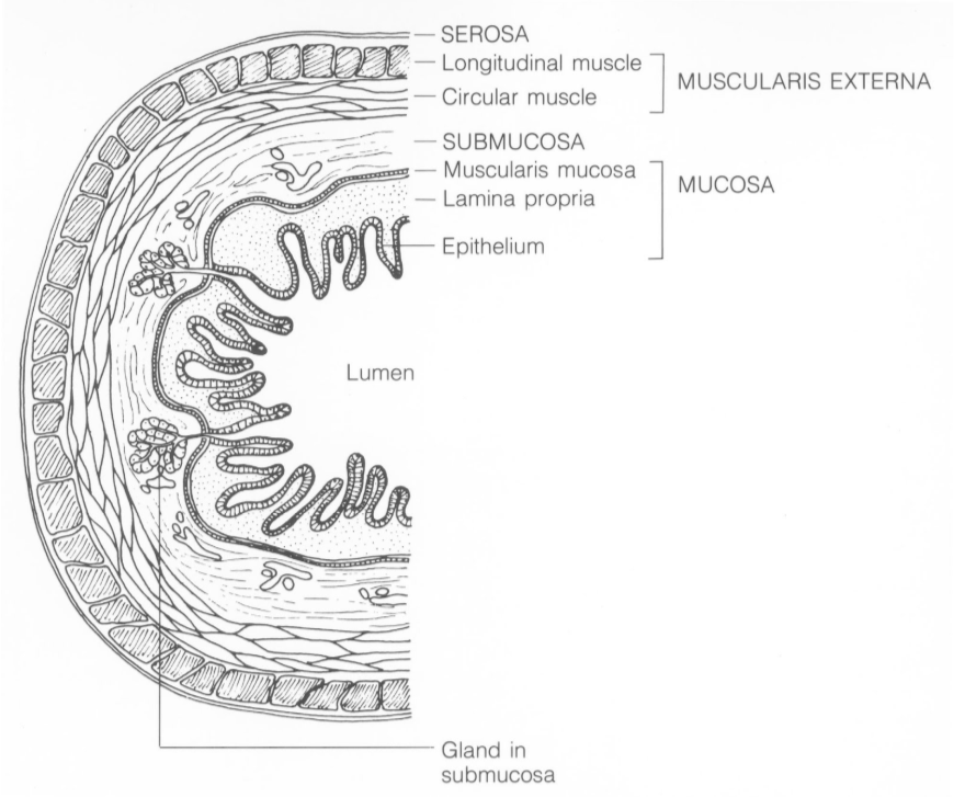
The small intestine consists of many layers, which can be seen in the cross section below.
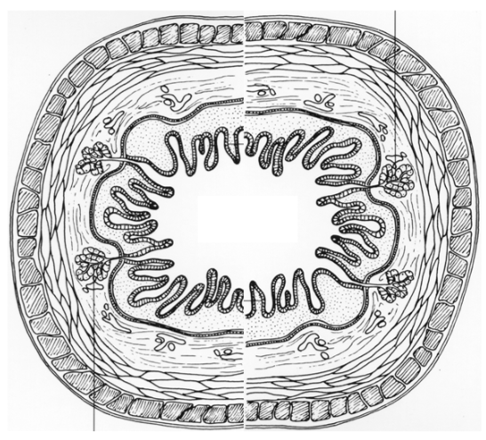
Examining these layers closer, we are going to focus on the epithelium, which comes into contact with the chyme and is responsible for absorption. The lumen is the name of the cavity that is considered “outside the body” that chyme moves through.
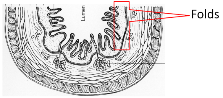
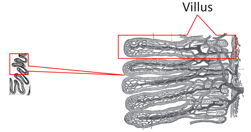
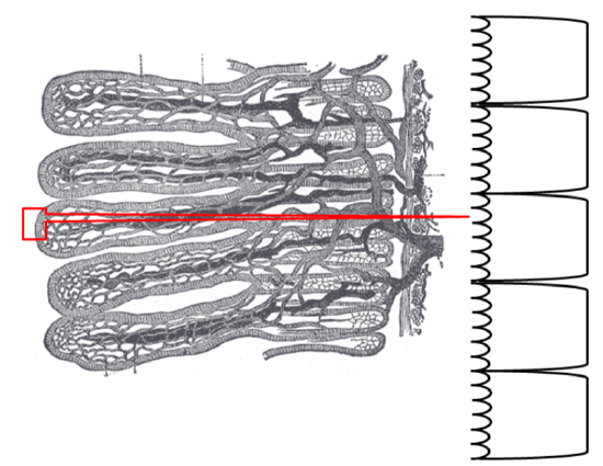
The organization of the small intestine is in such a way that it contains circular folds and finger-like projections known as villi. The folds and villi are shown in the next few figures.

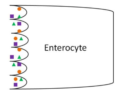
If we were to zoom in even closer, we would be able to see that enterocytes (small intestine absorptive cells) line villi as shown below.
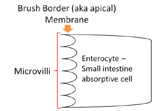
The side, or membrane, of the enterocyte that faces the lumen is not smooth either. It is lined with microvilli, and is known as the brush border (aka apical) membrane, as shown below.
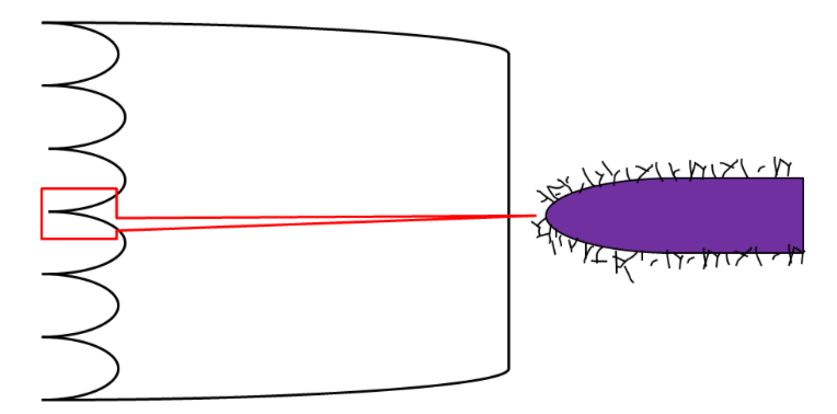
Together these features (folds + villi + microvilli) increase the surface area ~600 times versus if it was a smooth tube5. More surface area leads to more contact with the enterocytes and thus, increased absorption.
Going even closer, we discover that the surface of the microvilli is covered by the hair-like glycocalyx, which is made up of glycoproteins (proteins with carbohydrates attached to them) and carbohydrates as shown below.
Now that you have learned about the anatomy of the small intestine, the following subsections go through the different digestive processes that occur there.
Digestive Hormones, Accessory Organs & Secretions
Before we go into the digestive details of the small intestine, it is important that you have a basic understanding of the anatomy and physiology of the following digestion accessory organs: pancreas, liver, and gallbladder. Digestion accessory organs assist in digestion, but are not part of the gastrointestinal tract. How are these organs involved?
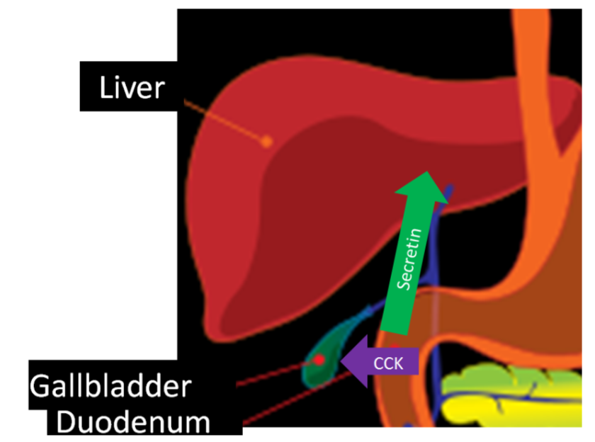
Upon entering the duodenum, chyme causes the release of two hormones from the small intestine: secretin and cholecystokinin (CCK, previously known as pancreozymin) in response to acid and fat, respectively. These hormones have multiple effects on different tissues. In the pancreas, secretin stimulates the secretion of bicarbonate (\(\ce{HCO3}\)), while CCK stimulates the secretion of digestive enzymes. The bicarbonate and digestive enzymes released together are collectively known as pancreatic juice, which travels to the small intestine, as shown below.
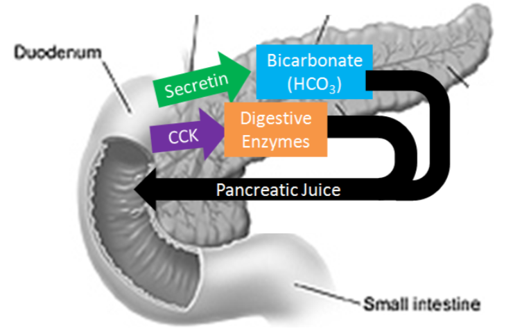
In addition, CCK also stimulates the contraction of the gallbladder causing the secretion of bile into the duodenum.
Pancreas
The pancreas is found behind the stomach and has two different portions. It has an endocrine (hormone-producing) portion that contains alpha and beta cells that secrete the hormones glucagon and insulin, respectively. However, the vast majority of the pancreas is made up of acini that are responsible for producing pancreatic juice. The following video does a nice job of showing and explaining the function of the different pancreatic cells.
Video: How the Body Works - The Pancreas.
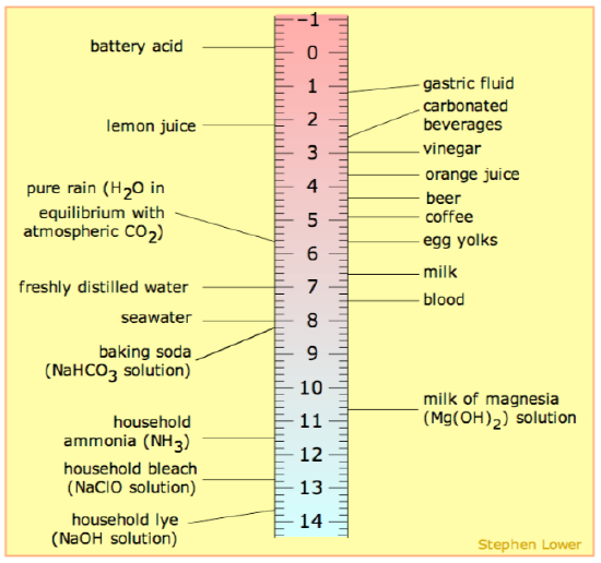
Bicarbonate is a base (high pH) meaning that it can help neutralize acid. You can find sodium bicarbonate (\(\ce{NaHCO3}\), baking soda) on the ruler below to get an idea of its pH.
The main digestive enzymes in pancreatic juice are listed in the table below. Their function will be discussed further in later subsections.
| Enzyme |
|---|
| Pancreatic alpha-amylase |
| Proteases |
| Pancreatic Lipase & Procolipase* |
| Phospholipase A2 |
| Cholesterol Esterase |
*Not an enzyme
Liver
The liver is the largest and most metabolically active internal organ in the body. The figure below shows the liver and the accessory organs position relative to the stomach.

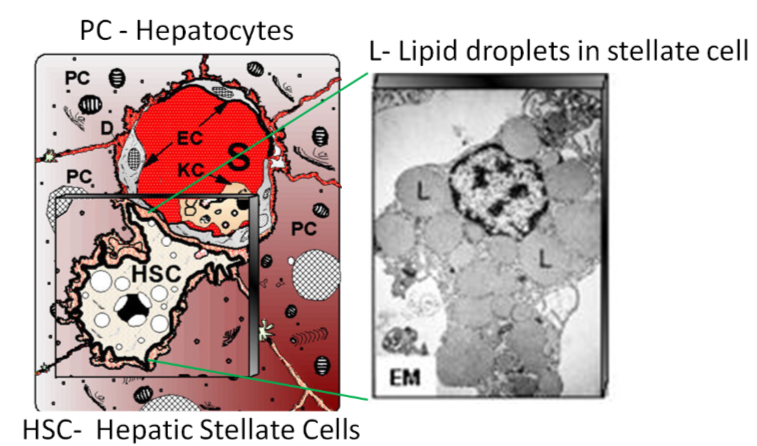
The liver is made up of two major types of cells. The primary liver cells are hepatocytes, which carry out most of the liver’s functions. Hepatic is another term for liver. For example, if you are going to refer to liver concentrations of a certain nutrient, these are often reported as hepatic concentrations. The other major cells are hepatic stellate (also known as Ito) cells. These are lipid-storing cells in the liver. These two cell types are depicted below.
The liver's major role in digestion is to produce bile. This is a greenish-yellow fluid that is composed primarily of bile acids, but also contains cholesterol, phospholipids, and the pigments bilirubin and biliverdin. Bile acids are synthesized from cholesterol. The two primary bile acids are chenodeoxycholic acid and cholic acid. In the same way that fatty acids are found in the form of salts, these bile acids can also be found as salts. These salts have an (-ate) ending, as shown below.
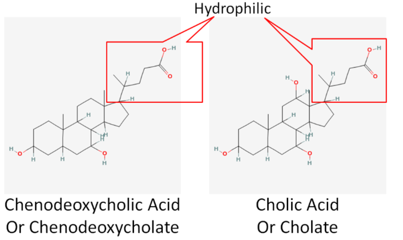
Bile acids, much like phospholipids, have a hydrophobic and hydrophilic end. This makes them excellent emulsifiers that are instrumental in fat digestion. Bile is then transported to the gallbladder.
Gallbladder
The gallbladder is a small sac-like organ found just off the liver (see figures above). Its primary function is to store and concentrate bile made by the liver. The bile is then transported to the duodenum through the common bile duct.
Why do we need bile?
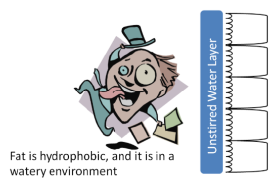
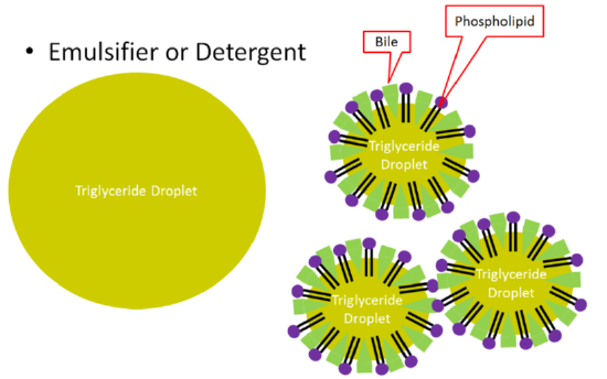
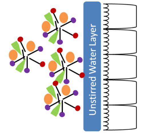
Bile is important because fat is hydrophobic and the environment in the lumen of the small intestine is watery. In addition, there is an unstirred water layer that fat must cross to reach the enterocytes in order to be absorbed.
Here triglycerides form large triglyceride droplets to keep the interaction with the watery environment to a minimum. This is inefficient for digestion because enzymes cannot access the interior of the droplet. Bile acts as an emulsifier. It, along with phospholipids, form smaller triglyceride droplets that increase the surface area that is accessible for triglyceride digestion enzymes, as shown below.
The following video does a nice job of showing and explaining what bile does.
Secretin and CCK also control the production and secretion of bile. Secretin stimulates the flow of bile from the liver to the gallbladder. CCK stimulates the gallbladder to contract, causing bile to be secreted into the duodenum, as shown below.
Carbohydrate Digestion in the Small Intestine
The small intestine is the primary site of carbohydrate digestion. Pancreatic alpha-amylase is the primary carbohydrate-digesting enzyme. Pancreatic alpha-amylase, like salivary amylase, cleaves the alpha 1-4 glycosidic bonds of carbohydrates, reducing them to simpler carbohydrates such as glucose, maltose, maltotriose, and dextrins (oligosaccharides containing 1 or more alpha 1-6 glycosidic bonds). Pancreatic alpha-amylase is also unable to cleave the branch point alpha 1-6 bonds10.
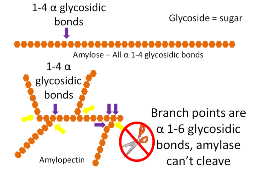
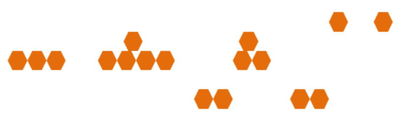
The pancreatic alpha-amylase products, along with the disaccharides sucrose and lactose, then move to the surface of the enterocyte. Here, there are disaccharidase enzymes (lactase, sucrase, maltase) on the outside of the enterocyte. Enzymes, like these, that are on the outside of cell walls, are referred to as ectoenzymes. Individual monosaccharides are formed when lactase cleaves lactose, sucrase cleaves sucrose, and maltase cleaves maltose. There is also another brush border enzyme, alpha-dextrinase. This enzyme cleaves alpha 1-6 glycosidic bonds in dextrins, primarily the branch point bonds in amylopectin. The products from these brush border enzymes are the single monosaccharides glucose, fructose, and galactose that are ready for absorption into the enterocyte10.

Protein Digestion in the Small Intestine
The small intestine is the major site of protein digestion by proteases (enzymes that cleave proteins). The pancreas secretes a number of proteases as zymogens into the duodenum where they must be activated before they can cleave peptide bonds10. This activation occurs through an activation cascade. A cascade is a series of reactions in which one step activates the next in a sequence that results in an amplification of the response. An example of a cascade is shown below.
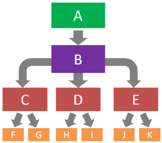
In this example, A activates B, B activates C, D, and E, C activates F and G, D activates H and I, and E activates J and K. Cascades also help to serve as control points for certain process. In the protease cascade, the activation of B is really important because it starts the cascade.
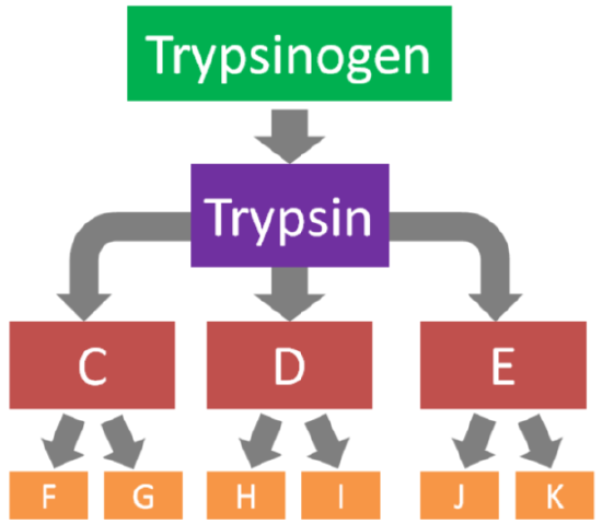
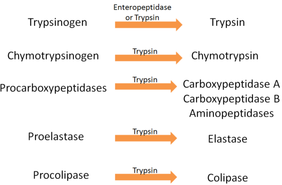
The protease/colipase activation scheme starts with the enzyme enteropeptidase (secreted from the intestinal brush border) that converts trypsinogen to trypsin. Trypsin can activate all the proteases (including itself) and colipase (involved in fat digestion)1 as shown in the 2 figures below.
The products of the action of proteases on proteins are dipeptides, tripeptides, and individual amino acids, as shown below.
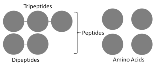
At the brush border, much like disaccharidases, there are peptidases that cleave some peptides down to amino acids. Not all peptides are cleaved to individual amino acid, because small peptides can be taken up into the enterocyte, thus, the peptides do not need to be completely broken down to individual amino acids. Thus the end products of protein digestion are primarily dipeptides and tripeptides, along with individual amino acids10.
Lipid Digestion in the Small Intestine
The small intestine is the major site for lipid digestion. There are specific enzymes for the digestion of triglycerides, phospholipids, and cleavage of esters from cholesterol. We will look at each in this section.
Triglycerides
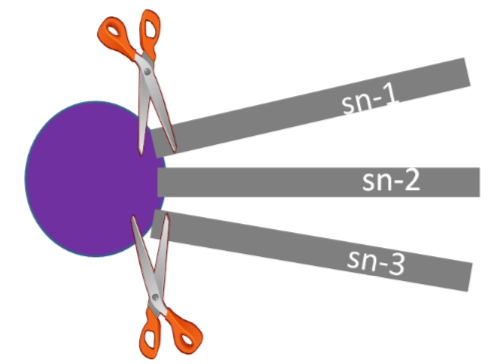
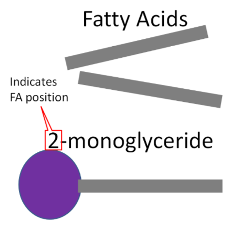
The pancreas secretes pancreatic lipase into the duodenum as part of pancreatic juice. This major triglyceride digestion enzyme preferentially cleaves the sn-1 and sn-3 fatty acids from triglycerides. This cleavage results in the formation of a 2-monoglyceride and two free fatty acids as shown below.
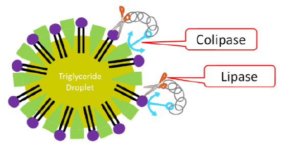
To assist lipase, colipase serves as an anchor point to help pancreatic lipase attach to the triglyceride droplet.
Phospholipids
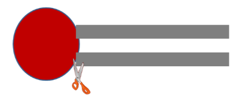
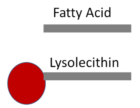
The enzyme phospholipase \(A_2\) cleaves the C-2 fatty acid of lecithin, producing lysolecithin and a free fatty acid.
Cholesterol Esters
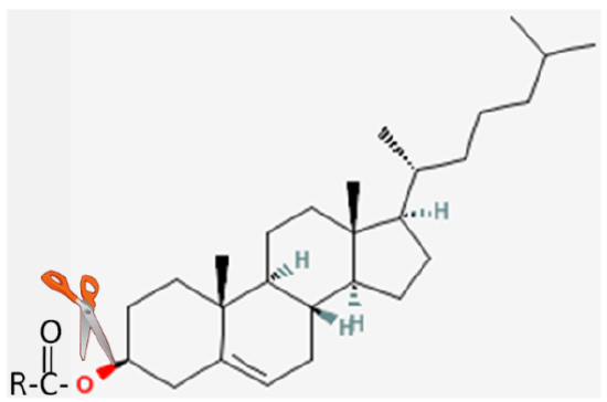
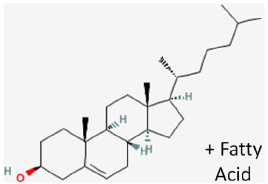
The fatty acid in cholesterol esters is cleaved by the enzyme cholesterol esterase, producing cholesterol and a fatty acid.
Formation of Mixed Micelles
If nothing else happened at this point, the 2-monoglycerides and fatty acids produced by pancreatic lipase would form micelles. The hydrophilic heads would be outward and the fatty acids would be buried on the interior. These micelles are not sufficiently water-soluble to cross the unstirred water layer to get to the brush border of enterocytes. Thus, mixed micelles are formed containing cholesterol, bile acids, and lysolecithin in addition to the 2-monoglycerides and fatty acids, as illustrated below10.
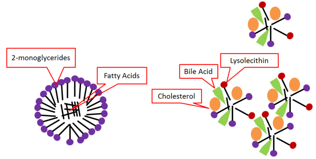
Mixed micelles are more water-soluble, allowing them to cross the unstirred water layer to the brush border of enterocytes for absorption.
References
- commons.wikimedia.org/wiki/Im...atal%C3%A0.png
- Author unknown, NCI, http://visualsonline.cancer.gov/deta...m?imageid=1781
- digestive.niddk.nih.gov/ddiseases/pubs/celiac/
- commons.wikimedia.org/wiki/Image:Gray1061.png
- Byrd-Bredbenner C, Moe G, Beshgetoor D, Berning J. (2009) Wardlaw's perspectives in nutrition. New York, NY: McGraw-Hill.
- Don Bliss, NCI, http://visualsonline.cancer.gov
- upload.wikimedia.org/wikipedi...6/PH_scale.png
- http://www.wpclipart.com/medical/ana..._page.png.html
- http://www.comparative-hepatology.com/content/6/1/7
- Gropper SS, Smith JL, Groff JL. (2008) Advanced nutrition and human metabolism. Belmont, CA: Wadsworth Publishing.