
4.2: Layers of the Skin
- Page ID
- 63383
- Identify the components of the integumentary system
- Describe the layers of the skin and the functions of each layer
- Identify and describe the hypodermis
- Describe the role of keratinocytes and their life cycle
- Describe the role of melanocytes in skin pigmentation
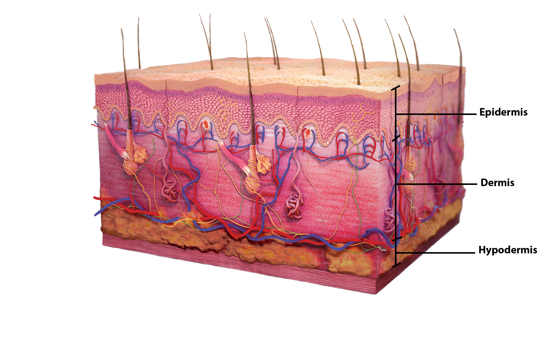
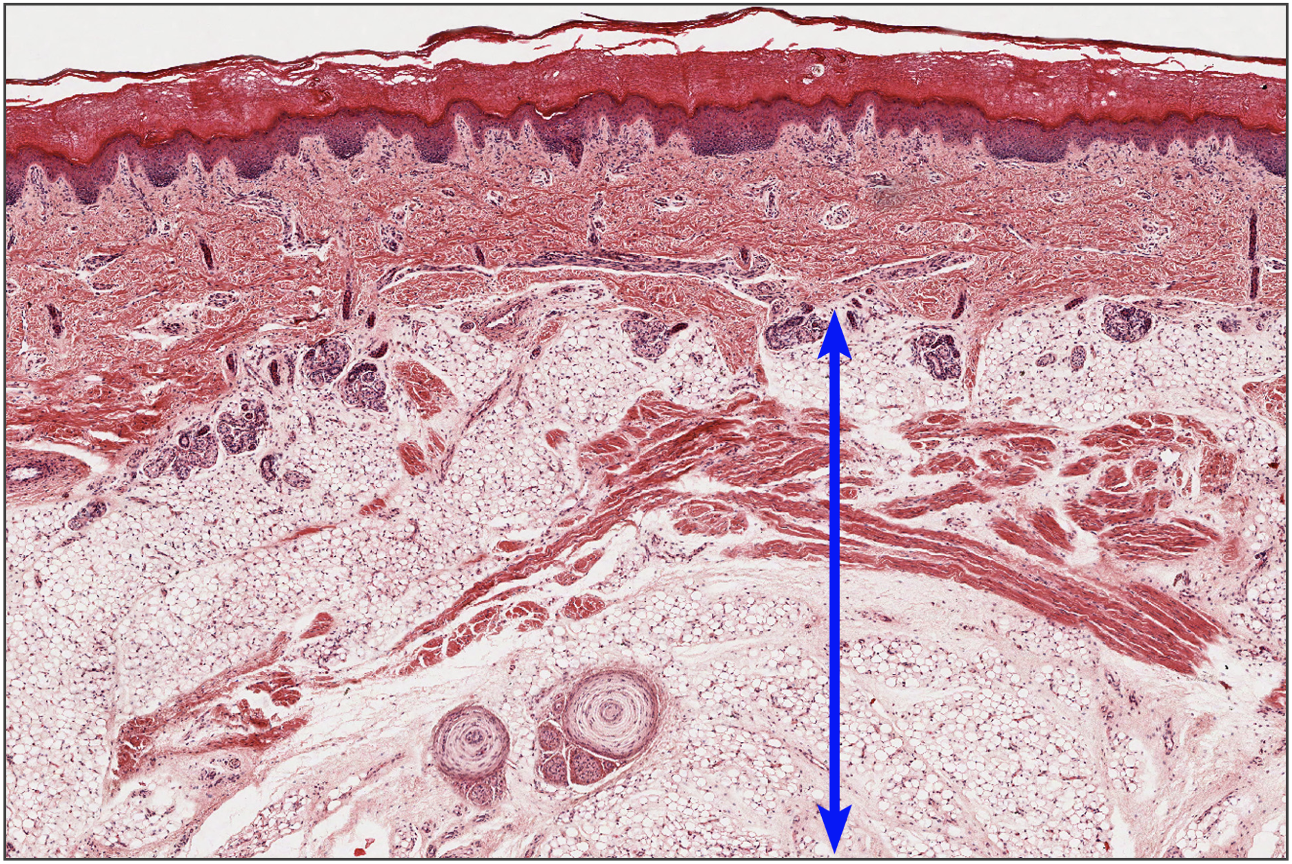
Although you may not typically think of the skin as an organ, it is in fact made of tissues that work together as a single structure to perform unique and critical functions. The skin and its accessory structures make up the integumentary system, which provides the body with overall protection. The skin is made of multiple layers of cells and tissues, which are held to underlying structures by connective tissue (Figure \(\PageIndex{1}\)). The superficial layer, known as the epidermis, is composed primarily of tightly pack epithelial cells. The deeper layer of skin, the dermis, is well vascularized (has numerous blood vessels) and is where several accessory structures, such as hair follicles, sweat glands, and oil glands, can be found. It also has numerous sensory, and autonomic and sympathetic nerve fibers ensuring communication to and from the brain. Below the dermis lies the superficial fascia, or hypodermis, which is not technically part of the skin, but serves to connect the skin to the underlying fascia. This layer is mainly composed of loose, areolar and adipose connective tissues.
The Epidermis
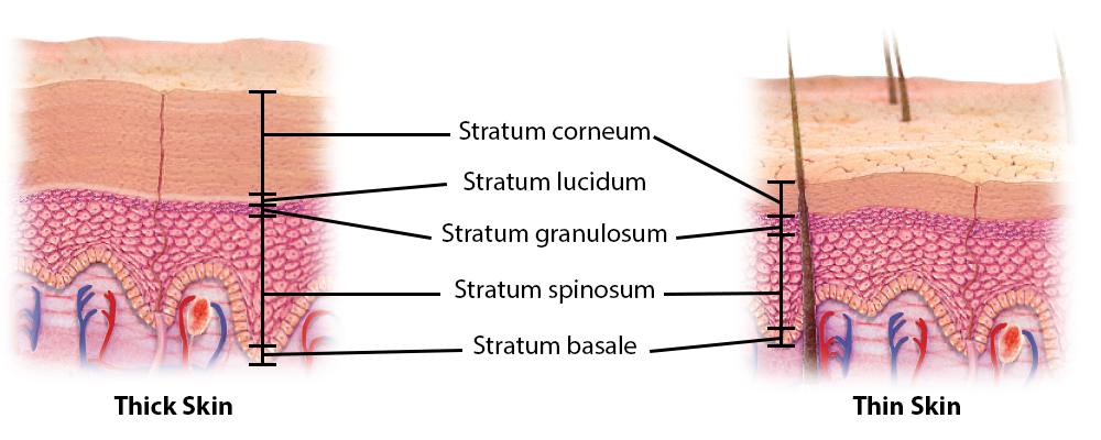
The epidermis is composed of keratinized, stratified squamous epithelium. It is made of four or five layers of epithelial cells, depending on its location in the body. It does not have any blood vessels within it (i.e., it is avascular). From deep to superficial, these layers are:
- the stratum basale (basal layer)
- stratum spinosum (spiny layer)
- stratum granulosum (grainy layer)
- stratum lucidum (clear layer)
- stratum corneum (horny layer)
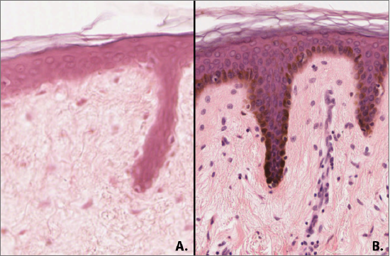
Most of the skin can be classified as thin skin. Thin skin measures between 0.07-0.15 mm from apical to basal side of the epithelium. It has four layers (no stratum lucidum) and has fewer layers of cells in the stratum corneum. “Thick skin” is found on the palms of the hands and the soles of the feet, where it measures approximately 1.5 mm deep. It has a fifth layer, called the stratum lucidum as well as more cells in the stratum corneum that contribute to extra depth. (Figure \(\PageIndex{2}\)).

.png?revision=1&size=bestfit&width=259&height=400)
The majority of cells in the epidermis are called keratinocytes. A keratinocyte is a cell that manufactures and stores the protein keratin. Keratin is an intracellular fibrous protein that gives hair, nails, and skin their hardness and water-resistant properties. The keratinocytes on the apical surface are dead and regularly slough away, being replaced by cells from the deeper layers.
Stratum Basale
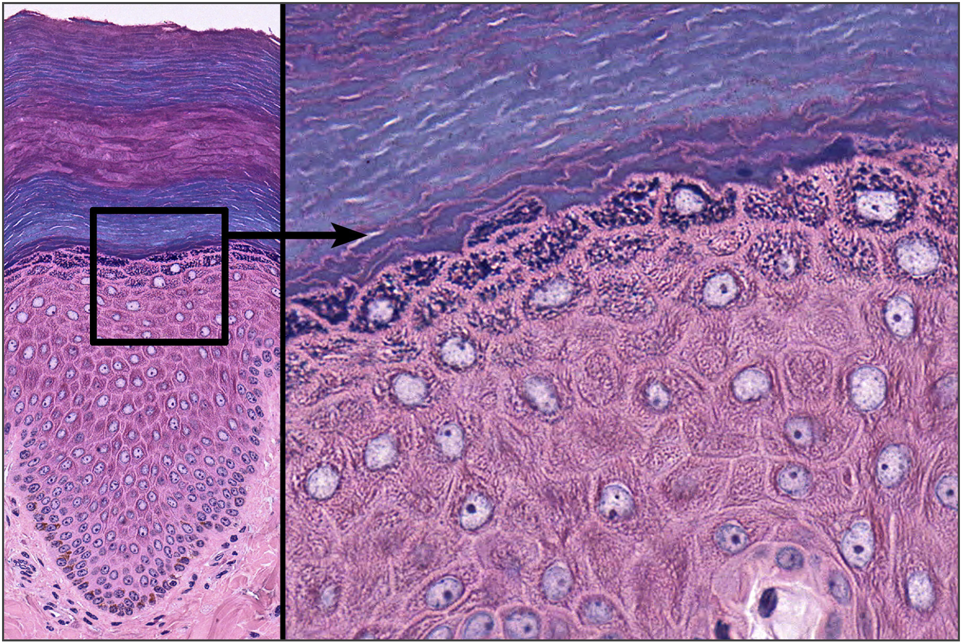
The stratum basale (also called the stratum germinativum) is the deepest epidermal layer and attaches the epidermis to the basal lamina, below which lie the layers of the dermis. The cells in the stratum basale bond to the dermis via intertwining collagen fibers, referred to as the basement membrane. A finger-like projection, or fold, known as the dermal papilla (plural = dermal papillae) is found in the superficial portion of the dermis. Dermal papillae increase the strength of the connection between the epidermis and dermis; the greater the surface area conveyed by folding, the stronger the connections made (Figure \(\PageIndex{3}\)).
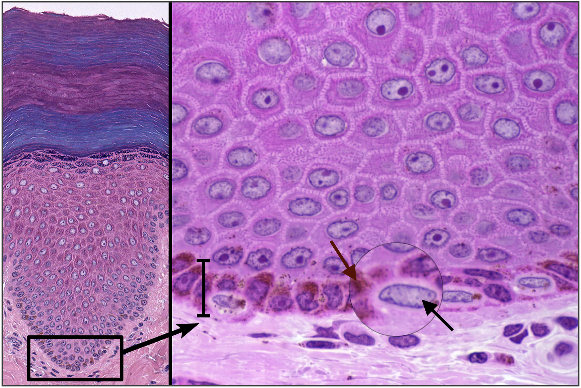
Figure \(\PageIndex{3}\): Stratum Basale. The stratum basale contains a layer of keratinocytes with melanocytes and Merkel cells dispersed throughout. In the enlargement you can see four melanocytes (at arrow - nuclei are more horizontal) and the reddish-brown melanin granules. LM @200x and LM @1000x (Image credit: "Stratum Basale" by Jennifer Lange, micrographs provided by Virginia Commonwealth University under CC-BY-NC-SA 4.0)
The stratum basale is a single layer of cells primarily made of basal cells. A basal cell is a cuboidal-shaped stem cell that is a precursor of the keratinocytes of the epidermis. All of the keratinocytes are produced from this single layer of cells, which are constantly going through mitosis to produce new cells. As new cells are formed, the existing cells are pushed superficially away from the stratum basale. Two other cell types are found dispersed among the basal cells in the stratum basale. The first is a Merkel cell, also known as a tactile cell, which functions as a receptor and is responsible for stimulating sensory nerves that the brain perceives as touch. These cells are especially abundant on the surfaces of the hands and feet. The second is a melanocyte, a cell that produces the pigment melanin. Melanin gives hair and skin its color, and also helps protect the living cells of the epidermis from ultraviolet (UV) radiation damage.
In a growing fetus, fingerprints form where the cells of the stratum basale meet the papillae of the underlying dermal layer (papillary layer), resulting in the formation of the ridges on your fingers that you recognize as fingerprints. Fingerprints are unique to each individual and are used for forensic analyses because the patterns do not change with the growth and aging processes.
Stratum Spinosum
As the name suggests, the stratum spinosum is spiny in appearance due to the protruding cell processes that join the cells via an anchoring junction common in epithelial tissues called a desmosome. The desmosomes interlock with each other and strengthen the bond between the cells. It is interesting to note that the “spiny” nature of this layer is an artifact of the staining process. Unstained epidermis samples do not exhibit this characteristic appearance. The stratum spinosum is composed of eight to ten layers of keratinocytes, formed as a result of cell division in the stratum basale (Figure \(\PageIndex{4}\)). The keratinocytes in the stratum spinosum begin the synthesis of keratin and release a water-repelling glycolipid that helps prevent water loss from the body, making the skin relatively waterproof. As new keratinocytes are produced atop the stratum basale, the keratinocytes of the stratum spinosum are pushed into the stratum granulosum. Wandering among the keratinocytes of this layer is a type of dendritic cell called the Langerhans cell which differentiates from and functions similarly to a macrophage (type of white blood cell) by engulfing bacteria, foreign particles, and damaged cells that occur in this layer.
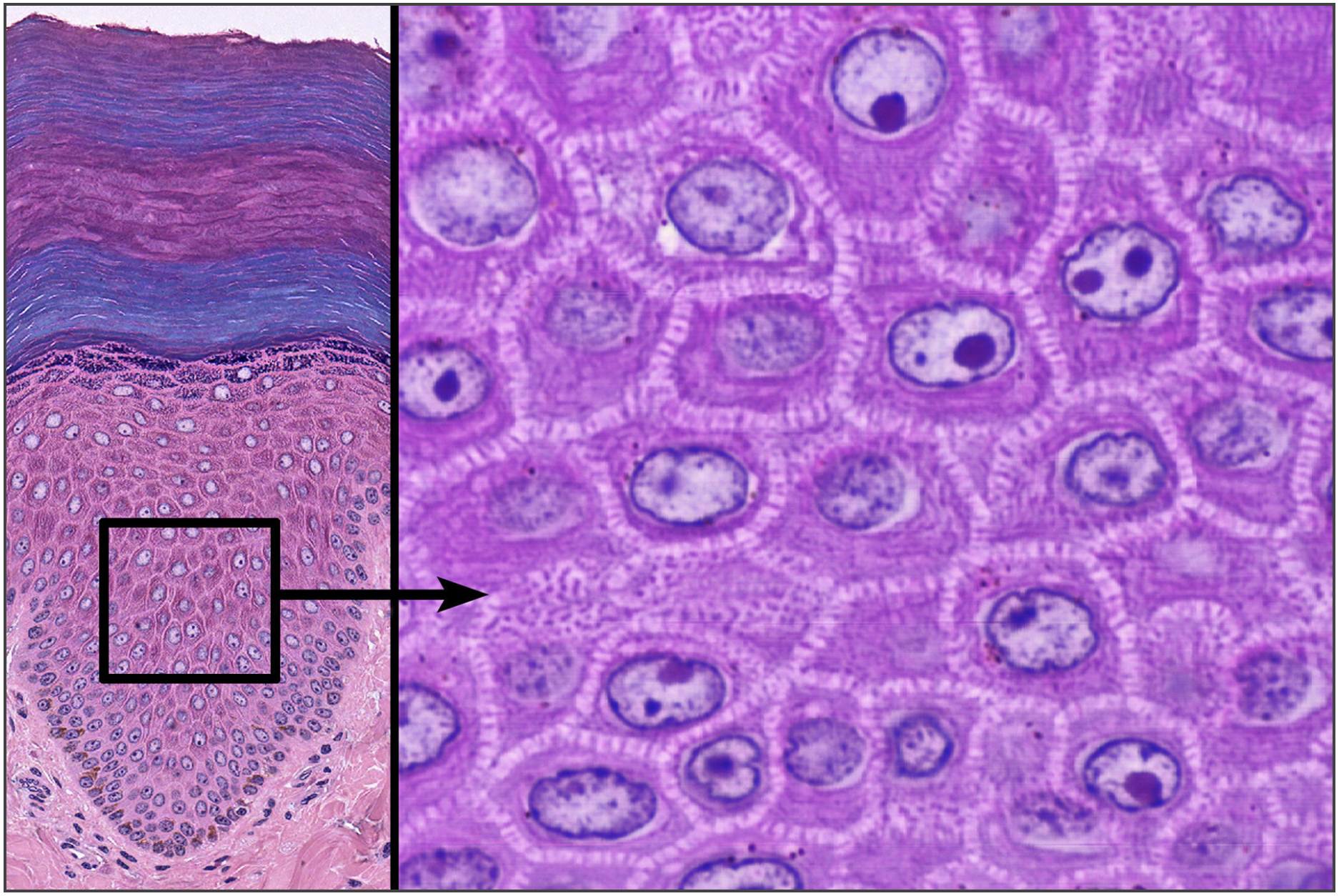
Figure \(\PageIndex{4}\): Intercellular Junctions in the Stratum Spinosum. The enlargement of this slide shows the filaments of the desmosomes anchoring each cell to its neighbors. These "spines" give this layer its name. LM @200x and @1000x. (Image credit: "Stratum Spinosum" micrographs provided by Virginia Commonwealth University under CC-BY-NC-SA 4.0)
Stratum Granulosum
The stratum granulosum has a grainy appearance due to further changes to the keratinocytes as they are pushed from the stratum spinosum. The cells here (three to five layers thick) become flatter and they contain large, dark purple staining (basophilic) granules that give the layer its grainy appearance (see Figure \(\PageIndex{5}\)). The keratohyalin granules contain keratin precursors that eventually aggregate, crosslink, and form bundles. The nuclei and other cell organelles degenerate as the cells die, leaving behind the keratin and lipid-coated cell membranes that will form the stratum lucidum, the stratum corneum, and the accessory structures of hair and nails.
Stratum Lucidum
The stratum lucidum is a smooth, seemingly translucent layer of the epidermis located just above the stratum granulosum and below the stratum corneum. This thin layer of cells is found only in the thick skin of the palms, soles, and digits. The keratinocytes that compose the stratum lucidum are dead and flattened (see Figure \(\PageIndex{6}\)). These cells are densely packed with eleiden, a clear protein rich in lipids, derived from keratohyalin, which gives these cells their transparent (i.e., lucid) appearance and provides a barrier to water.
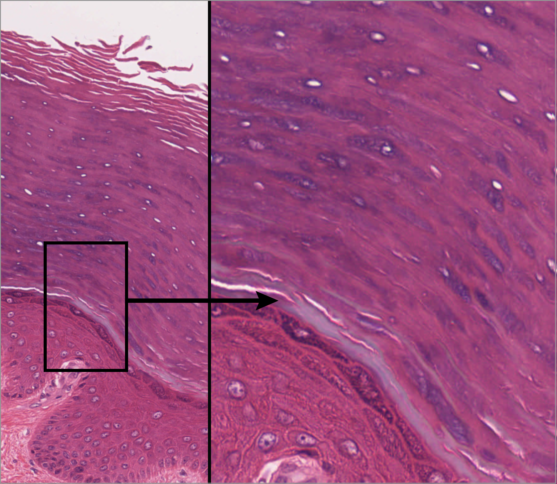
Stratum Corneum
The stratum corneum is the most superficial layer of the epidermis and is the layer exposed to the outside environment (see Figure \(\PageIndex{6}\)). The increased keratinization (also called cornification) of the cells in this layer gives it its name. There are usually 10 to 30 layers (or 35 in thick skin) of anucleate cells in the stratum corneum. This dry, dead layer helps prevent the penetration of microbes and the dehydration of underlying tissues, and provides a mechanical protection against abrasion for the more delicate, underlying layers. Cells in this layer are shed periodically and are replaced by cells pushed up from the stratum granulosum (or stratum lucidum in the case of the palms and soles of feet). The entire layer is replaced during a period of about 4 weeks. Cosmetic procedures, such as microderm abrasion, help remove some of the dry, upper layer and aim to keep the skin looking “fresh” and healthy.
Dermis
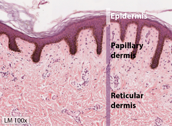
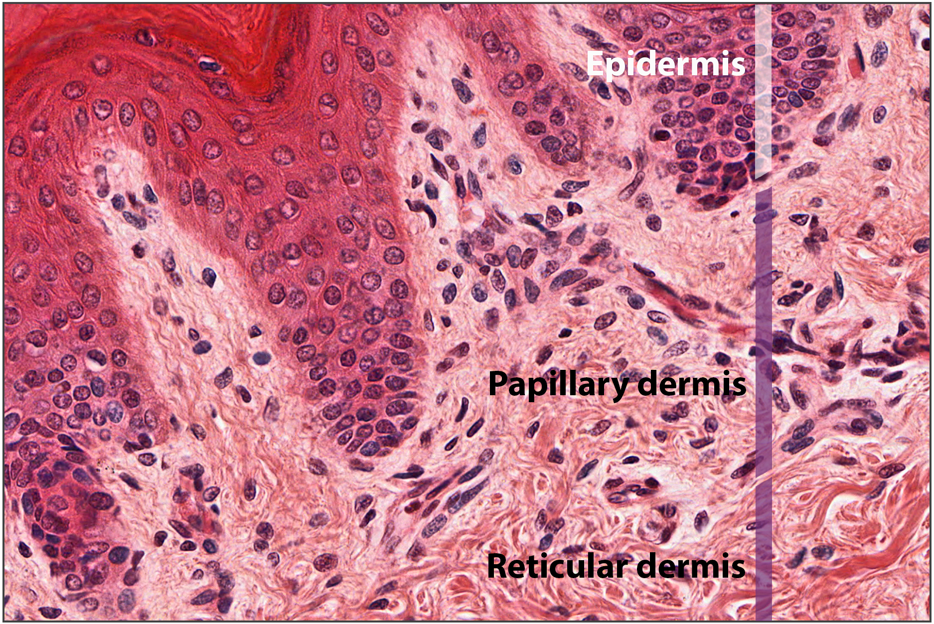
The dermis might be considered the “core” of the integumentary system (derma- = “skin”), as distinct from the epidermis (epi- = “upon” or “over”) and hypodermis (hypo- = “below”). It contains blood and lymph vessels, nerves, and other structures, such as hair follicles and sweat glands. The dermis is made of two layers of connective tissue, the superficial papillary layer and deep reticular layer, that compose an interconnected mesh of elastic and collagen fibers, produced by fibroblasts (Figure \(\PageIndex{7}\)).
Papillary Layer
The papillary layer is made of loose, areolar connective tissue, which means the fibers of this layer form a loose mesh. This superficial layer of the dermis projects up under the stratum basale of the epidermis to form finger-like dermal papillae. Within the papillary layer are fibroblasts, a small number of fat cells (adipocytes), an abundance of small blood vessels, and phagocytes, defensive cells that help fight bacteria or other infections that have breached the epidermis. This layer also contains lymphatic capillaries, nerve fibers, and touch receptors called the Meissner corpuscles. Also known as tactile corpuscles, Meissner corpuscles can be found located in the dermal papillae, and are responsible for sensing light touch. More details on the sensory functions of skin are covered later in this chapter.
Reticular Layer
Underlying the papillary layer is the much thicker reticular layer, composed of dense irregular connective tissue. This layer is well vascularized and has a rich sensory and sympathetic nerve supply. The reticular layer appears reticulated (net-like) due to a tight meshwork of fibers with small amounts of ground substance. Elastic fibers provide some elasticity to the skin, enabling movement. Collagen fibers provide structure and tensile strength, with strands of collagen extending into both the papillary layer and the hypodermis. In addition, collagen binds water to keep the skin hydrated. Collagen injections and Retin-A creams help restore skin turgor by either introducing collagen externally or stimulating blood flow and repair of the dermis, respectively.
Hypodermis
The hypodermis (also called the subcutaneous layer) is a layer directly deep to the dermis that serves to connect the skin to the underlying fascia (fibrous tissue) of the bones and muscles. It is not strictly a part of the skin, although some of the accessory organs of the integument as well as sensory receptors are located in the hypodermis. The hypodermis consists of well-vascularized, areolar connective tissue and adipose tissue that function as a mode of fat storage and provides insulation and cushioning for the skin.
Lipid Storage
The hypodermis is home to most of the fat that concerns people when they are trying to keep their weight under control. Adipose tissue present in the hypodermis consists of fat-storing cells called adipocytes. This stored fat can serve as an energy reserve, insulate the body to prevent heat loss, and act as a cushion to protect underlying structures from trauma.
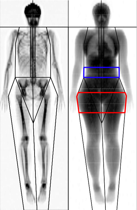
This full body scan highlights the different locations of body fat deposition in males (outlined in blue) and females (outlined in red). (Image credit: "Typical Full Body DXA Scan" from ResearchGate.net is licensed under CC BY 2.0)
Where the fat is deposited and accumulates within the hypodermis depends on hormones (testosterone, estrogen, insulin, glucagon, leptin, and others), as well as genetic factors. Fat distribution changes as our bodies mature and age. Men tend to accumulate fat in different areas (neck, arms, lower back, and abdomen) than do women (breasts, hips, thighs, and buttocks). The body mass index (BMI) is often used as a measure of fat, although this measure is, in fact, derived from a mathematical formula that compares body weight (mass) to height. Therefore, its accuracy as a health indicator can be called into question in individuals who are extremely physically fit.
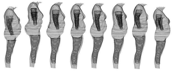
Body Mass Index (BMI) only looks at the ratio of body weight to height, not how that weight is distributed in the body. Each of these eight women have a BMI of 30. (Image credit: Richard2902 at English Wikipedia, Public domain, via Wikimedia Commons)
In many animals, there is a pattern of storing excess calories as fat to be used in times when food is not readily available. In much of the developed world, insufficient exercise coupled with the ready availability and consumption of high-calorie foods have resulted in unwanted accumulations of stored lipids in adipose tissue in many people. Although periodic accumulation of excess fat may have provided an evolutionary advantage to our ancestors, who experienced unpredictable bouts of famine, it is now becoming chronic and considered a major health threat. Recent studies indicate that a distressing percentage of our population is overweight and/or clinically obese. Not only is this a problem for the individuals affected, but it also has a severe impact on our healthcare system. Changes in lifestyle, specifically in diet and exercise, are the best ways to control body fat accumulation, especially when it reaches levels that increase the risk of heart disease and diabetes.
Pigmentation
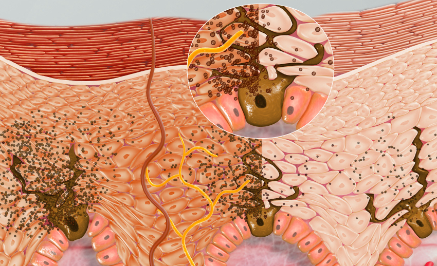
The color of skin is influenced by a number of pigments, including melanin, carotene, and hemoglobin. Recall that melanin is produced by cells called melanocytes, which are found scattered throughout the stratum basale of the epidermis. The melanin is transferred into the keratinocytes via a cellular vesicle called a melanosome (Figure \(\PageIndex{3}\)) and (Figure \(\PageIndex{9}\)).
Melanin occurs in two primary forms: eumelanin exists as black and brown whereas pheomelanin provides a red color. Multiple genes are involved in determining how much and which type of melanin are produced, with dark-skinned individuals produce more melanin than those with pale skin. Exposure to the UV rays of the sun or a tanning salon causes melanin to be manufactured and built up in keratinocytes, as sun exposure stimulates keratinocytes to secrete chemicals that stimulate melanocytes. The accumulation of melanin in keratinocytes results in the darkening of the skin, or a tan. This increased melanin accumulation is protection from UV radiation, which could damage the DNA of epidermal cells and breakdown folic acid, a nutrient necessary for our health and well-being, circulating through the bloodstream in the dermis. In contrast, too much melanin can interfere with the production of vitamin D, an important nutrient involved in calcium absorption, since UV radiation is required for its production. Thus, the amount of melanin present in our skin is dependent on a balance between available sunlight and folic acid destruction, and protection from UV radiation and vitamin D production.
It requires about 10 days after initial sun exposure for melanin synthesis to peak, which is why pale-skinned individuals tend to suffer sunburns of the epidermis initially. Dark-skinned individuals can also get sunburns, but are more protected than are pale-skinned individuals. Melanosomes are temporary structures that are eventually destroyed by fusion with lysosomes; this fact, along with melanin-filled keratinocytes in the stratum corneum sloughing off, makes tanning impermanent.
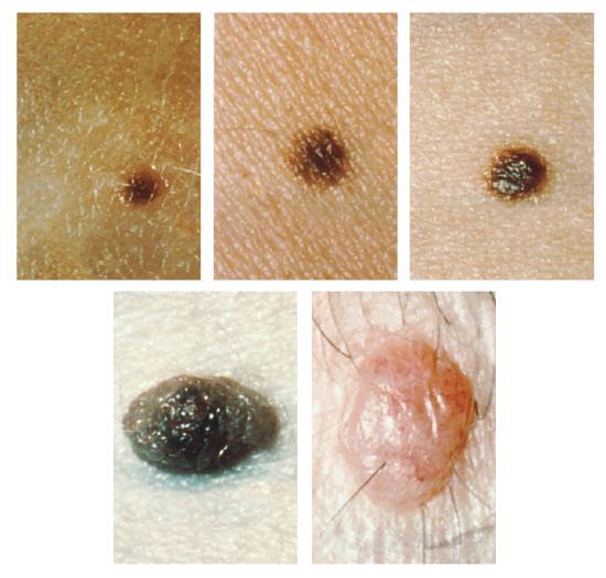
Too much sun exposure can eventually lead to wrinkling due to the destruction of the cellular structure of the skin, and in severe cases, can cause sufficient DNA damage to result in skin cancer. When there is an irregular accumulation of melanocytes in the skin, freckles appear. Moles are larger masses of melanocytes, and although most are benign, they should be monitored for changes that might indicate the presence of cancer (Figure \(\PageIndex{11}\)).
Carotene, an orange/red pigment found in many vegetables such as carrots, can also have an affect on skin pigmentation. Once consumed, carotene is converted by the human body into vitamin A, which in turn is used for vision, immune system function, and to stimulate fibroblasts in the skin. Recall that fibroblasts produce the elastic and collagen fibers of the dermis, so having active fibroblast helps with the firmness of skin. However, the over intake of carotene can cause the skin to turn yellowish-orange in color, as a result of the excess being stored just under the skin.
Another factor that can affect the appearance of skin is the activity of hemoglobin in the blood stream. Hemoglobin is a pigment found in red blood cells that is responsible for carrying oxygen throughout the body. A sudden drop in oxygenation can affect skin color, causing the skin to initially turn ashen (white). With a prolonged reduction in oxygen levels, dark red deoxyhemoglobin (hemoglobin not carrying oxygen) becomes dominant in the blood, making the skin appear blue, a condition referred to as cyanosis (kyanos is the Greek word for “blue”). This happens when the oxygen supply is restricted, as when someone is experiencing difficulty in breathing because of asthma or a heart attack. However, in these cases the effect on skin color has nothing do with the skin’s pigmentation.
Integumentary System
The first thing a clinician sees is the skin, and so the examination of the skin should be part of any thorough physical examination. Most skin disorders are relatively benign, but a few, including melanomas, can be fatal if untreated. A couple of the more noticeable disorders, albinism and vitiligo, affect the appearance of the skin and its accessory organs. Although neither is fatal, it would be hard to claim that they are benign, at least to the individuals so afflicted.
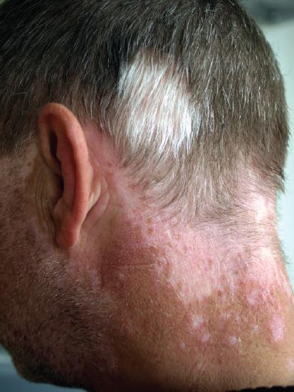
Albinism is a genetic disorder that affects (completely or partially) the coloring of skin, hair, and eyes. The defect is primarily due to the inability of melanocytes to produce melanin. Individuals with albinism tend to appear white or very pale due to the lack of melanin in their skin and hair. Recall that melanin helps protect the skin from the harmful effects of UV radiation. Individuals with albinism tend to need more protection from UV radiation, as they are more prone to sunburns and skin cancer. They also tend to be more sensitive to light and have vision problems due to the lack of pigmentation on the retinal wall. Treatment of this disorder usually involves addressing the symptoms, such as limiting UV light exposure to the skin and eyes. In vitiligo, the melanocytes in certain areas lose their ability to produce melanin, possibly due to an autoimmune reaction. This leads to a loss of color in patches (Figure \(\PageIndex{12}\)). Neither albinism nor vitiligo directly affects the lifespan of an individual.
Other changes in the appearance of skin coloration can be indicative of diseases associated with other body systems. Liver disease or liver cancer can cause the accumulation of bile and the yellow pigment bilirubin, leading to the skin appearing yellow or jaundiced (jaune is the French word for “yellow”). Tumors of the pituitary gland can result in the secretion of large amounts of melanocyte-stimulating hormone (MSH), which results in a darkening of the skin. Similarly, Addison’s disease can stimulate the release of excess amounts of adrenocorticotropic hormone (ACTH), which can give the skin a deep bronze color.
Concept Review
The skin is composed of two major layers: a superficial epidermis and a deeper dermis. The epidermis consists of several layers beginning with the innermost (deepest) stratum basale (germinatum), followed by the stratum spinosum, stratum granulosum, stratum lucidum (when present), and ending with the outermost layer, the stratum corneum. The topmost layer, the stratum corneum, consists of dead cells that shed periodically and is progressively replaced by cells formed from the basal layer. The stratum basale also contains melanocytes, cells that produce melanin, the pigment primarily responsible for giving skin its color. Melanin is transferred to keratinocytes in the stratum spinosum to protect from UV rays.
The dermis connects the epidermis to the hypodermis, and provides strength and elasticity due to the presence of collagen and elastic fibers. It has only two layers: the papillary layer composed of loose connective tissue with papillae that extend into the epidermis and the lower, reticular layer composed of dense irregular connective tissue. The hypodermis, deep to the dermis of skin, is the connective tissue that connects the dermis to underlying structures; it also harbors adipose tissue for fat storage and protection.
Review Questions
Q. The papillary layer of the dermis is most closely associated with which layer of the epidermis?
A. stratum spinosum
B. stratum corneum
C. stratum granulosum
D. stratum basale
- Answer
-
Answer: D
Q. Langerhans cells are commonly found in the ________.
A. stratum spinosum
B. stratum corneum
C. stratum granulosum
D. stratum basale
- Answer
-
Answer: A
Q. The papillary and reticular layers of the dermis are composed mainly of ________.
A. melanocytes
B. keratinocytes
C. connective tissue
D. adipose tissue
- Answer
-
Answer: C
Q. Collagen lends ________ to the skin.
A. elasticity
B. structure
C. color
D. UV protection
- Answer
-
Answer: B
Q. Which of the following is not a function of the hypodermis?
A. protects underlying organs
B. helps maintain body temperature
C. source of blood vessels in the epidermis
D. a site to long-term energy storage
- Answer
-
Answer: C
Critical Thinking Questions
Q. What determines the color of skin, and what is the process that darkens skin when it is exposed to UV light?
- Answer
-
A. The pigment melanin, produced by melanocytes, is primarily responsible for skin color. Melanin comes in different shades of brown and black. Individuals with darker skin have darker, more abundant melanin, whereas fair-skinned individuals accumulate less of a lighter shade of melanin. Exposure to UV irradiation stimulates the melanocytes to produce and secrete more melanin.
Q. Cells of the epidermis derive from stem cells of the stratum basale. Describe how the cells change as they become integrated into the different layers of the epidermis.
- Answer
-
A. As the cells move into the stratum spinosum, they begin the synthesis of keratin and extend cell processes, desmosomes, which link the cells. As the stratum basale continues to produce new cells, the keratinocytes of the stratum spinosum are pushed into the stratum granulosum. The cells become flatter, their cell membranes thicken, and they generate large amounts of the proteins keratin and keratohyalin. The nuclei and other cell organelles disintegrate as the cells die, leaving behind the keratin, keratohyalin, and cell membranes that form the stratum lucidum and the stratum corneum. The keratinocytes in these layers are mostly dead and flattened. Cells in the stratum corneum are periodically shed.
Glossary
- albinism
- genetic disorder that affects the skin, in which there is no melanin production
- basal cell
- type of stem cell found in the stratum basale and in the hair matrix that continually undergoes cell division, producing the keratinocytes of the epidermis
- dermal papilla
- (plural = dermal papillae) extension of the papillary layer of the dermis that increases surface contact between the epidermis and dermis
- dermis
- layer of skin between the epidermis and hypodermis, composed mainly of connective tissue and containing blood vessels, hair follicles, sweat glands, and other structures
- desmosome
- structure that forms an impermeable junction between cells
- elastic fibers
- fibers made of the protein elastin that increase the elasticity of the dermis
- eleiden
- clear protein-bound lipid found in the stratum lucidum that is derived from keratohyalin and helps to prevent water loss
- epidermis
- outermost tissue layer of the skin
- hypodermis
- connective tissue connecting the integument to the underlying bone and muscle
- integumentary system
- skin and its accessory structures
- keratin
- type of structural protein that gives skin, hair, and nails its hard, water-resistant properties
- keratinocyte
- cell that produces keratin and is the most predominant type of cell found in the epidermis
- keratohyalin
- granulated protein found in the stratum granulosum
- Langerhans cell
- specialized dendritic cell found in the stratum spinosum that functions as a macrophage
- melanin
- pigment that determines the color of hair and skin
- melanocyte
- cell found in the stratum basale of the epidermis that produces the pigment melanin
- melanosome
- intercellular vesicle that transfers melanin from melanocytes into keratinocytes of the epidermis
- Merkel cell
- receptor cell in the stratum basale of the epidermis that responds to the sense of touch
- papillary layer
- superficial layer of the dermis, made of loose, areolar connective tissue
- reticular layer
- deeper layer of the dermis; it has a reticulated appearance due to the presence of abundant collagen and elastin fibers
- stratum basale
- deepest layer of the epidermis, made of epidermal stem cells
- stratum corneum
- most superficial layer of the epidermis
- stratum granulosum
- layer of the epidermis superficial to the stratum spinosum
- stratum lucidum
- layer of the epidermis between the stratum granulosum and stratum corneum, found only in thick skin covering the palms, soles of the feet, and digits
- stratum spinosum
- layer of the epidermis superficial to the stratum basale, characterized by the presence of desmosomes
- vitiligo
- skin condition in which melanocytes in certain areas lose the ability to produce melanin, possibly due an autoimmune reaction that leads to loss of color in patches
Contributors and Attributions
OpenStax Anatomy & Physiology (CC BY 4.0). Access for free at https://openstax.org/books/anatomy-and-physiology